IJOCR
ORIGINAL RESEARCH
Hard Tissue Response to Gutta-percha, Resilon and
Epiphany - A Comparative Histopathological Evaluation in
Animal Model
J. Sreeja1, Mini K. John2, C. U. Vivek Chand3, E. Aparna Mohan4, F. Rejula5, K. Madhavadas6
ABSTRACT
Aims and Objectives: The aim of the present study is to assess the biocompatibility of the resin-based Resilon/Epiphany obtu-ration system and to compare it with the reference material gutta-percha (GP) by intraosseous implantation in rabbit femur. The main objectives of the study are as follows: (1) To eval-uate the hard tissue response to the following materials after intraosseous implantation in rabbit femur at different time peri-ods of 1, 4, and 12 weeks. (a) Resilon,(b) Resilon/Epiphany system,(c) The gold standard of obturation materials, GP.(2) To compare the hard tissue response to the above-mentioned materials among each other after intraosseous implantation in rabbit femur at different time periods of 1, 4, and 12 weeks. Materials and Methods: The study was planned on 12 rab-bit models. They were divided into two groups of six animals each - Group 1 and Group 2. In Group 1, three samples of 6 × 2 mm Resilon were implanted into the right side femur (test), and on the left side, three similar dimension samples of control, GP was kept. Similarly, in Group 2, three samples of Resilon coated with Epiphany sealer were kept in the right side (test) and control, GP in the left side. Animals were sacri-ficed at 1, 4, and 12 weeks intervals and bone specimens from femur with implants obtained from all sites. The specimens were processed to obtained sections of 60 mm thickness and stained for microscopic observation. Local tissue reaction and bone-material interface were evaluated histologically in all specimens. The observations were charted as per parameters
1,2,5Associate Professor, 3Endodontist, 4Senior Resident, 6Private Practitioner
1Department of Conservative Dentistry, Government Dental
College, Alappuzha, Kerala, India
2Department of Conservative Dentistry, Government Dental
College, Kottayam, Kerala, India
3Department of Endodontist, Lavender Smiles Dental Practice,
Trivandrum, Kerala, India
4Department of Conservative Dentistry, Government Dental
College, Alappuzha, Kerala, India
5Department of Conservative Dentistry, Government Dental
College, Trivandrum, Kerala, India
6Private Practitioner (Endodontist), Smile Craft Speciality
Dental Clinic, Kesavadasapuram, Trivandrum, Kerala, India Corresponding Author: Dr. Sreeja J, Associate Professor, Department of Conservative Dentistry, Government Dental College, Alappuzha, Kerala, India. Phone: +91-9446551581. e-mail: [email protected]
to statistically analyze the implanted materials and to deter-mine their compatibility.
Results: The results of this study are summarized as follows:(1) All the three specimens of this study, the control, GP, and the test materials, Resilon and Resilon + Epiphany sealer seem to retain their inflammation potential even at 12 weeks.(2) Resilon, Resilon + Epiphany SE sealer, and GP showed no statistically significant difference in inflamma-tory reaction in all three time periods of observation, though a slightly better mean value is given by Resilon followed by GP and then Resilon + Epiphany.(3) With regard to new bone formation, both Resilon and GP showed positive results at all time periods of observations. However, Resilon + Epiphany combination seems to be not having any osteogenic potential within this study period, as mean value of bone formation is 0 by the 12th week.(4) On comparing Resilon and Epiphany
sealer within the limitations of this study, Epiphany, though not provoking severe inflammation, seems to hinder the osteo-genic potential of Resilon.
Conclusion: Within the limitations of the present study, it is con-cluded that the new resin-based Resilon/Epiphany obturation system is comparable to the gold standard, GP as far as local inflammatory reaction is concerned. However, when it comes to osteogenesis, the new solid core/sealer combination seems to be not so promising. This may be due to the sealer Epiphany’s effect, as Resilon alone was found to be highly osteogenic. Keywords: Animal model, Epiphany, Gutta-percha, Hard tis-sue response, Histopathological evaluation, Resilon.
How to cite this article: Sreeja J, John MK, Chand CUV, Mohan EA, Rejula F, Madhavadas K. Hard Tissue Response to Gutta-percha, Resilon and Epiphany - A Comparative Histopathological Evaluation in Animal Model. Int J Oral Care Res 2018;6(2):S1-6.
Source of support: Nil Conflicts of interest: None
INTRODUCTION
Endodontic treatment comprises three main proce-dures: Cleaning and shaping, disinfection, and obtura-tion of the root canal space. The objective of the final phase, obturation, is to eliminate all the pathways between the periodontium and the root canal by sealing the root canal completely with a condensed, bioinert fill-ing material.[1] A number of filling materials have been
ings, due to its inertness, biocompatibility, dimensional stability, compatibility, plasticity when warmed, and ease of removal for post-placement and retreatment.[2,3]
However, one of its disadvantages is lack of true adhe-sion to dentin by itself or through a sealer.
The design and development of a new or novel end-odontic material involves extensive material property testing as well as the evaluation of its biocompatibility. In general, the biocompatibility of a root canal obturat-ing material and its sealer is assessed by a three-step approach. The first step is to screen a new material using a series of in vitro cytotoxicity assays (primary tests). If the material is found to be a non-cytotoxic and non-mutagenic invitro, it can be implanted in subcu-taneous or intraosseous tissues of small animals such as rats and rabbits and the local tissue reaction eval-uated (secondary tests). Finally, the invivo reaction of the target tissues with the material must be evaluated in higher animals or human beings (usage tests).[4] The
present study is conducted to evaluate the reaction of bone to Resilon obturating material and its sealer Epiphany and to compare it with the most accepted root filling material, GP by intraosseous implantation in rabbit femur.
Aims and Objectives
The aim of the present study is to assess the biocompat-ibility of the resin-based Resilon/Epiphany obturation system and to compare it with the reference material GP by intraosseous implantation in rabbit femur.
materials after intraosseous implantation in rabbit femur at different time periods of 1, 4, and 12 weeks. a. Resilon.
b. Resilon/Epiphany system.
c. The gold standard of obturation materials, GP. 2. To compare the hard tissue response to the
above-mentioned materials among each other after intraosseous implantation in rabbit femur at differ-ent time periods of 1, 4, and 12 weeks.
MATERIALS AND METHODS
In the present study, three materials are used for evalu-ating biocompatibility. They are as follows:
1. GP (control)
2. Epiphany points or pellets (resilon) (test material) 3. Epiphany sealer (test material).
0.04 taper GP and Resilon points of #40 sizes were
selected and standard sizes of test materials were made by sectioning 6 mm. The size and shape of specimen are as per the ISO10993recommendations.[5] Ethylene
trioxide sterilized GP and Resilon points were used. Epiphany SE sealer was mixed under aseptic condi-tions according to manufacturer’s instruccondi-tions and was coated directly onto Resilon points before implantation.
Methods of the Study
The experiment was done in the Biomedical Technology Wing, Sree Chitra Institute for Medical Sciences and Technology, Poojappura, Trivandrum, Kerala, India.
Hard tissue response to Gutta-percha IJOCR
The experiment was divided into two parts: 1. Animal implantation and autopsy
2. Preparation of the slides and histopathological evaluation.
Animal Implantation
The experimental protocol consisted of rabbit models for dental implants as per the ISO 10993-6, 1994(E).[5]
Femur implants were done. This model was selected as per F 981 revision.
Experimental Animal Features
A total of 12 young adult albino rabbits weighing not <2.0 kg were chosen. Animals were grouped into two each consisting of six animals/group (one group for testing Resilon and other for Resilon + Sealer). Animals were caged in anodized aluminum fabricated cages and
fed on commercial rabbit feed. Studies were conducted during the 1st, 4th, and 12th weeks.[5]
Study Design
The 12 animals for the study were divided into two groups of six animal models per group.
• Group 1: Resilon as test and GP as control material. • Group 2: Resilon + Epiphany SE sealer as test and
GP as control.
Both Groups 1 and 2 are further divided into three groups corresponding to three time periods of testing, as T1, T2, and T3, i.e., 1, 4, and 12 weeks’ time periods.[6]
Each time period groups had two animal models in it. Each animal received three test implants on the right leg femur bone and three control material on the left leg femur. Hence, total of six implant sites of test and con-trol materials can be identified at each time period in one animal [Figure 3].
Surgical Procedure
The implantation procedure was carried out under aseptic conditions. Rabbits were anesthetized using Atropine (0.5 mg/kg), Diazepam (0.6 mg/kg), and Ketamine (90 mg/kg) + Xylazine (5 mg/kg body weight).[7] The skin of the leg of anesthetized rabbits
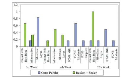
was lightly swabbed using 70% alcohol followed by betadine solution. Incision was made with a No. 15 BP blade and by blunt dissection cortex region of femoral bone is exposed. Then, drill three holes in each femur of 2 mm size were made using tapered fissure burs. GP (control) was implanted snugly into each of the three Figure 2: Comparison of bone-material interface between Resilon +Sealer and Gutta-percha at different weeks
placed, and in other six models of Group 2, Resilon + Epiphany SE sealer kept and wounds sutured [Figure 3]. After the surgical procedure, all the animals were given post-operative care.
Autopsy
At each time intervals 1, 4, and 12 weeks, four ani-mals were euthanized by administering an overdose of anesthetic agent. The femur bone was dislocated at both proximal and distal joints. Bone was cleared of soft tissues and implant sites identified. These sites were macroscopically examined for any evidence of tissue reaction.
Bones with implants were fixed in 10% buffered for-malin. Sections of bone were cut with a high-speed pre-cision saw to obtain blocks of bone with implant. The blocks were dehydrated in ascending grades of alcohol, defatted in acetone-alcohol mixture, and embedded in polymethylmethacrylate medium. 60 mm thick cross sections were cut, ground, and polished. Sections were stained with Stevenel’s blue and examined by light microscopy and the histological features were recorded. Images were captured using a digital camera.[8]
Statistical Analysis
Data were analyzed using computer software, Statistical Package for the Social Sciences version 10. Data are expressed in its frequency and percentage as well as mean and standard deviation. To elucidate the associ-ations and comparisons between different parameters, Chi-square test was used as non-parametric test. To compare different groups each other, non-parametric Mann–Whitney’s U-test was employed. Kruskal–Wallis ANOVA was also employed for comparing different observations.
Histopathological Observations
Sections were qualitatively evaluated for the location and morphology of implant. The morphology of both implants differs (Groups A and B). Group A implants are grayish and pasty with irregular contour resembling soft material. Group B implants are dark solid with defi-nite contour.
Inflammation is present around the implants in all cases in both groups at the 1st week. Healing of margins
of the cavity in host bone is evident from 4 weeks in both Groups A and B with new woven bone being pres-ent along the host bone in Group 1, whereas in Group 2, healing was shown only in B group.
Group 1
Soft tissue reaction
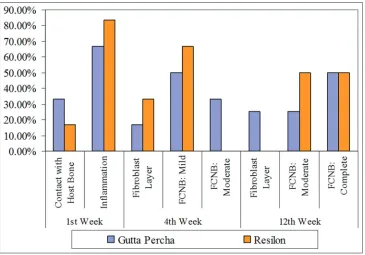
Comparison of soft tissue inflammation grades between Resilon and GP at the 1st week, 4th week, and 12th week
was statistically not significant (P > 0.05).
Bone reaction
Comparison of scores obtained on analyzing the bone-implant material interface shows that in both test and control materials, at the 1st week, 4th week, and
12th week, are statistically not significant by Chi-square
test (P > 0.05) [Figures 1, 4 and 5].
Group 2 Tissue reaction
The Resilon + Epiphany sealer (R+E) showed a simi-lar local tissue reaction as GP at the 1st week and the
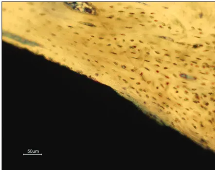
Figure 4: Implant material Resilon after 12 weeks
Hard tissue response to Gutta-percha IJOCR
reaction is not statistically significant. At the 4th week
time period, the observations are statistically significant with P < 0.05. At 12 weeks, inflammation is not statisti-cally significant.
Bone reaction
On assessing the bone-material interface for new bone formation, at the 1st week time, R+E samples show
min-imum contact with host bone, along with inflamma-tion, while GP showed fibroblast with P < 0.01 (highly significant). At T2 and T3 reactions were statistically significant with minimum FCNB for the test materials [Figures 2 and 6].
The above observations can be further analyzed using other statistical tests such as Kruskal–Wallis H and Mann–Whitney U-tests.
Multivariate comparison within Group I (GP and Resilon), comparing weeks and material
Kruskal–Wallis H-test is used for comparing inflam-mation at three time periods of T1, T2, and T3. Resilon shows a statistically significant inflammatory reaction which decreases by the 4th week and then again shows
slight increase. Whereas for GP, not much variation in inflammation is there as time progress.
Kruskal–Wallis H-test comparing bone-material inter-face at three time periods gives a statistically very highly significant value of <0.001 for Resilon, whereas GP group gives a statistically highly significant value of <0.01.
For comparing groups Resilon and GP, Mann– Whitney U-test is used and P values obtained. Accordingly, inflammatory reaction comparison gives a statistically highly significant value at the 4th week
(Resilon being mild), which gives a different result from Chi-square P value. At all other two time periods (T1 and T3), test and control give almost similar results, which are statistically not significant.
The Mann–Whitney U values for assessing bone-ma-terial interface of both Resilon and GP give similar results which are statistically not significant.
Multivariate comparison within Group II (GP and Resilon + Sealer), comparing weeks and material
Kruskal–Wallis H-test is used for comparing inflamma-tion at three time periods of T1, T2, and T3 in A (test) and B (control) groups and P values obtained. Group A gives mean values in the range of mild-to-moderate inflammatory reaction. For both groups, P values are statistically not significant (>0.05).
Kruskal–Wallis H-test comparing bone-material interface at three time periods gives a statistically sig-nificant value of <0.05 for R+E group and a statistically highly significant value of <0.01 for GP. For R+E group, the mean values at three time periods are 0.33, 1.17, and 0.00, which signify that there is no focal contact with new bone on sides at the end of 12 weeks. Whereas for GP, there is new woven bone formation.
For comparing groups such as Resilon + Epiphany and GP at the same time period, Mann–Whitney U-test is used and P values obtained. Accordingly, inflammatory reaction comparison shows that there is no statistically sig-nificant difference between the two groups at all the three time periods. This observation is different from Chi-square
P value at the 4th week, where it is statistically significant.
Comparison of groups for changes in bone-mate-rial interface gives a statistically significant P < 0.01. At 4 weeks and 12 weeks, this becomes statistically highly significant (Chi-square P < 0.05).
Multivariate Comparison between Tests in Group I A and II A (Resilon and Resilon + Sealer), Comparing Weeks and Material
As in the previous multivariate comparison, here, the test materials of Group 1A and Group 2A are compared. Using Kruskal–Wallis H-test, P values are obtained to compare results of inflammatory reaction of Resilon and Resilon + Epiphany sealer. As per the above table, Resilon shows sta-tistically significant variation with advance of time, whereas R+E gives P > 0.05, which is not statistically significant.
For Resilon, P < 0.001, which is statistically very highly significant, and for R+E, P < 0.05, which is sta-tistically significant, though there is no new bone for-mation at the end of the study period. For Resilon, the mean values go on increasing from 0.83 to 4.5 as time advances to 12 weeks. However, for R+E, the mean value at 1 week is 0.33 which tends to increase slightly up to 1.17 in 4 weeks which signifies inflammation only, and then, in the 12th week, it subsides to just contact
period. At 1 week, inflammation is not statistically significant. Similar results were obtained with T2 & T3. However, comparison of bone-material interface changes shows a statistically highly significant differ-ence between both groups at 12-week time period. In T1 and T2, it is not statistically significant.
DISCUSSION
The present animal study was done for the evaluation of the biocompatibility of the resin-based obturation system (Resilon/Epiphany) in comparing with GP. In this study, GP was the control material to which the test material (Resilon/Epiphany) was compared.
Local Tissue Response
Regarding the local tissue response of the material tested, it had been found that inflammation caused by GP and Resilon showed no significant difference at 1- and 12-week time period. The inflammatory reaction of the tested mate-rial was comparable with each other. This result corrobo-rates with study regarding local tissue response of Resilon and GP.[9,10]With progression of the time, the local response
of tissue toward the material is merely milder in nature and the difference among the materials was not significant.[11]
Analyzing the data of local tissue response of the GP and Resilon-Epiphany sealer, it showed that moderate inflammatory reaction was comparable among GP and Resilon-Epiphany group. Even with progression of time period, tissue response was not statistically significant among the groups.[12]
Bone-Material Interface
In this study, it was observed that both GP and Resilon group promotes new bone formation around the mate-rial and was evident from 12-week time period. When Resilon-Epiphany group was observed, it was found that there is no new bone formation which shows that Epiphany had hindering effect on the new bone forma-tion. With multivariate comparison of both Resilon and Resilon-Epiphany group, the inflammatory reaction in both group has no statistical difference (P > 0.05) at T1, T2, and T3 time periods. Moreover, it had been shown that Epiphany group has sustained inflammatory poten-tial when compared to Resilon group.[13] However,
the inflammatory response is not significant among the group but when the osteogenic potential of the groups was compared it showed that Epiphany group has no ability to form new bone and it was statistically
nature.
The biological basis of root canal therapy is lagging behind the impressive technological advances in end-odontics. However, although required before being pro-moted for clinical use, the majority of the materials lack even basic safety testing, of which the biocompatibility testing of a material is crucial for its use in clinical sce-nario, no single test is used alone for the purpose. To date, only few studies have been conducted to histologically analyze the effect of Resilon-Epiphany system implanta-tion in bone. Hence, the present study to a certain extent helps to evaluate histologically the properties of the novel resin-based root canal filling material, Resilon-Epiphany system compared to the conventional, well-established gold standard, GP. More clinical studies are yet to be per-formed on this resin-based obturation system.
REFERENCES
1. Grossman L, Oliet S, Rio CD. Endodontic Practice. 11th ed.
India: Varghese Publishing House; 1988. p. 242-68.
2. Cohen S, Burns RC. Pathways of the Pulp. 8th ed. New Delhi:
Harcourt India Pvt Ltd. Mosby; 2002. p. 293-364.
3. Schilder H. Filling root canals in three dimensions. Dent Clin North Am 1967;11:723-44.
4. John M. Powers, Ronald LS. Craig’s Restorative Dental Materials. 11th ed. Houstan, Texas, Portland: Elsevier; 2006.
p. 97-130.
5. Gad SC. Safety Evaluation of Medical Devices. Vol. 2. New York: Marcel Dekker, Inc.; 2002. p. 189-98.
6. ISO 10993-6:1994(E) Biological Evaluation of Medical Devices-Part 6: Tests for local effects after implantation; clause 3.2.1, 3.2.2 and 3.2.3
7. Vogel HG. Drugs Discovery and Evaluation. New York: Springer; 1997. p. 736.
8. Bancroft JD, Stevens A. Theory and Practice of Histological Techniques. 3rd ed. New York: Churchil Livingstone; 1990.
p. 309-43.
9. Key JE, Rahemtulla FG, Eleazer PD. Cytotoxicity of a new root canal filling material on human gingival fibroblasts. J Endod 2006;32:756-8.
10. Merdad K, Pascon AE, Kulkarni G, Santerre P, Friedman S. Short-term cytotoxicity assessment of components of the epiphany resin-percha obturating system by indirect and direct contact millipore filter assays. J Endod 2007;33:24-7. 11. Bodrumlu E, Muglali M, Sumer M, Guvenc T. The response
of subcutaneous connective tissue to a new endodontic filling material. J Biomed Mater Res B Appl Biomater 2008;84:463-7. 12. Sousa CJ, Montes CR, Pascon EA, Loyola AM, Versiani MA.
Comparison of the intraosseous biocompatibility of AH plus, endoREZ, and epiphany root canal sealers. J Endod 2006;32:656-62.