Orhan U. Zorba
1, A–D, Sema Sirma
2, C, D, Gulay Ozgon
2, A, D, Emre Salabas
3, C, D,
Ugur Ozbek
2, E, Ates Kadioglu
3, A, B, E, FComparison of Apoptotic Gene Expression Profiles
Between Peyronie’s Disease Plaque and Tunica
Albuginea*
Porównanie profili ekspresji genów apoptotycznych płytki włóknistej
występującej w chorobie Peyroniego i osłonki białawej
1 Department of Urology, Rize University, Rize Medical Faculty, Rize, Turkey
2 Department of Genetics, Institute for Experimental Medical Research (DETAE), Istanbul University, Istanbul,
Turkey
3 Department of Urology, Istanbul University, Istanbul Medical Faculty, Istanbul, Turkey
A – research concept and design; B – collection and/or assembly of data; C – data analysis and interpretation;
D – writing the article; E – critical revision of the article; F – final approval of article; G – other
Abstract
Background. The fibrotic plaques of Peyronie’s disease and other localized fibrotic conditions have been consid-ered to be the result of an abnormal wound healing process. The potential role of regulatory disorders of apoptosis in abnormal wound healing may also play a role in the development of Peyronie’s disease.
Objectives. To examine the phenomenon of apoptosis in Peyronie’s disease, authors quantified differential levels of gene expression of apoptotic proteins, Fas, Fas Ligand, Bcl-2, p53, Caspase 3 and 8 in Peyronie’s plaque and tunica albuginea.
Material and Methods. Eight patients with Peyronie’s disease undergoing surgical correction of the curvature had biopsy specimens taken from both the Peyronie’s plaque and normal tunica albuginea. Messenger RNA expression of the apoptotic proteins in the plaque and normal tunica was measured by reverse transcriptase PCR.
Results. Apoptotic gene expression was lower than the housekeeping gene`s in half of the tunica albuginea samples and two thirds of the plaque samples. Overall mRNA expressions in the plaque were not significantly different from the normal tunica albuginea.
Conclusions. The fibrotic plaques of Peyronie’s disease and other localized fibrotic conditions have been consid-ered to be the result of an abnormal wound healing process. The potential role of regulatory disorders of apoptosis in abnormal wound healing may also play a role in the development of Peyronie’s disease. In this study, the lower expression of apoptotic genes may cause the persistence of collagen producing cells which were up-regulated for unknown reasons and consequently result in plaque formation. Similar expression levels of apoptotic genes in both tunica albuginea and Peyronie’s plaques may be due to the generalized physiopathologic alterations in tunica albuginea that lead to plaque formation at a vulnerable region subjected to recurrent traumas (Adv Clin Exp Med 2012, 21, 5, 607–614).
Key words: apoptosis, fibrosis, Peyronie’s disease, messenger RNA, caspase.
Streszczenie
Wprowadzenie. Płytka włóknista występująca w chorobie Peyroniego i inne zaburzenia zwłókniające miejscowo zostały uznane za wynik nieprawidłowego procesu gojenia ran. Regulacja zaburzeń apoptozy w nieprawidłowym gojeniu się ran może również odgrywać rolę w rozwoju choroby Peyroniego.
Adv Clin Exp Med 2012, 21, 5, 607–614 ISSN 1899–5276
ORIGINAL PAPERS
© Copyright by Wroclaw Medical University
Peyronie’s disease (PD) is a localized connec-tive tissue disorder leading to the formation of fi-brous plaques in tunica albuginea (TA) and erectile tissue. These plaques contain excessive amounts of collagen and fibroelastic proliferation with a swell-ing of the extracellular matrix that leads to differ-ent degrees of curvature and narrowing as well as penile pain and erectile dysfunction [1]
The etiology of PD is poorly understood. The favorite hypothesis is penile trauma. The bend-ing stresses happenbend-ing in the coitus are believed to result in delamination of the tunical fibers and consequently in microhemorrhages, acute, and subsequently chronic inflammation and finally scar formation [2]. The fibrous plaque of PD is as-sumed to develop from an inelastic scar tissue, the result of an abnormal healing process, resembling its counterparts in the other parts of the body [3]. The development of fibrotic plaque involves both the proliferation of myofibroblasts from the fibro-blast population normally present in penile TA
and stimulation of collagen synthesis [4].The role
of myofibroblasts in wound healing is to approxi-mate the edges of the wound and then they are re-moved by apoptosis. Their prolonged existence is associated with abnormal wound healing and scar formation [5–7].
Apoptosis is a physiological phenomenon that balances proliferation and cell death. The dysreg-ulation of apoptotic cell death is thought to play an important role in the abnormal wound healing process. There are too many genes to differentiate the inducing and inhibiting ones in the apoptotic process.
Alterations in the expression of some selected genes associated with the apoptotic mechanism in the formation of Peyronie plaques were noted in some studies [8–10].
In this preliminary study, the gene expressions of proteins associated with apoptosis (Fas, Fas Li-gand, Bcl-2, p53, Caspase 3 and 8) were compared in Peyronie plaque and healthy TA.
Material and Methods
Patients
Eight patients (mean age: 49.3 ± 12.3 years) were included in the study. The curvatures had been stable for at least 1 year. The degree of the curvature was calculated after combined intracav-ernous injection of papaverine (60 mg) and phen-tolamine (1 mg) and sexual stimulation (CIS) by the same physician (Ateş Kadıoğlu). None of the patients had diabetes mellitus or hypertension. Erectile dysfunction was seen in 2 patients and managed with oral medication and penile prosthe-sis implantation. The international index of erec-tile function (IIEF) scores of these patients were 7 and 8.
After a circumcising incision, the penis was degloved. With an artificial erection, the loca-tion and severity of the curvature was established. Buck’s fascia along with the dorsal neurovascular bundle were mobilized off of the underlying tunica albuginea with a lateral approach. The plaque was incised at the maximal erectile curvature during an artificial erection. The plaque incision was carried around to the lateral corporal body and to release tension with an inward pointing V on either end. A venous patch was placed into the tunical defect. Artificial erection was performed after the graft was closed in a watertight fashion. Any remain-ing curvature seen after the venous patch was cor-rected with a Nesbitt procedure.
Cel pracy. W celu zbadania zjawiska apoptozy w chorobie Peyroniego autorzy ilościowo porównali stężenie eks-presji genów apoptotycznych białek Fas, FasL, Bcl-2, p53, kaspazy 3 i 8 w płytce włóknistej w chorobie Peyroniego i osłonce białawej.
Materiał i metody. Od ośmiu pacjentów z chorobą Peyroniego, u których przeprowadzono chirurgiczną korektę krzywizny prącia, pobrano wycinki zarówno płytki Peyroniego, jak i prawidłowej osłonki białawej. Ekspresję prze-kaźnikowego RNA białek apoptotycznych płytki i prawidłowej osłonki mierzono za pomocą metody PCR z odwrot-ną transkryptazą.
Wyniki. Ekspresja genów apoptotycznych była mniejsza niż genów metabolizmu podstawowego w połowie próbek osłonki białawej i dwóch trzecich próbek płytki. Ogólna ekspresja mRNA na płytce nie różni się istotnie od ekspresji na osłonce białawej.
Wnioski. Płytka włóknista występująca w chorobie Peyroniego i inne zaburzenia miejscowo zwłókniające zostały uznane za wynik nieprawidłowego procesu gojenia ran. Regulacja zaburzeń apoptozy w nieprawidłowym gojeniu się ran może również odgrywać rolę w rozwoju choroby Peyroniego. W tym badaniu mniejsza ekspresja genów apoptozy mogła spowodować przetrwanie komórek wydzielających kolagen, których liczba zwiększała się z niezna-nych powodów, a tym samym spowodowała tworzenie się płytki. Przyczyną podobnego stężenia ekspresji genów apoptotycznych zarówno białawej osłonki, jak i płytki w chorobie Peyroniego może być ogólna patofizjologiczna zmiana osłonki białawej, która prowadzi do tworzenia płytki we wrażliwej części ciała podatnej na nawracające urazy (Adv Clin Exp Med 2012, 21, 5, 607–614).
Ethic committee approval was received and conforms to the provisions of the Declaration of Helsinki.
Tissue Harvesting, Storage
and RNA Isolation
Excisional biopsies of the Peyronie’s plaque and normal TA were performed during surgery. After excision, tissues were fresh frozen in liquid nitrogen and immediately transferred to cryovials
preserved at –80oC.
The tissues removed were pulverized into a fine powder under liquid nitrogen by using mor-tar and pestle.
Reverse Transcription-Polymerase
Chain Reaction (RT-PCR) Analysis
RNA was extracted from the frozen tissue us-ing an RNeasy fibrous tissue kit (Qiagen, Mary-land, USA). cDNA was synthesized from 1 µg of total RNA using random hexamers [11].
Validation of Relative Gene
Expression by Quantitative
Fluorescent PCR
PCR was carried out with LightCycler, a rapid thermal cycling instrument by Roche (Roche Diag-nostics GmbH, Germany) in capillary glass tubes. Fas, FasL, Bcl-2, Caspase 3, Caspase 8, p53 and
be-ta2-microglobulin (housekeeping gene) products were generated by separate PCR reactions using primers. For each reaction, a 2 μl of cDNA sample was used. Also a 10 μl PCR reaction mix including
3mM MgCl2 5μM of each primer and 1 μl
mas-ter mix (LightCycler FastStart DNA Masmas-ter SYBR Green 1 Roche, Mannheim, Germany) were used with and without 10 pmol of the primers. PCR am-plifications were performed using cDNA specific primers that anneal to sequences in exons on both sides of an intron in order to exclude amplifica-tion of contaminating genomic DNA. Different sets of primers were tested to optimize the cDNA amplification so that the Fas, FasL, Bcl-2, Caspase 3, Caspase 8, p53 and beta 2-microglobulin were amplified under the same PCR conditions.
The amplification program consisted of 1 cycle
at 95oC with a 60-second hold, followed by 45
cy-cles at 95oC with a 10-second hold, annealing
tem-perature at 55oC with a 10-second hold and 72oC
with a 20-second hold. Amplification was followed by melting curve analysis using the program run
for one cycle at 95oC with a 0-second hold, 65oC
with a 10 second hold and 95oC with a 0-second
hold in the step acquisition mode.
Each PCR run included 6 points of the stan-dard curve (5-fold serial dilutions of human blood cDNA), a non-template control with water and unknown cDNA samples.
The concentration of each gene was deter-mined on the basis of a kinetic approach using the LightCycler software. PCR data was analyzed using the LightCycler Software (version 3.4). The
Table 1. Demographic characteristics of the patients with Peyronie’s disease who underwent surgical interventions
Tabela 1. Charakterystyka demograficzna pacjentów z chorobą Peyroniego poddanych zabiegom chirurgicznym Patient
No. (Nr pacjenta)
Age – years (Wiek – lata)
Side, angle of the curvature (Strona, kąt krzywizny)
Comorbidity (Choroby ogól-noustrojowe)
ED Surgery
(Operacje chirur-giczne)
Duration of the curvature – years (Czas występo-wania krzywizny – lata)
1 54 D 70 – – IVP 1
2 46 R 50 – – IVP 2
3 61 V 80 – + IVP 2
4 54 D 90 – – IVP+Plic 3
5 42 L 30, V 20 – – IVP+Plic 3
6 63 R 90, D 45 – + Prost+RFG 3
7 51 L 50 – – IVP+Nesbitt 2
8 24 L 30, V 10 – – IVP+Plic 7
D: Dorsal, V: Ventral, L: Left, R: Right, Comorbidities: Hypertension, Diabetes mellitus, ED: Erectile Dysfunction, IVP: Incisional venous patch, Plic: Plication, Prost: Prosthesis, RFG: Rectus fascia graft.
values for each PCR product were normalized against beta2-microglobulin mRNA to compare their expressions in normal TA and Peyronie’s plaque. Quantitative PCR assays were conducted in duplicate for each sample and a mean value was used to calculate mRNA levels.
Statistical Analysis
A Mann-Whitney-U test was performed to de-termine the difference between gene expressions in TA and Peyronie’s plaque. Statistical calculations were performed using PASW version 18 (SPSS Inc., Chicago, IL, USA). The null hypothesis was rejected if p was < 0.05.
Results
Apoptotic Gene Expressions
in Tunica Albuginea
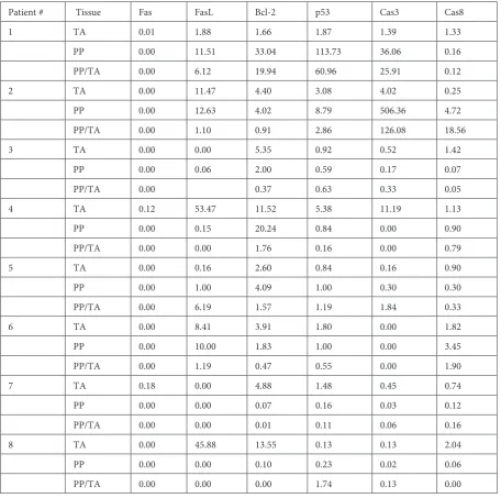
Fas expression was observed in the TA biopsy specimens of 3 patients. The FasL gene was not ap-parently expressed in 2 patients. The median value for FasL expression was found to be 5.14. Expres-sions of Bcl-2, p53 and Caspase 8 genes were de-tected in all patients with corresponding median values of 4.63, 1.64, and 1.23, respectively. The Caspase 3 gene was expressed in all but one patient with a median value of 0.48 (Tables 2 and 3). Table 2. Comparative gene expressions in tunica albuginea (TA) and Peyronie’s plaque (PP). Cas3: Caspase 3, Cas8: Caspase 8
Tabela 2. Porównanie ekspresji genów w białawej osłonce (TA) i płytce włóknistej Peyroniego (PP). Cas3: kaspaza 3, Cas8: kaspaza 8
Patient # Tissue Fas FasL Bcl-2 p53 Cas3 Cas8
1 TA 0.01 1.88 1.66 1.87 1.39 1.33
PP 0.00 11.51 33.04 113.73 36.06 0.16
PP/TA 0.00 6.12 19.94 60.96 25.91 0.12
2 TA 0.00 11.47 4.40 3.08 4.02 0.25
PP 0.00 12.63 4.02 8.79 506.36 4.72
PP/TA 0.00 1.10 0.91 2.86 126.08 18.56
3 TA 0.00 0.00 5.35 0.92 0.52 1.42
PP 0.00 0.06 2.00 0.59 0.17 0.07
PP/TA 0.00 0.37 0.63 0.33 0.05
4 TA 0.12 53.47 11.52 5.38 11.19 1.13
PP 0.00 0.15 20.24 0.84 0.00 0.90
PP/TA 0.00 0.00 1.76 0.16 0.00 0.79
5 TA 0.00 0.16 2.60 0.84 0.16 0.90
PP 0.00 1.00 4.09 1.00 0.30 0.30
PP/TA 0.00 6.19 1.57 1.19 1.84 0.33
6 TA 0.00 8.41 3.91 1.80 0.00 1.82
PP 0.00 10.00 1.83 1.00 0.00 3.45
PP/TA 0.00 1.19 0.47 0.55 0.00 1.90
7 TA 0.18 0.00 4.88 1.48 0.45 0.74
PP 0.00 0.00 0.07 0.16 0.03 0.12
PP/TA 0.00 0.00 0.01 0.11 0.06 0.16
8 TA 0.00 45.88 13.55 0.13 0.13 2.04
PP 0.00 0.00 0.10 0.23 0.02 0.06
Apoptotic Gene Expressions
in Peyronie’s Plaque
The Fas gene was not expressed in any of the 8 Peyronie’s plaques. FasL was expressed in all but 2 patients, with a median value of 0.57. The Bcl-2 gene was expressed in all patients with a median value of 3. In one patient the expression of the p53 gene in his Peyronie’s plaque was increased approximately 62 fold. This level of increased p53 expression (median: 0.91) was not detected in the remainder of the patients. Although Caspase 3 was expressed at an extremely higher level in one pa-tient (126 fold), when considering all the papa-tients with Peyronie’s plaque, its expression remained at a relatively lower level. In 2 patients its expression was not observed (median value: 0.09). Caspase 8 gene was expressed in all patients with a median value of 0.23 (Table 2 and 3).
An increase of more than 1.5 fold was detected in 10 and 13 out of 40 apoptotic genes in Peyronie’s plaque and TA relatively in comparison. However 17 genes were similarly expressed in both tissues.
As for gene expressions in the plaque, 3 of 10 genes which were expressed in higher amounts were detected in patients #1 and 2, while in patient #5, two genes and in patients #6 and 8, one gene was expressed more than 1.5-fold
Comparative Evaluation of
Apoptotic Gene Expressions
in Tunica Albuginea, and
Peyronie’s Plaque
A statistically significant difference was not detected between the expression of the above-mentioned genes in Peyronie’s plaque and TA (Table 3).
Discussion
Similarities between the pathophysiologic fea-tures of Peyronie’s disease and abnormal wound healing have suggested the possible role of mecha-nisms of abnormal wound healing in the develop-ment of Peyronie’s plaque [2].
During normal wound healing, scar tissue forms as a result of a decrease in the cellularity of hyper-cellular granulation tissue. A decrease in hyper-cellularity due to apoptosis has been thought to have a major role during the transition between granulation tis-sue and scar [12]. Fas, Fas ligand, caspase cascade and the p53 and BCL2 gene families play important roles in the genetic regulation of apoptosis.
Although similarities between the pathophysi-ological mechanisms of Peyronie’s disease and abnormal wound healing have been established, there are only a limited number of studies evaluat-ing the apoptotic processes in Peyronie’s disease. Still, a limited number of studies have investigated Table 3. Expressions of apoptotic genes inPeyronie’s plaque (PP), and tunica albuginea (TA). Cas3: Caspase 3, Cas8: Caspase 8
Tabela 3. Ekspresja genów apoptotycznych białawej osłonki (TA) i płytki włóknistej Peyroniego (PP). Cas3: kaspaza 3, Cas8: kaspaza 8
N Min Max Mean SD Median P
Fas (TA) 8 0 0.1800 0.0382 0.0709 0 0.234
Fas (PP) 8 0 0 0 0 0
FasL (TA) 8 0 53.4662 15.1582 21.8160 5.1425 0.574
FasL (PP) 8 0 12.6326 4.4187 5.8175 0.5738
Bcl-2 (TA) 8 1.6570 13.5548 5.9829 4.2517 4.6391 0.442
Bcl-2 (PP) 8 0.0725 33.0384 8.1718 11.9946 3.0086
p53 (TA) 8 0.1323 5.3761 1.9371 1.6405 1.6413 0.574
p53 (PP) 8 0.1570 113.7304 15.7911 39.6775 0.9180
Cas 3 (TA) 8 0 11.1917 2.2330 3.8535 0.4855 0.505
Cas 3 (PP) 8 0 506.3645 67.8663 177.6266 0.0975
Cas 8 (TA) 8 0.2543 2.0351 1.2027 0.5791 1.2306 0.234
expressions of apoptotic genes in Peyronie’s dis-ease.
Present findings have revealed that the Fas re-ceptor gene involved in the first step of extrinsic apoptosis is not expressed (or expressed in scarce amounts) in TA or Peyronie’s plaque. Increased Fas gene expression induces proliferation, and its decreased levels trigger apoptosis of fibroblasts [13, 14]. In these studies, Fas ligand levels have not been investigated.
In this study, Fas ligand was expressed in rela-tively higher amounts in TA when compared with its receptor (Fas). Although not to the extent seen in TA, expression of Fas ligand also increased in Peyronie’s plaque relative to its receptor. In a study performed on mice, higher rates of Fas, FasL and apoptosis were detected in the infarct region dur-ing the acute post-myocardial infarction (MI) pe-riod (1st week). In mice devoid of Fas receptors,
apoptosis could not be detected during the post-MI period despite the presence of FasL genes [15]. Despite higher levels of Fas L expression in tumor cells of various types of cancers, in many individu-als these tumors have been demonstrated to be re-sistant to apoptosis, and protect themselves from the counter-effects of the immune system by in-ducing apoptosis in immune cells [16–21]. In light of the above-mentioned information, the data au-thors have obtained suggests that in the absence of Fas receptor, Fas L is not able to induce apoptosis which might lead to plaque formation.
With its proapoptotic, antiapoptotic members, the Bcl-2 gene family has an important role in the regulation of intrinsic apoptotic processes. Bcl-2 is an antiapoptotic member of the gene family. In Bcl-2 expression analyses conducted in cultures of myofibroblasts, recruited from hypertrophic scar and normal wound healing sites, relatively higher rates of expression have been found in the hyper-trophic scar tissue [22]. In 19 of 20 keloid tissues where analysis of Bcl-2 was conducted, higher ex-pression of the Bcl-2 gene was detected in keloid
tissues [23].Like the studies mentioned above, in
this study increased levels of antiapoptotic Bcl-2 gene expression in normal TA and plaque tissues were detected.
The p53 protein detects DNA lesions such as nucleotide mismatches, fractures of DNA spirals and DNA lesions induced by chemotherapy. It also suppresses cell cycles and induces intrinsic apoptosis leading to cellular death. p53 is the primary media-tor of intrinsic apoptosis. In malignant cases where the p53 gene is lacking, genomic instability and in-hibition of apoptosis have been noted [24]. Aberrant p53 functions might lead to cellular proliferation and cell immortality resulting in the development of malignity. Under appropriate conditions,
dete-rioration in the functionality of p53 might lead to cellular proliferation, and non-malignant
fibroma-tosis [25]. Dysfunctional p53 genes and increases
in the expression of Bcl-2 have been associated with higher rates of cellular proliferation and decreased
cell death rates [26].Similar to the results of
stud-ies about keloids, in this study, expressions of p53 in Peyronie’s plaques were found to be decreased while those of the antiapoptotic Bcl-2 gene were in-creased, suggesting enhanced cellular proliferation. Higher levels of p53 gene expression were detected in 5 patients’ TA. However no significant difference between the expressions of p53 in plaques and TA were observed.
Serine proteases called caspases constitute the last phase of the cell death signal in apoptosis. Cas-pases play key roles in the inhibition and induc-tion of apoptosis. Activainduc-tion of caspase is associ-ated with the induction and its inhibition is linked with the arrest of apoptosis.
In the molecular assessment of apoptosis, the evaluation of caspase 3, which constitutes the final step in the intrinsic and extrinsic pathways, is ex-tremely important. Activation of caspase 3 is the uniting point of the intrinsic and extrinsic apop-tosis pathways. Following induction of apopapop-tosis in normal and keloidal fibroblasts with stauro-sporine, caspase 3 activation was detected only
in normal fibroblasts [27].In this study, caspase
3 expression was detected at a lower rate in the normal TA of 5 patients and plaque formation of 6 patients, which suggests the general presence of apoptotic inhibition in the tunica albuginea of Peyronie’s patients.
Caspase 8 is the first activated caspase in ex-trinsic apoptosis and plays a role in the activation of caspase 3. When compared with expressions in the TA, the expression of caspase 8 in Peyronie’s plaque was found to be decreased in all but 2 cases. As supported by expressions of Fas and Fas ligand, the extrinsic apoptotic activity in Peyronie’s plaque seems to be at minimal level.
While the role of the inhibition of apoptosis in the pathophysiology of an abnormal wound heal-ing process is almost completely acknowledged, re-sults supporting the possible role of the inhibition of apoptosis could not be obtained in all studies
related to apoptotic gene expression [28].In this
was performed after maturation of the plaque or retrieval of biopsy material during alleviation of the influence of paracrinal and hormonal factors. In their biomolecular study of apoptosis activation in PD, Loreto et al. also found extrinsic apoptosis pathway induced, which could underpin plaque stabilization and the halting of fibrosis progression in maturated plaque.
In 2 patients the authors detected that 6 of 10 apoptotic genes were expressed at higher rates (> 1.5 fold) in Peyronie’s plaque relative to normal tunica albuginea. In these 2 patients, a pathologic process specific to the plaque region rather than an apoptotic defect in the entire tunica albuginea might be considered. However when the whole patient population was taken into consideration similar gene expressions in both plaque and tunica
albuginea suggest a disorder affecting the entire tu-nica albuginea rather than a localized condition.
Similar expressions in Peyronie’s plaque and tunica albuginea suggest that Peyronie’s plaque is not a localized disease of tunica albuginea, rather it develops from the vulnerable regions of TA ex-posed to deleterious factor(s), trauma being the prominent one.
In spite of the numerous studies emphasiz-ing the association between Peyronie’s disease and abnormal wound healing, only a small number of studies have addressed the role of apoptosis inhi-bition in this process. In conclusion, Peyronie’s disease might be the result of defective apoptosis affecting tunica albuginea. For definite conclu-sions, detailed investigations with larger patient populations should be performed.
References
[1] BrockG, Hsu GL, Nunes L, von Heyden B, Lue TF: The anatomy of the tunica albuginea in the normal penis and Peyronie’s disease. J Urol 1997, 157, 276–281.
[2] Mulhall JP: Expanding the paradigm for plaque development in Peyronie’s disease. Int J Impot Res 2003, 15 Suppl 5, S93–102.
[3] Ehrlich HP: Scar contracture: cellular and connective tissue aspects in Peyronie’s disease. J Urol 1997, 157, 316– 319.
[4] Vernet D, Ferrini GM, Valente GE, Magee TR, Bou-Gharios G, Rajfer J, Gonzalez-Cadavid NF: Effect of nitric oxide on the differentiation of fibroblasts into myofibroblasts in the Peyronie’s fibrotic plaque and its rat model. Nitric Oxide 2002, 7(4), 262–276.
[5] Powei DW, Mifflin RC, Valentich JD, Ceowe SE, Saada JI, West AB: Myofibroblasts: paracrine cells important in health and disease. Am J Physiol 1999, 277, C1–C19.
[6] Walker GA, Guerrero IA, Leinwand LA: Myofibroblasts: Molecular crossdressers, Curr Top Dev Biol 2001, 51, 91–107.
[7] Serini G, Gabbiani G: Mechanisms of myofibroblast activity and phenotypic modulation. Exp Cell Res 1999, 250, 273–283.
[8] Magee TR, Qian A, Rajfer J, Sander CF, Levine AL, Gonzalez-Cadavid DF: Gene expression profiles in the Peyronie’s disease plaque. Urology 2002, 59, 451–457.
[9] Olofsson B: Rho guanine dissociation inhibitors: pivotal molecules in cellular signaling. Cell Signal 1999, 11, 545–554.
[10] Qian A, Meals RA, Rajfer J, Gonzalez-Cadavid NF: Comparison of gene expression profiles between Peyronie’s disease and Dupuytren’s contracture. Urology 2004, 64, 399–404.
[11] Kusec R, Laczika K, Knöbl P, Fried J, Grenix H, Khals P, Linkesch W, Schwarzinger I, Mitterbauer G, Purtscher B, Haas OA, Lechner K, Jaeger U: AML1/ETO fusion mRNA can be detected in remission blood samples of all patients with t(8;21) acute myeloid leukemia after chemotherapy or autologous bone marrow trans-plantation. Leukemia 1994, 8, 735–739.
[12] Desmouliere A, Redard M, Darby I, Gabbiani G: Apoptosis mediates the decrease in cellularity during the transi-tion between granulatransi-tion tissue and scar. Am J Pathol 1995, 146, 56–66.
[13] Freiberg R, Spencer D, Chaote K, Duh H, Schreiber S: Fas signal transduction triggers either proliferation or apoptosis in human fibroblasts. J Invest Dermatol 1997, 108, 215–219.
[14] Aggarwal BH, Singh S, LaPushin R, Totpal K: Fas antigen signals proliferation of normal human diploid fibro-blast and its mechanism is different from tumor necrosis factor receptor. FEBS 1995, 364, 5–8.
[15] Li Y, Takemura G, Kosai K, Takahashi T, Okada H, Miyata S, Yuge K, Nagano S, Esaki M, Khai NC, Goto K, Mikami A, Maruyama R, Minatoguchi S, Fujiwara T, Fujiwara H: Critical Roles for the Fas/Fas Ligand System in Postinfarction Ventricular Remodeling and Heart Failure. Circ Res 2004, 95, 627–634.
[16] Okada K, Komuta K, Hashimoto S, Matsuzaki S, Kanematsu T, Koji T: Frequency of apoptosis of tumor-infil-trating lymphocytes induced by Fas counterattack in human colorectal carcinoma and its correlation with prog-nosis. Clin Cancer Res 2000, 6, 3560–3564.
[17] Zheng HC, Sun JM, Wei ZL, Yang XF, Zhang YC, Xin Y: Expression of Fas ligand and caspase-3 contributes to formation of immune escape in gastric cancer. World J Gastroenterol 2003, 9, 1415–1420.
[19] Saas P, Walker PR, Hahne M, Quiquerez AL, Schnuriger V, Perrin G, French L, Van Meir EG, de Tribolet N, Tschopp J: Fas ligand expression by astrocytoma in vivo: maintaining immune privilege in the brain? J Clin Invest 1997, 99, 1173–1178.
[20] Bennett MW, O’Connell J, O’Sullivan GC, Brady C, Roche D, Collins JK, Shanahan F: The Fas counterattack
in vivo: apoptotic depletion of tumor-infiltrating lymphocytes associated with Fas ligand expression by human
esophageal carcinoma. J Immunol 1998, 160, 5669–5675.
[21] Hahne M, Rimoldi D, Schroter M, Romero P, Schreier M, French LE, Schneider P, Bornand T, Fontana A, Lienard D: Melanoma cell expression of Fas(Apo-1/CD95) ligand: implications for tumor immune escape. Science 1996, 274, 1363–1366.
[22] Moulin V, Larochelle S, Langlois C, Thibault I, Lopez-Valle CA, Roy M: Normal skin wound and hypertrophic scar myofibroblasts have differential responses to apoptotic inductors. J Cell Physiol 2004, 198(3), 350–358.
[23] Ladin D, Hou Z, Patel D: p53 and apoptosis alterations in keloids and keloid fibroblasts. Wound Rep Reg 1998, 6, 28–37.
[24] Ghobrial IM, Witzig TE, Alex A, Adjei C: Targeting Apoptosis Pathways in Cancer Therapy. CA Cancer J Clin 2005, 55, 178–194.
[25] JP Mulhall, MS Anderson, T Lubrano, TV Shankey: Peyronie’s disease cell culture models: phenotypic, geno-typic and functional analyses. Int J Imp Res 2002, 14, 397–405.
[26] Ladin D, Hou Z, Patel D: p53 and apoptosis alterations in keloids and keloid fibroblasts. Wound Rep Reg 1998, 6, 28–37.
[27] Chodon T, Sugihara T, Igawa H, Funayama E, Furukawa H: Keloid-derived fibroblasts are refractory to Fas-mediated apoptosis and neutralization of autocrine transforming growth factor-β1 can abrogate this resistance. Am J Pathol 2000, 157, 1661–1669.
[28] Liu Y, Ren LS, Cen Y: Experimental study of Bcl-2 and Fas gene expression in fibroblast of scar. Zhongguo Xiu Fu Chong Jian Wai Ke Za Zhi 2001, 15(6), 351–353.
[29] Wassermann RJ, Polo M, Smith P, Wang X, Ko F, Robson MC: Differential Production of Apoptosis-Modulating Proteins in Patients with Hypertrophic Burn Scar. J Surg Res 1998, 75, 74–80.
[30] Loreto C, Barbagli G, Djinovic R, Vespasiani G, Carnazza ML, Miano R, Musumeci G, Sansalone S: Tumor necrosis factor-related apoptosis-inducing ligand (trail) and its death receptor (dr5) in Peyronie’s disease. A Biomolecular Study of Apoptosis Activation. J Sex Med 2011, 8, 109–115.
Address for correspondence:
Orhan Unal ZorbaIslampasa Mah. Iskender Sok. No:6/12 Rize 53000
Turkey
E-mail: [email protected] Tel.: 00 90 533 463 48 73
Conflict of interest: None declared