R E S E A R C H
Open Access
Next-generation sequencing identifies
contribution of both class I and II HLA
genes on susceptibility of multiple sclerosis
in Japanese
Kotaro Ogawa
1,2, Tatsusada Okuno
2, Kazuyoshi Hosomichi
3, Akiko Hosokawa
2,4, Jun Hirata
1,5, Ken Suzuki
1,
Saori Sakaue
1, Makoto Kinoshita
2, Yoshihiro Asano
6, Katsuichi Miyamoto
7, Ituro Inoue
8, Susumu Kusunoki
7,
Yukinori Okada
1,9*and Hideki Mochizuki
2Abstract
Background:The spectrum of classical and non-classical HLA genes related to the risk of multiple sclerosis (MS) and neuromyelitis optica spectrum disorder (NMOSD) in the Japanese population has not been studied in detail. We conducted a case-control analysis of classical and non-classical HLA genes.
Methods:We used next-generation sequencing (NGS)-based HLA genotyping methods for mapping risk for 45 MS
patients, 31 NMOSD patients, and 429 healthy controls. We evaluated the association of the HLA variants with the risk of MS and NMOSD using logistic regression analysis and Fisher’s exact test.
Results:We confirmed that HLA-DRB1*15:01 showed the strongest association with MS (P= 2.1 × 10⁻⁵; odds ratio [OR] = 3.44, 95% confidence interval [95% CI] = 1.95–6.07). Stepwise conditional analysis identified HLA-DRB1*04:05, HLA-B*39:01, and HLA-B*15:01 as being associated with independent MS susceptibility (PConditional< 8.3 × 10⁻⁴). With respect to amino acid polymorphisms in HLA genes, we found that phenylalanine at HLA-DQβ1 position 9 had the strongest effect on MS susceptibility (P= 3.7 × 10⁻⁸, OR = 3.48, 95% CI = 2.23–5.43). MS risk at HLA-DQβ1 Phe9 was independent of HLA-DRB1*15:01 (PConditional= 1.5 × 10⁻⁵, OR = 2.91, 95% CI = 1.79–4.72), while HLA-DRB1*15:01 was just significant when conditioned on HLA-DQβ1 Phe9 (PConditional= 0.037). Regarding a case-control analysis for NMOSD, HLA-DQA1*05:03 had a significant association with NMOSD (P= 1.5 × 10⁻⁴, OR = 6.96, 95% CI = 2.55–19.0).
Conclusions:We identified HLA variants associated with the risk of MS and NMOSD. Our study contributes to the understanding of the genetic architecture of MS and NMOSD in the Japanese population.
Keywords:Multiple sclerosis, Neuromyelitis optica spectrum disorder, HLA, Next-generation sequencing
Background
The major histocompatibility complex (MHC) region at 6p21.3 is associated with a wide range of complex human diseases and quantitative traits [1–3]. In auto-immune diseases, such as rheumatoid arthritis, psoriasis,
and type 1 diabetes, fine-mapping studies have identified strong genetic risk variants in the MHC region [4–6].
Multiple sclerosis (MS) is an immune-mediated de-myelinating disease of the central nervous system, which affects approximately 2.3 million people worldwide [7]. Genome-wide association studies (GWAS) have reported that the MHC region is the most significant locus
asso-ciated with MS [8–12]. In Europeans, previous studies
have reported a relationship between classical HLA genes and MS, and found that HLA-DRB1*15:01 had the strongest effect on MS susceptibility [13]. In 2013, Patsopoulos, et al. reported that both class II HLA genes
© The Author(s). 2019Open AccessThis article is distributed under the terms of the Creative Commons Attribution 4.0 International License (http://creativecommons.org/licenses/by/4.0/), which permits unrestricted use, distribution, and reproduction in any medium, provided you give appropriate credit to the original author(s) and the source, provide a link to the Creative Commons license, and indicate if changes were made. The Creative Commons Public Domain Dedication waiver (http://creativecommons.org/publicdomain/zero/1.0/) applies to the data made available in this article, unless otherwise stated. * Correspondence:[email protected]
1
Department of Statistical Genetics, Osaka University Graduate School of Medicine, 2-2 Yamadaoka, Suita 565-0871, Japan
9Laboratory of Statistical Immunology, Immunology Frontier Research Center
(WPI-IFReC), Osaka University, Suita 565-0871, Japan
and class I HLA genes independently confer susceptibil-ity to MS [14].
These researchers used an HLA imputation
method, which computationally imputed HLA vari-ants from single nucleotide polymorphisms (SNP) genotype data obtained from GWAS. They analyzed MS associations with eight classical HLA genes and found HLA-A*02:01 had an independent protective effect on MS. In 2018, high-resolution HLA analysis of MS by using next-generation sequencing (NGS) was conducted in Europeans [15]. The researchers found that specific class I HLA polymorphisms, such as HLA-A*02:01, HLA-C*03:04, and HLA-B*40:01 were protective for MS, independently from HLA-DRB1*15:01. In a Japanese population, Yoshimura et al. reported in 2012 that HLA-DRB1*04:05 and HLA-DPB1*03:01 were significantly associated with MS susceptibility [16]. The authors also reported that MS patients with HLA-DRB1*04:05 have a younger onset of disease. In 2016, Nakamura et al. conducted HLA analysis and found that HLA-DRB1*04:05 and HLA-DRB1*15:01 were risk factors for MS in Japanese individuals [17]. They also found that living at higher latitude and HLA-DRB1*04:05 independently affect the severity of MS.
Neuromyelitis optica spectrum disorder (NMOSD) is an immune-mediated disorder of the central nervous system that mainly affects the optic nerve and spinal cord [18]. The MHC region also contributes to the
genetic architecture of NMOSD like MS [18, 19]. In
Europeans, HLA-DRB1*03:01 has been reported to be significantly associated with NMOSD from a whole-genome sequencing study [19]. In the Japanese popula-tion, HLA-DRB1*16:02 and HLA-DPB1*05:01 have been reported to be positively associated with NMOSD susceptibility, and HLA-DRB1*09:01 was negatively associated with NMOSD susceptibility [20]. These Japanese studies used sequence-specific oligonucleotide hybridization [21], and only two classical HLA genes (HLA-DRB1, HLA-DPB1) were genotyped [16, 20, 21]. Thus, the classical and non-classical HLA genes affect-ing MS and NMOSD risk in the Japanese population have not been studied in detail. In this research, we ex-plored the MHC region comprehensively in the Japanese population. We utilized NGS-based HLA genotyping for
fine-mapping of risk [22–24] for 45 MS patients, 31
NMOSD patients, and 429 controls.
Methods Sample collection
We enrolled 45 MS patients and 31 NMOSD patients. MS and NMOSD cases were collected from three medical insti-tutes in Japan (Osaka General Medical Center, Kindai University Hospital, and Osaka University Hospital) and
assessed in a retrospective manner. All subjects were of Japanese origin and provided written informed consent that was approved by the ethics committee of each hospital. All participants lived in the Kinki region which is located in the central part of Honshu Island, Japan. The Kinki region is lo-cated at latitude 35° north and longitude 135° east. MS was diagnosed by the McDonald 2010 criteria for the diagnosis of MS [25]. NMOSD was diagnosed using the international consensus diagnostic tests for NMOSD [26]. We collected clinical data of patients including sex, age, disease onset, disease duration, MS subtype, Kurtzke’s Expanded Disabil-ity Status Scale (EDSS), and anti-AQP4 antibody (Table1). Case genomic DNA was obtained from peripheral blood.
We extracted DNA using QIAGEN’s QIAampBlood Midi
(Mini) Kit. This is one of the integrated DNA extraction manuals recommended by the HLA typing Quality Work-shop of the Japanese Society for Histocompatibility and Im-munogenetics. We used HLA genotyping data of 429 healthy individuals which were used in a previous HLA study from the Japan Biological Informatics Consortium
(JBIC), as described elsewhere [6, 27]. Control genomic
DNA was obtained from Epstein-Barr virus-transformed B-lymphoblast cell lines of unrelated individuals. Control genomic DNA was extracted using phenol-chloroform ex-traction method. All samples were checked to ensure the quality was sufficient for DNA sequencing. (OD260nm/
OD280nm = 1.8 to 2.0). We received DNA samples from
JBIC.
This study was approved by the ethical committee of Osaka University Graduate School of Medicine.
NGS-based HLA genotyping of classical and non-classical HLA genes
We conducted HLA genotyping (4-digit) of the 16 HLA genes in the subjects, 9 of which were classical HLA
genes (class I: HLA-A, HLA-B, and HLA-C, class II:
DRA, DRB1, DQA1, DQB1,
HLA-DPA1, and HLA-DPB1), seven were non-classical HLA
genes (HLA-E, HLA-F, HLA-G, HLA-DOA, HLA-DOB,
HLA-DMA, andHLA-DMB). HLA gene sequencing was
Table 1Clinical features of multiple sclerosis and neuromyelitis optica spectrum disorder patients
MS (n= 45) NMOSD (n= 31)
Sex (male/female) 10/35 7/24
Age (years) 43.6 (20–65) 52.7 (17–82)
Age at onset (years) 34.3 (5–58) 47.4 (15–81)
Disease duration (years) 9.3 (1–38) 5.3 (0–18)
EDSS 3.1 (0–8) 4.6 (0–9)
IgG index 0.73 (n= 34) 0.57 (n= 13)
Oligo clonal band 21/38 –
carried out using sequence capture technology based upon hybridization between DNA of an adapter-ligated library (KAPA Hyper Prep Kit, Roche, CA, USA) and a biotinylated DNA probe (SepCap EZ choice kit, Roche, CA, USA), designed based upon target sequences of the genes in the MHC region [24]. Paired-end sequence reads were obtained using a MiSeq DNA sequencer (Illumina, the USA). Typing of 4-digit HLA alleles was conducted using Omixon Target Software (Omixon, Hungary) using the IPD-IMGT/HLA Database.
In order to supplement the HLA allele information, which was specific to the Japanese population, and was not correctly implemented in the Omixon, we updated the HLA typing results based on those obtained from a sequence-based typing method. We downloaded the GRCh37 reference genome from Ensembl, a genome browser for vertebrate genomes. GRCh37 reference gen-ome was created by the Gengen-ome Reference Consortium (GRC) in 2009. The sequence reads were aligned to the GRCh37 of the MHC region using BWA (version 0.7.15) [28]. We obtained HLA allele sequences from the IPD and IMGT/HLA databases [29]. We used the GATK Unified Genotyper and Haplotypecaller for variant call-ing [30]. Details of the HLA genotypcall-ing protocol is described in our previous study [31,32].
The statistical framework for analysis
We evaluated the association of the HLA variants with the risk of MS and NMOSD using a logistic regression model, which assumed additive effects of allele dosages on a log-odds scale using the R statistical software (ver-sion 3.4.1) when the number of cases was more than five. Otherwise, we used Fisher’s exact test. We analyzed HLA variants that were more than 0.1% both in case and control samples. We excluded HLA amino acid polymorphisms that were monomorphic in cases or controls. We assessed the significance level of the associ-ation study by applying a Bonferroni correction accord-ing to the number of the assessed variants (adjustedp< 0.05). We conducted conditional association analysis of the HLA variants by including the top-associated HLA
variants as covariates. An omnibus p value was
calcu-lated for each HLA amino acid position which had more than two amino acid residues based on a log-likelihood ratio test. We assessed the significance of the improve-ment in the fit by calculating the deviance for the posi-tions with n residues, which followed a χ2 distribution withn-1 degrees of freedom.
HLA amino acid 3D-structure models
We modeled the 3D-structure of HLA molecules and amino acids using the UCSF Chimera software, which is widely used for the visualization of molecular structures of proteins and three-dimensional ribbon models [33].
We downloaded the protein structure of HLA-DQ mole-cules from the Protein Data Bank in Europe entires 1jk8.
Results
NGS-based genotyping of the HLA genes in the Japanese MS and NMOSD patients and controls
For the 45 MS patients, 31 NMOSD patients, and 429 healthy controls, we conducted NGS-based genotyping of 16 HLA genes with 4-digit-level allele resolution (Additional file1: Table S1, Table S2). Among the 16 se-quenced HLA genes, 9 were classical HLA genes (class I: HLA-A, HLA-B, and HLA-C, class II: HLA-DRA, HLA-DRB1, HLA-DQA1, HLA-DQB1, HLA-DPA1, and HLA-DPB1) and 7 were non-classical HLA genes ( HLA-E, HLA-F, HLA-G, HLA-DOA, HLA-DOB, HLA-DMA, and HLA-DMB). We used a target capture technique and sequencing with relatively long read lengths to get high accuracy, as described in our previous study [27].
Associations of HLA alleles with MS susceptibility
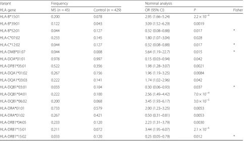
We evaluated the associations of the 4-digit HLA alleles with MS risk and found the most significant association to be with HLA-DRB1*15:01 (P = 2.1 × 10⁻⁵, odds ratio
[OR] = 3.44, 95% confidence interval [95% CI] = 1.95–
6.07; Table 2). HLA-DQB1*06:02, which was in linkage
disequilibrium with HLA-DRB1*15:01 (r2 = 0.94 in the
controls), was also significantly associated with MS (P= 3.0 × 10⁻⁵, OR = 3.45, 95% CI = 1.93–6.17; Table 2). We found that HLA-B*15:01 was significantly associated with MS (P = 2.2 × 10⁻⁴, OR = 2.95, 95% CI = 1.66–5.24;
Table 2). Previous studies in Japanese and European
in-dividuals reported the strongest association of
HLA-DRB1*15:01 with MS [13, 17], and our study replicated
these results.
We then conducted stepwise conditional analysis with re-spect to the top-associated HLA alleles. Conditional
ana-lysis of HLA-DRB1*15:01 revealed an independent
association with HLA-DRB1*04:05 (P = 2.2 × 10⁻⁴, OR = 2.83, 95% CI = 1.63–4.90; Additional file1: Table S1). Sub-sequent conditional analysis regarding HLA-DRB1*15:01 and HLA-DRB1*04:05 revealed an independent association with HLA-B*39:01 (P = 8.3 × 10⁻⁴, OR = 3.51, 95% CI =
1.68–7.34; Additional file 1: Table S1). Conditional
analysis of HLA-DRB1*15:01, HLA-DRB1*04:05, and HLA-B*39:01 revealed an independent association
with HLA-B*15:01 (P = 2.3 × 10⁻⁴, OR = 3.10, 95%
CI = 1.70–5.65; Additional file 1: Table S1). After con-ditioning DRB1*15:01, DRB1*04:05, HLA-B*39:01, and HLA-B*15:01, no significant association
locus was observed (Additional file 1: Table S1).
independently associated with MS (HLA-DRB1*15:01 for
P = 5.7 × 10⁻⁶ and OR = 4.06, HLA-DRB1*04:05 for P =
1.2 × 10⁻⁴and OR = 3.02, and HLA-B*39:01 for P = 1.9 × 10⁻⁴ and OR = 4.13, and HLA-B*15:01 for P = 2.3 × 10⁻⁴ and OR = 3.10; Table 3). Multivariate analysis confirmed that theHLA-Balleles were associated with MS susceptibil-ity independently of theHLA-DRB1alleles. As reported in a previous European study [14], we identified independent contributions of the class I HLA alleles to MS risk in Japanese subjects.
HLA alleles with MS susceptibility and MS clinical phenotype
We identified four HLA alleles associated with MS sus-ceptibility. We then focused on whether these HLA
alleles (HLA-DRB1*15:01, HLA-DQB1*06:02,
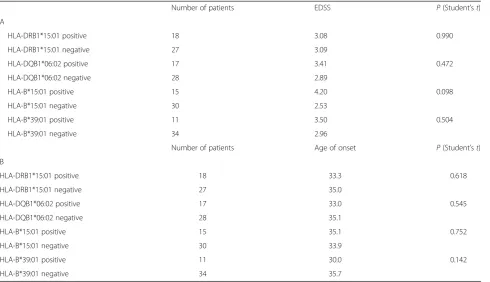
HLA-B*15:01, and HLA-B*39:01) influence the clinical course of MS. We checked the clinical feature of HLA alleles
associated with MS in our research. We focused on the onset of age and EDSS score, that is widely used for dis-ease severity and measured in all participants in this study. The results showed that no HLA alleles have a
significant association with EDSS score (P > 0.09;
Table4A) and the onset of age (P> 0.14; Table4B).
Association of HLA amino acid polymorphisms with MS susceptibility
We then analyzed associations of the HLA amino acid polymorphisms with respect to MS susceptibility. We
se-lected HLA-DRB1, HLA-DQB1, and HLA-B, which were
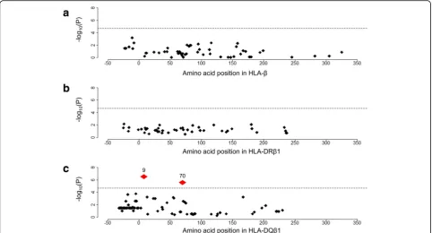
associated with MS in our HLA allele-based study, for ana-lysis (Additional file 1: Table S3–1). We found that the leading risk variant was the presence of phenylalanine at
the HLA-DQβ1 position 9 (HLA-DQβ1 Phe9; P = 3.7 ×
10−8, OR = 3.48, 95%CI = 2.23–5.43; Additional file1: Table S3–1, Fig. 1). We also found significant associations with
Table 2HLA association analysis of multiple sclerosis in the Japanese population
Variant Frequency Nominal analysis
HLA gene MS (n= 45) Control (n= 429) OR (95% CI) P Fisher
HLA-B*15:01 0.200 0.078 2.95 (1.66–5.24) 2.2 × 10⁻⁴
HLA-B*39:01 0.122 0.043 3.09 (1.52–6.29) 0.0019
HLA-B*52:01 0.044 0.127 0.32 (0.08–0.88) 0.017 *
HLA-C*07:02 0.233 0.145 1.80 (1.07–3.04) 0.028
HLA-C*12:02 0.044 0.127 0.32 (0.08–0.88) 0.017 *
HLA-DMB*01:07 0.044 0.008 5.64 (1.19–22.7) 0.015 *
HLA-DOA*01:01 0.978 0.997 0.15 (0.03–0.94) 0.042
HLA-DPB1*05:01 0.522 0.356 1.98 (1.28–3.07) 0.0021
HLA-DQA1*01:02 0.267 0.156 1.96 (1.19–3.25) 0.0084
HLA-DQA1*03:03 0.222 0.141 1.74 (1.02–2.96) 0.042
HLA-DQB1*03:01 0.033 0.104 0.30 (0.06–0.93) 0.037 *
HLA-DQB1*04:01 0.222 0.100 2.56 (1.49–4.42) 7.0 × 10⁻⁴
HLA-DQB1*06:02 0.200 0.068 3.45 (1.93–6.17) 3.0 × 10⁻⁵
HLA-DRA*01:01 0.733 0.579 2.00 (1.23–3.25) 0.0053
HLA-DRA*01:02 0.267 0.421 0.50 (0.31–0.81) 0.0053
HLA-DRB1*04:05 0.233 0.120 2.23 (1.31–3.79) 0.0030
HLA-DRB1*15:01 0.211 0.072 3.44 (1.95–6.07) 2.1 × 10⁻⁵
HLA-DRB1*15:02 0.033 0.120 0.25 (0.05–0.79) 0.012 *
*Fisher’s exact test was used for association analysis, otherwise logistic regression was used
Table 3HLA conditional analysis of multiple sclerosis in the Japanese population
Variant Frequency Conditional association
DRB1*15:01, DRB1*04:05, B*39:01, and B*15:01
HLA gene MS (n= 45) Control (n= 429) OR (95% CI) P
HLA-DRB1*15:01 0.211 0.072 4.06 (2.22–7.45) 5.7 × 10⁻⁶
HLA-DRB1*04:05 0.233 0.120 3.02 (1.72–5.31) 1.2 × 10⁻⁴
HLA-B*39:01 0.122 0.043 4.13 (1.96–8.71) 1.9 × 10⁻⁴
the presence of arginine at HLA-DQβ1 position 70 (P = 2.7 × 10⁻⁶, OR = 0.34, 95% CI = 0.22–0.54; Additional file1: Table S3–1), leucine at HLA-DQβ1 position−5 (P= 8.9 × 10⁻⁶; OR = 3.54, 95% CI = 2.03–6.20; Additional file1: Table S3–1), and valine at HLA-DQβ1 position−18 (P = 2.1 × 10⁻⁴, OR = 2.80, 95% CI = 1.63–4.84; Additional file 1: Table S3–1). We assessed the significance of each amino acid position at HLA-DQβ1 using stepwise conditional analysis. After conditioning HLA-DQβ1 Phe9, no amino acid at HLA-DQβ1 was nominally associated
with MS (Additional file1: Table S3–2). We then
con-ducted an omnibus test for each HLA amino acid position that had polymorphisms based on a
log-likelihood ratio test [31, 32]. The HLA-DQβ1 position
9 showed the strongest association with MS (P= 3.6 ×
10−7; Fig. 2), although its association was less signifi-cant than that of the binary test of HLA-DQβ1 Phe9.
HLA-DQβ1 Phe9 independently confers MS risk with HLA-DRB1*15:01
Our study identified a significant MS risk associated with DRB1*15:01 and phenylalanine 9 at HLA-DQβ1, but it was not clear which of the variants caused these effects. We therefore conducted an ana-lysis to investigate these effects. When conditioning HLA-DRB1*15:01, HLA-DQβ1 Phe9 was significantly
associated with MS susceptibility (P = 1.5 × 10⁻⁵,
OR = 2.91, 95% CI = 1.79–4.72; Additional file 1:
Table 4HLA association analysis with the clinical course of MS
Number of patients EDSS P(Student’st)
A
HLA-DRB1*15:01 positive 18 3.08 0.990
HLA-DRB1*15:01 negative 27 3.09
HLA-DQB1*06:02 positive 17 3.41 0.472
HLA-DQB1*06:02 negative 28 2.89
HLA-B*15:01 positive 15 4.20 0.098
HLA-B*15:01 negative 30 2.53
HLA-B*39:01 positive 11 3.50 0.504
HLA-B*39:01 negative 34 2.96
Number of patients Age of onset P(Student’st)
B
HLA-DRB1*15:01 positive 18 33.3 0.618
HLA-DRB1*15:01 negative 27 35.0
HLA-DQB1*06:02 positive 17 33.0 0.545
HLA-DQB1*06:02 negative 28 35.1
HLA-B*15:01 positive 15 35.1 0.752
HLA-B*15:01 negative 30 33.9
HLA-B*39:01 positive 11 30.0 0.142
HLA-B*39:01 negative 34 35.7
*Student’sttest was used for association analysis
Table S3–2). Conversely, when conditioning HLA-DQβ1 Phe9, HLA-DRB1*15:01 was just nominally significant (P = 0.037, OR = 1.94, 95% CI = 1.04–3.63). While further validation study should be warranted, our results suggested that HLA-DQβ1 Phe9 is a causal driver of HLA variants for MS risk in the Japanese population.
Associations of the HLA alleles with NMOSD susceptibility
We assessed the risk of NMOSD with 4-digit HLA al-leles and found the most significant association to be at
HLA-DQA1*05:03 (P = 1.5 × 10⁻⁴, OR = 6.96, 95% CI =
2.55–19.0; Table 5). We conducted conditional analysis controlling for the top HLA alleles associated with NMOSD. After conditioning for HLA-DQA1*05:03, no
Fig. 2Multiple sclerosis risk-associated amino acid positions of the HLA genes. Diamonds represent the–log10(P) of the amino acid positions of
the tested HLA genes. Labeled red diamonds represent the–log10(P) values of the amino acid positions significantly associated with MS. The
horizontal lines represent the associationPvalue of HLA-DRB1*15:01.aNo amino acid polymorphisms ofHLA-Bwas significantly associated with
MS.bNo amino acid polymorphisms ofHLA-DRB1is significantly associated with MS.cThe HLA-DQβ1 position 9 and 70 were associated with
MS more significantly than HLA-DRB1*15:01
Table 5HLA association analysis of neuromyelitis optica spectrum disorder in the Japanese population
Variant Frequency Nominal analysis
HLA gene NMOSD (n= 31) Control (n= 429) OR (95% CI) P Fisher
HLA-A*26:03 0.065 0.020 3.40 (0.81–10.9) 0.047 *
HLA-B*07:02 0.129 0.059 2.34 (1.06–5.19) 0.036
HLA-DPB1*05:01 0.516 0.356 1.93 (1.15–3.24) 0.012
HLA-DQA1*01:01 0.129 0.058 2.39 (1.08–5.30) 0.031
HLA-DQA1*03:02 0.048 0.147 0.30 (0.06–0.93) 0.035 *
HLA-DQA1*05:03 0.097 0.015 6.96 (2.55–19.0) 1.5 × 10⁻⁴
HLA-DQB1*03:01 0.210 0.104 2.29 (1.20–4.39) 0.012
HLA-DQB1*03:03 0.048 0.149 0.29 (0.06–0.91) 0.024 *
HLA-DRB1*01:01 0.129 0.058 2.39 (1.08–5.30) 0.031
HLA-DRB1*09:01 0.016 0.135 0.10 (0.003–0.62) 0.0027 *
HLA-DRB1*14:06 0.081 0.012 7.40 (1.92–24.8) 0.0021 *
significant association was observed after Bonferroni correction, based on the number of alleles tested (Pcorr> 0.05; Additional file 1: Table S2). Our results indicated that HLA-DQA1*05:03 is a newly identified HLA allele associated with NMOSD in the Japanese population.
Associations of the HLA amino acid polymorphisms with NMOSD susceptibility
We analyzed the association of the HLA amino acid poly-morphisms with NMOSD susceptibility. We selected HLA-DQA1, which was associated with NMOSD in our HLA allele-based association study. We found no amino acid variants with significant risk after Bonferroni correc-tion, based on the number of amino acids tested (Pcorr> 0.05; Additional file1: Table S4).
Discussion
In this study, we performed HLA analysis of MS/ NMOSD in Japanese individuals. We replicated results indicating that HLA-DRB1*15:01 is the allele most strongly associated with MS susceptibility, a finding which was comparable with those of previous Japanese
and European populations [13, 17]. HLA-B*39:01 and
HLA-B*15:01 also appeared to be associated with MS. Previous European studies have reported an associ-ation of class I HLA alleles with MS susceptibility [14]. These results suggest a relationship between class I HLA alleles and MS susceptibility, as well as class II HLA alleles, in both Europeans and Japanese.
We also conducted an association study involving HLA amino acid polymorphisms and found that HLA-DQβ1 amino acid polymorphisms led to a significantly increased risk of MS. Previous studies have reported the
importance of HLA-DRB1 with respect to MS [14, 34],
but the role ofHLA-DQB1 has not been studied in
de-tail. In our study, even conditioning HLA-DRB1*15:01, HLA amino acid polymorphisms at HLA-DQβ1 were as-sociated with increased risk of MS. This result indicates that HLA-DQβ1 polymorphisms could independently confer risk of MS. The association of HLA-DQB1 with MS has not been reported well. MHC class II molecules are heterodimers of an alpha and a beta chain, and have a peptide-binding site for presenting antigenic peptides to T cells [35]. In this study, we found that two amino acid positions associated with MS (HLA-DQβ1 position 9 and 70). Previous studies showed that HLA-DQβ1 position 9 is in the peptide-binding groove of HLA-DQ molecules and HLA-DQβ1 position 70 residues in the putative T cell receptor (TCR) [36]. Within the central nervous system, antigen-presenting cells such as micro-glia and astrocytes work as defenders against infections and inflammation [37]. Changes in antigen-presenting function of peptide binding amino acid changes could in-fluence the susceptibility to MS. Animal model studies
have found that susceptibility to myelin oligodendrocyte-binding protein or proteolipid protein were determined by
HLA-DQB1*06:02 [38, 39]. These findings also suggest
the possibility thatHLA-DQB1plays an important role in susceptibility to MS.
HLA-DQA1*05:03 was identified as being associated with
NMOSD in our study.HLA-DQA1has been reported to be
associated with immune-mediated diseases such as celiac disease and type 1 diabetes [40,41]. In Europeans, a recent
GWAS identified rs28383224, which is located in
HLA-DQA1, as having the strongest association with NMOSD
susceptibility [19]. Although the role of theHLA-DQA1risk SNP has not been investigated in detail, the importance of HLA-DQA1in NMOSD pathophysiology was suggested.
A previous study in Japan reported that HLA-DRB1*16: 02 and HLA-DPB1*05:01 presented a significantly elevated risk of NMOSD, while HLA-DRB1*09:01 reduced the risk. [22] In our analysis, HLA-DRB1*09:01 and HLA-DPB1*05:
01 were nominally significant (HLA-DRB1*09:01; P =
0.0027, OR = 0.10, 95% CI = 0.003–0.62, HLA-DPB1*05:01; P= 0.012, OR = 1.93, 95% CI = 1.15–3.24; Additional file1 Table S2), but after performing Bonferroni correction, they were not significantly associated with NMOSD. This result may be because our study sample size was smaller than that of the previous study in Japan. HLA-DRB1*16:02 was not significant in our study (P = 0.39, OR = 2.32, 95% CI =
0.05–19.6). These findings may indicate that the HLA
alleles associated with NMOSD are localized in Japan. Further studies are needed to identify the details of the vari-ability of HLA alleles in the Japanese population.
Conclusions
In summary, through NGS-based HLA typing of the MS and NMOSD patients and controls of Japanese ancestry, we analyzed the variants of the 16 HLA genes. We newly identified the risk HLA alleles associated with MS and NMOSD risk. We also identified that the amino acid poly-morphisms at HLA-DQβ1 are more strongly associated with MS susceptibility than HLA alleles. Our study con-tributes to the understanding of the genetic background of MS and NMOSD in Japanese.
Additional file
Additional file 1: Table S1 Results of the association analysis of HLA alleles (multiple sclerosis). Table S2 Results of the association analysis of
HLA alleles (neuromyelitis optica spectrum disorder). Table S3–1 Results
of the HLA amino acid association analysis (multiple sclerosis). Table S3–2
Results of the conditional HLA DQβ1 association analysis (multiple
sclerosis). Table S4 Results of the HLA amino acid association analysis (neuromyelitis optica spectrum disorder). (PDF 542 kb)
Abbreviations
NMOSD: Neuromyelitis optica spectrum disorder; SNP: Single-nucleotide polymorphisms
Acknowledgements Not applicable.
Authors’contributions
YO supervised the study. KO, TO, YO, and HM wrote the manuscript. KO, TO, KH, AH, JH, KS, SS, YA, and YO conducted the data analysis. TO, AH, MK, KM, SK, and HM collected the samples. KH and II conducted the experiments. All authors read and approved the final manuscript.
Funding
This research was supported by Bioinformatics Initiative of Osaka University Graduate School of Medicine, and Integrated Frontier Research for Medical Science Division, Institute for Open and Transdisciplinary Research Initiatives, Osaka University. Y.O. was supported by the Japan Society for the Promotion of Science (JSPS) KAKENHI (15H05911 and 19H01021), AMED (19gm6010001h0004, 19ek0410041h0003, and 19ek0109413h0001). J.H. is an employee of Teijin Pharma Limited.
Availability of data and materials
The datasets used and analyzed during the current study are available from the corresponding author on reasonable request.
Ethics approval and consent to participate
This study was approved by the ethical committee of Osaka University Graduate School of Medicine. All participants were of Japanese origin and provided written informed consent that was approved by the ethical committee of each hospital.
Consent for publication Not applicable.
Competing interests
The authors declare that they have no competing interests.
Author details 1
Department of Statistical Genetics, Osaka University Graduate School of Medicine, 2-2 Yamadaoka, Suita 565-0871, Japan.2Department of Neurology,
Osaka University Graduate School of Medicine, Suita 565-0871, Japan.
3Department of Bioinformatics and Genomics Graduate School of Advanced
Preventive Medical Sciences, Kanazawa University, Kanazawa 920-8640, Japan.
4Department of Neurology, Suita Municipal Hospital, Suita 564-8567, Japan. 5
Pharmaceutical Discovery Research Laboratories, Teijin Pharma Limited, Hino 191-8512, Japan.6Department of Cardiovascular Medicine, Osaka University
Graduate School of Medicine, Suita 565-0871, Japan.7Department of Neurology, Kindai University Faculty of Medicine, Osaka-Sayama 589-8511, Japan.8Division of Human Genetics, National Institute of Genetics, Shizuoka 411-8540, Japan.9Laboratory of Statistical Immunology, Immunology Frontier
Research Center (WPI-IFReC), Osaka University, Suita 565-0871, Japan.
Received: 28 March 2019 Accepted: 22 July 2019
References
1. Trowsdale J, Knight JC. Major histocompatibility complex genomics and
human disease. Annu Rev Genomics Hum Genet. 2013;14:301–23.
2. Dendrou CA, Petersen J, Rossjohn J, et al. HLA variation and disease. Nat
Rev Immunol. 2018;18(5):325–39.
3. Kanai M, Akiyama M, Takahashi A, et al. Genetic analysis of quantitative traits
in the Japanese population links cell types to complex human diseases. Nat
Genet. 2018;50(3):390–400.
4. Silman AJ, Pearson JE. Epidemiology and genetics of rheumatoid arthritis.
Arthritis Res. 2002;4(Suppl 3):S265–72.
5. Noble JA. Immunogenetics of type 1 diabetes: a comprehensive review. J
Autoimmun. 2015;64:101–12.
6. Hirata J, Hirota T, Ozeki T, et al. Variants at A, C, and
HLA-DQB1 confer risk of psoriasis vulgaris in Japanese. J Invest Dermatol.
2018;138(3):542–8.
7. Browne P, Chandraratna D, Angood C, et al. Atlas of multiple sclerosis 2013:
a growing global problem with widespread inequity. Neurology. 2014;
83(11):1022–4.
8. International Multiple Sclerosis Genetics C, Hafler DA, Compston A, et al.
Risk alleles for multiple sclerosis identified by a genomewide study. N Engl J
Med. 2007;357(9):851–62.
9. Australia, New Zealand Multiple Sclerosis Genetics C. Genome-wide
association study identifies new multiple sclerosis susceptibility loci on
chromosomes 12 and 20. Nat Genet. 2009;41(7):824–8.
10. International Multiple Sclerosis Genetics C, Wellcome Trust Case Control C,
Sawcer S, et al. Genetic risk and a primary role for cell-mediated immune
mechanisms in multiple sclerosis. Nature. 2011;476(7359):214–9.
11. International Multiple Sclerosis Genetics C, Beecham AH, Patsopoulos NA,
et al. Analysis of immune-related loci identifies 48 new susceptibility
variants for multiple sclerosis. Nat Genet. 2013;45(11):1353–60.
12. Ogawa K, Okada Y. Statistical genetics and its application to
neuroimmunology. Clin Exp Neuroimmunol. 2018;9(1):7–12.
13. Ramagopalan SV, Knight JC, Ebers GC. Multiple sclerosis and the major
histocompatibility complex. Curr Opin Neurol. 2009;22(3):219–25.
14. Patsopoulos NA, Barcellos LF, Hintzen RQ, et al. Fine-mapping the genetic
association of the major histocompatibility complex in multiple sclerosis: HLA and non-HLA effects. PLoS Genet. 2013;9(11):e1003926.
15. Mack SJ, Udell J, Cohen F, et al. High resolution HLA analysis reveals
independent class I haplotypes and amino-acid motifs protective for
multiple sclerosis. Genes Immun. 2018;20:308–26.
16. Yoshimura S, Isobe N, Yonekawa T, et al. Genetic and infectious profiles of
Japanese multiple sclerosis patients. PLoS One. 2012;7(11):e48592.
17. Nakamura Y, Matsushita T, Sato S, et al. Latitude and HLA-DRB1*04:05
independently influence disease severity in Japanese multiple sclerosis: a cross-sectional study. J Neuroinflammation. 2016;13(1):239.
18. Arellano B, Hussain R, Miller-Little WA, et al. A single amino acid
substitution prevents recognition of a dominant human aquaporin-4 determinant in the context of HLA-DRB1*03:01 by a murine TCR. PLoS One. 2016;11(4):e0152720.
19. Estrada K, Whelan CW, Zhao F, et al. A whole-genome sequence
study identifies genetic risk factors for neuromyelitis optica. Nat Commun. 2018;9(1):1929.
20. Yoshimura S, Isobe N, Matsushita T, et al. Distinct genetic and
infectious profiles in Japanese neuromyelitis optica patients according to anti-aquaporin 4 antibody status. J Neurol Neurosurg Psychiatry.
2013;84(1):29–34.
21. Matsushita T, Matsuoka T, Isobe N, et al. Association of the HLA-DPB1*0501
allele with anti-aquaporin-4 antibody positivity in Japanese patients with idiopathic central nervous system demyelinating disorders. Tissue Antigens.
2009;73(2):171–6.
22. Hosomichi K, Jinam TA, Mitsunaga S, et al. Phase-defined complete
sequencing of the HLA genes by next-generation sequencing. BMC Genomics. 2013;14:355.
23. Hosomichi K, Mitsunaga S, Nagasaki H, et al. A bead-based normalization for
uniform sequencing depth (BeNUS) protocol for multi-samples sequencing exemplified by HLA-B. BMC Genomics. 2014;15:645.
24. Hosomichi K, Shiina T, Tajima A, et al. The impact of next-generation
sequencing technologies on HLA research. J Hum Genet.
2015;60(11):665–73.
25. Polman CH, Reingold SC, Banwell B, et al. Diagnostic criteria for
multiple sclerosis: 2010 revisions to the McDonald criteria. Ann Neurol.
2011;69(2):292–302.
26. Wingerchuk DM, Banwell B, Bennett JL, et al. International consensus
diagnostic criteria for neuromyelitis optica spectrum disorders.
Neurology. 2015;85(2):177–89.
27. Hirata J, Hosomichi K, Sakaue S, et al. Genetic and phenotypic landscape of
the major histocompatibilty complex region in the Japanese population.
Nat Genet. 2019;51(3):470–80.
28. Li H, Durbin R. Fast and accurate short read alignment with
Burrows-Wheeler transform. Bioinformatics. 2009;25(14):1754–60.
29. Robinson J, Soormally AR, Hayhurst JD, et al. The IPD-IMGT/HLA
database—new developments in reporting HLA variation. Hum
Immunol. 2016;77(3):233–7.
30. Pirooznia M, Kramer M, Parla J, et al. Validation and assessment of
31. Okada Y, Momozawa Y, Ashikawa K, et al. Construction of a
population-specific HLA imputation reference panel and its application to Graves’
disease risk in Japanese. Nat Genet. 2015;47(7):798–802.
32. Okada Y, Suzuki A, Ikari K, et al. Contribution of a non-classical HLA
gene, HLA-DOA, to the risk of rheumatoid arthritis. Am J Hum Genet.
2016;99(2):366–74.
33. Pettersen EF, Goddard TD, Huang CC, et al. UCSF chimera—a
visualization system for exploratory research and analysis. J Comput
Chem. 2004;25(13):1605–12.
34. Brum DG, Barreira AA, dos Santos AC, et al. HLA-DRB association in
neuromyelitis optica is different from that observed in multiple sclerosis.
Mult Scler. 2010;16(1):21–9.
35. Call MJ. Small molecule modulators of MHC class II antigen presentation:
mechanistic insights and implications for therapeutic application. Mol
Immunol. 2011;48(15–16):1735–43.
36. Jones EY, Fugger L, Strominger JL, et al. MHC class II proteins and disease: a
structural perspective. Nat Rev Immunol. 2006;6(4):271–82.
37. Chastain EM, Duncan DS, Rodgers JM, et al. The role of antigen presenting
cells in multiple sclerosis. Biochim Biophys Acta. 2011;1812(2):265–74.
38. Kaushansky N, Altmann DM, Ascough S, et al. HLA-DQB1*0602
determines disease susceptibility in a new“humanized”multiple
sclerosis model in HLA-DR15 (DRB1*1501;DQB1*0602) transgenic mice. J
Immunol. 2009;183(5):3531–41.
39. Kaushansky N, Altmann DM, David CS, et al. DQB1*0602 rather than
DRB1*1501 confers susceptibility to multiple sclerosis-like disease induced by proteolipid protein (PLP). J Neuroinflammation. 2012;9:29.
40. Megiorni F, Pizzuti A. HLA-DQA1 and HLA-DQB1 in Celiac disease
predisposition: practical implications of the HLA molecular typing. J Biomed Sci. 2012;19:88.
41. El-Amir MI, El-Feky MA, Laine AP, et al. Risk genes and autoantibodies in
Egyptian children with type 1 diabetes - low frequency of autoantibodies in carriers of the HLA-DRB1*04:05-DQA1*03-DQB1*02 risk haplotype. Diabetes
Metab Res Rev. 2015;31(3):287–94.
Publisher’s Note