R E S E A R C H
Open Access
CCAAT/enhancer-binding protein delta
regulates the stemness of glioma stem-like
cells through activating PDGFA expression
upon inflammatory stimulation
Shao-Ming Wang
1†, Hong-Yi Lin
2†, Yen-Lin Chen
3, Tsung-I Hsu
1,4,5, Jian-Ying Chuang
1,5, Tzu-Jen Kao
1and
Chiung-Yuan Ko
1,5*Abstract
Background:The small population of glioma stem-like cells (GSCs) contributes to tumor initiation, malignancy, and recurrence in glioblastoma. However, the maintenance of GSC properties in the tumor microenvironment remains unclear. In glioma, non-neoplastic cells create an inflammatory environment and subsequently mediate tumor progression and maintenance. Transcriptional factor CCAAT/enhancer-binding protein delta (CEBPD) is suggested to regulate various genes responsive to inflammatory cytokines, thus prompting us to investigate its role in regulating GSCs stemness after inflammatory stimulation.
Methods:Stemness properties were analyzed by using spheroid formation. Oncomine and TCGA bioinformatic databases were used to analyze gene expression. Western blotting, quantitative real-time polymerase chain reaction, luciferase reporter assay, and chromatin immunoprecipitation assay were used to analyze proteins and gene transcript levels. The glioma tissue microarrays were used for CEBPD and PDGFA expression by immunohistochemistry staining.
Results: We first found that IL-1β promotes glioma spheroid formation and is associated with elevated CEBPD expression. Using microarray analysis, platelet-derived growth factor subunit A (PDGFA) was confirmed as a CEBPD-regulated gene that mediates IL-1β-enhanced GSCs self-renewal. Further analysis of the genomic database and tissue array revealed that the expression levels between CEBPD and PDGFA were coincident in glioma patient samples.
Conclusion: This is the first report showing the activation of PDGFA expression by CEBPD through IL-1β treatment and a novel CEBPD function in maintaining the self-renewal feature of GSCs.
Keywords: Glioblastoma multiforme, Glioma stem-like cell, CEBPD, PDGFA, Inflammation, Interleukin-1β
Background
As one of the most common and lethal brain tumor types, glioblastoma multiforme (GBM) contains neo-plastic and non-neoneo-plastic cells, including glioma stem-like cells (GSCs), microglia, astrocytes, and
endothelial cells [1–3]. Glioma stem-like cells contrib-ute to the tumor-initiating activity and tumor recur-rence through the stem cell-like characteristics, including their self-renewal ability and the expression of neural stem cell markers [4–6]. Several studies report that the microenvironment serves as one of the key factors to promote cancer progression and malignancy in glioma [1,3,7]. In brain tumors, the majority of stromal cells are brain-intrinsic microglia/macrophages, which support tumor cell growth and invasion [3]. Moreover, glioma cells can release several chemotactic factors, which attract
© The Author(s). 2019Open Access This article is distributed under the terms of the Creative Commons Attribution 4.0 International License (http://creativecommons.org/licenses/by/4.0/), which permits unrestricted use, distribution, and reproduction in any medium, provided you give appropriate credit to the original author(s) and the source, provide a link to the Creative Commons license, and indicate if changes were made. The Creative Commons Public Domain Dedication waiver (http://creativecommons.org/publicdomain/zero/1.0/) applies to the data made available in this article, unless otherwise stated.
* Correspondence:[email protected]
†Shao-Ming Wang and Hong-Yi Lin are contributed equally to this work as
the first authors.
microglia/macrophages to the tumor bulk, and these microglia/macrophages can further produce several inflam-matory factors to promote glioma cell invasion [3,8]. How-ever, whether these inflammatory cytokines can promote GSCs feature remains unclear.
CCAAT/enhancer-binding protein delta belongs to the CCAAT/enhancer-binding protein (C/EBP) family. The expression of CEBPD protein is regulated by a va-riety of extracellular stimuli, such as IL-1β and tumor necrosis factor (TNF)-α [9, 10]. The biological func-tions of CEBPD include cell migration, anti-apoptosis, ROS formation, and differentiation [10–13]. In previous cancer studies, CEBPD has been suggested to induce cell growth arrest and apoptosis in hepatocellular car-cinoma and prostate cancer and thus function as a tumor suppressor [14, 15]. On the other hand, recent studies imply that CEBPD plays a pro-tumorigenic role in drug resistance and cell invasion in bladder cancer [16, 17]. According to GEO profile data (GSE2223, GSE4290, and GSE4536) [18–20], CEBPD is expressed in GBM at a higher level when comparing with that in normal brain tissues. However, the functions of CEBPD in glioma and inflammation-induced GSC stemness re-main to be clarified.
The activation of PDGF/PDGFR signaling can trigger cancer progression and malignancy [2, 21]. PDGFs are secretory factors, and four members have been identi-fied, including PDGFA, B, C, and D [22]. Among them, PDGFA is found in 70% of gliomas and is highly expressed in GBM [2, 22]. Previous studies indicate that PDGFA can promote glioma proliferation, invasion, and self-renewal [2, 23], thus suggesting that PDGFA regu-lates GSC stemness and promotes glioma progression. However, whether GSCs express PDGFA in an inflam-matory environment is still unknown, and the molecular mechanism driving the change of PDGFA expression in GSCs remains unclear.
In this study, we propose that CEBPD plays a regula-tory role in inflammaregula-tory responses and contributes to GSC stemness in GBM. To test this hypothesis, we in-vestigate the function of CEBPD in cultured glioma spheroids in vitro. We first show that IL-1β is highly expressed in GBM, promotes glioma spheroid formation, and is associated with the elevated CEBPD expression. Focusing on GSC stemness, we then show that CEBPD plays a functional role of inducing the stem-like feature in the inflammatory environment. We further demon-strate that PDGFA is responsive to CEBPD induction in glioma spheroid cells, and CEBPD regulates PDGFA transcription by binding to thePDGFApromoter region. Combined, our findings indicate that CEBPD is required for IL-1β-driven self-renewal of GSCs, and the PDGFA expression regulated by CEBPD is a key mechanism for GSCs self-renewal in the inflammatory environment.
Methods
Cell culture and transfection
The human U373MG cell line (human glioblastoma astrocytoma) and human T98G cell line (human glioblas-toma multiforme) were purchased from the American Type Culture Collection (ATCC) and were cultured in Dulbecco’s modified Eagle medium (DMEM, Thermo Fisher Scientific, MA, USA) containing 10% fetal bovine serum (FBS, Thermo Fisher Scientific, MA, USA), 100 units/mL penicillin, and 100μg/mL streptomycin (Thermo Fisher Scientific, MA, USA). We confirmed the authentica-tion of all cell lines by short tandem repeat (STR) analyses of cell DNA alleles. Cells were incubated in a humidified incubator at 37 °C with 5% CO2. The siRNA knockdown for siControl (4390843, Ambion, CA, USA) and siCEBPD (si2895: 5′-UUCUCUCGCAGUUUAGUGGTG-3′; si2896: 5′-AUUGCUGUUGAAGAGGUCGGC-3′) was purchased from Thermo (Ambion, CA, USA). The cells were transfected with siRNA using specific siRNA Lipofectamine reagent (RNAiMAX reagent, Thermo Fisher Scientific, MA, USA).
Isolation and culture of primary glioblastoma cells
The use of human specimens was approved by the In-stitute Review Board (IRB)/Ethics Committee from the of-fice of human research in Taipei Medical University (Taipei, Taiwan). The consent of the patient was obtained and approved by Taipei Medical University IRB protocols (N201006011). PT#3 glioblastoma cells were isolated from the glioblastoma tissue of a male patient. The patient was treated and taken care in the Taipei Medical University Hospital (Taipei, Taiwan). The freshly resected tissues were digested by 0.05% collagenase type IV (Sigma-Al-drich, St. Louis, MO, USA) and 5 units/mL DNase I (Sigma-Aldrich, St. Louis, MO, USA) at 37 °C for 2 h. After removing undigested tissues by centrifugation, the supernatant was mixed with DMEM supplemented with 10% fetal bovine serum (FBS, Thermo Fisher Scientific, MA, USA) and transferred to 6-well culture plates. Cells were then maintained in DMEM-10% FBS.
GEO database
The Oncomine database was used to assess the gene expression in normal and GBM tissues. The accession numbers of GEO datasets used in this study are Bredel dataset (GSE2223), Sun dataset (GSE4290), and Lee dataset (GSE4536).
Tumor spheroid formation assay
USA), 20 ng/mL EGF (Invitrogen, Carlsbad, CA, USA), and 20 ng/mL FGF (Invitrogen, Carlsbad, CA, USA).
Real-time quantitative reverse transcriptase PCR
Total RNA was extracted using the Direct-zol™ RNA MiniPrep kit (Zymo Research, CA, USA) from TRIsure RNA extraction reagent. The synthesis of complementary DNA (cDNA) was completed with reverse transcription (RT) reactions using the PrimeScript™ RT reagent Kit (TAKARA, JPN). Real-time PCR was conducted using iTaq Universal SYBR Green Supermix. PCR was con-ducted using StepOne Plus™real-time PCR systems (ABI) with the following pairs of specific primers: human CEBPD (S): 5-′GCCATGTACGACGACGAGAG-3′ and CEBPD (AS): 5-′TGTGATTGCTGTTGAAGAGGTC-3′; PDGFA (S): 5-′AAGTCCAGGTGAGGTTAG-3′ and PDGFA (AS): 5-′TCCTCTTCCCGATAATCC-3′; and GAPDH (S): 5-′GTCTCCTCTGACTTCAACAGCG-3′ and GAPDH (AS): 5-′ ACCACCCTGTTGCTGTAGC-CAA-3′. All reactions were performed in duplicates with
“no reverse transcriptase”as the control, and all data are mean ± SEM of at least three independent biological repli-cates. The relative expression levels were measured using the relative quantitation (RQ)ΔΔCt method and normal-ized to the housekeeping gene glyceraldehyde 3-phosphate dehydrogenase (GAPDH).
Western blot analysis
Cells were harvested and lysed with Pierce RIPA buffer (Thermo, MA, USA). Lysates were resolved on a sodium dodecyl sulfate gel containing 10 or 12% polyacrylamide, and then transferred to a polyvinylidene difluoride (PVDF) nylon membrane and hybridized with primary antibodies against CEBPD (sc-636x, Santa Cruz, CA, USA), PDGFA (sc-9974, Santa Cruz, CA, USA), CD133 (18470-1-AP, Proteintech, HB, CHN), or α-tubulin (T9026, Sigma-Aldrich, MO, USA) at 4 °C overnight. Specific proteins were detected by peroxidase-conjugated secondary anti-bodies incubated at room temperature for 1 h. Signals were revealed by the enhanced chemiluminescence West-ern blot system from ChemiDoc Touch™Imaging System (BIO-RAD, CA, USA).
Establishment of stably knockdown clones
Virus was produced from Phoenix cells by cotransfection of the various small hairpin RNA expression vectors in combination with pMD2.G and psPAX2. After determining the viral infection efficiency, 10 multiplicity of infection of lentivirus containing shβ-galactosidase (shLacZ, for sham control) or shCEBPD (shC7) were used to infect U373MG or T98G cells for 96 h. Cells were further selected and maintained in 2μg/mL puromycin (Gibco, MA, USA). In all lentiviral experiments, the medium containing un-infected viruses was removed before further assays were
conducted. The small hairpin RNA sequences in lentiviral expression vectors were as follows: shβ-galactosidase: 5′ -
CCGGTGTTCGCATTATCCGAACCATCTCGA-GATGGTTCGGATAATGCGAACATTTTTG-3′ and
shCEBPD (shC7): 5′ -CCGGGCTGTCGGCTGAGAAC-
GAGAACTCGAGTTCTCGTTCTCAGCCGA-CAGCTTTTT-3′. The lentiviral knockdown expression vectors were obtained from the National RNAi Core Fa-cility located at the Genomic Research Center of Institute of Molecular Biology, Academia Sinica (Taiwan).
Microarray analysis
Total RNAs were harvested from U373MG cells trans-fected with siC or siCEBPD for 24 h. Samples were vali-dated with Agilent Human Whole Genome Oligo 4 × 44 K Microarray (Welgene Biotech. Co., Taipei, Taiwan) fol-lowing the manufacturer’s protocols. All processes were performed by Welgene Biotech Company (Taipei, Taiwan). Good quality signals were obtained by filtering for scores of pvalue < 0.05 in all replicates,M value > 6 in all signals, and > 1.5-fold change.
Chromatin immunoprecipitation assay
In brief, cells were treated with 1% formaldehyde for 10 min, and the nuclear proteins were extracted. The cross-linked chromatin was then prepared and sonicated to an average length between 200 bp and 1000 bp. The DNA fragments were immunoprecipitated with specific anti-bodies recognizing CEBPD (sc-636x, Santa Cruz, CA, USA) or control rabbit immunoglobulin G (IgG) (sc-2027, Santa Cruz, CA, USA) at 4 °C for 16 h. After rever-sal of the crosslinking between proteins and genomic DNA, the precipitated DNA was amplified by PCR with primers related to the specific regions on the genomic loci of target genes. The following primers were used: PDGFA-I forward 5′-GACTCCCCCTCCTTTTATGG-3′ and PDGFA-I reverse 5′ -AGTGCCAGCTGCAATCCT-3′, and PDGFA-II forward 5′ -GGGGCTTTGATGGATT-TAGC-3′ and PDGFA-II reverse 5′-GGCGGGGA GAGGGTTATAG-3′.
Immunohistochemistry (IHC) analysis
The tissue microarrays were purchased from Biomax (GL481, Rockville, MD, USA). IHC staining was per-formed using a Ventana BenchMark XT automated stainer (Ventana, Tucson, AZ, USA) and specific primary anti-bodies against CEBPD (ab65081, Cambridge, MA, USA) and PDGFA (sc-9974, Santa Cruz, CA, USA). The H-scores ranging from 0 to 300 were calculated by multiply-ing the stainmultiply-ing intensity by the percentage of each core.
Statistical analysis
Two-tailed unpaired ttest and one-way ANOVA followed by Tukey’s multiple comparison test were used. The correl-ation analysis was determined by the Pearson correlcorrel-ation test. All experiments were repeated three times. Statis-tically significant differences are indicated by *p< 0.05, **p< 0.01, and ***p< 0.001.
Results
IL-1βinduces the spheroid formation of glioma stem cells and is associated with the elevated CEBPD expression
In GBM, inflammatory cytokines or growth factors, in-cluding IL-1β, IL-6, TGF-β, and EGF, are abundantly produced by microglia/macrophages [3]. From the GEO databases (GSE2223 and GSE4536), we found higher IL-1βexpression levels compared with other factors in GBM (Fig.1a). We thus used IL-1βto mimic the inflammatory environment. To know whether IL-1β stimulation con-tributes to GSC spheroid formation, glioma cells were subjected to the neurosphere formation assay to test the self-renewal properties of stem cells, upon treatment of IL-1β. Following IL-1βtreatment, the numbers of glioma spheroids significantly increased compared to those in untreated controls (Fig. 1b, c). In addition, we found increased expressions of CEBPD gene and stemness-related factors (CD133, SOX2, OCT4, and NANOG) following IL-1β stimulation in U373MG, T98G, and primary patient culture (PT#3)-driving spheroid cells (Fig.1d). These observations thus indicate that IL-1β pro-motes GSC spheroids, which may be associated with the upregulated CEBPD expression.
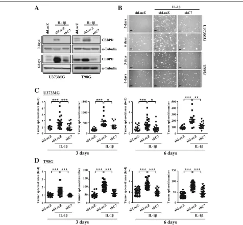
CEBPD contributes to the glioma stem cell spheroid formation under IL-1βtreatment
Previous studies showed that GSCs were the key drivers of glioma malignancy and progression [1, 2, 4, 5]. According to the GEO database, CEBPD was found expressed in GBM at higher levels compared with those in the normal brain tissue, which is in line with our results showing the association of increased CEBPD ex-pression with GSCs spheroid formation upon IL-1β treatment. To test this idea of CEBPD involvement in the GSCs spheroid formation induced by IL-1β, we gen-erated U373MG and T98G cells that had stable knock-down of CEBPD by lentiviral transduction of shCEBPD. Cells transduced with shLacZ were served as the sham control (Fig. 2a and Additional file 1: Figure S1a). As shown in Fig.2b–d and Additional file1: Figures S1 and S2, the knockdown of CEBPD in U373MG and T98G cells significantly reduced the area and number of tumor spheroids after IL-1β treatment. Similar results were observed in primary patient cells (Additional file 1: Figure S3). These data thus suggest that the expression of CEBPD is critical in glioma spheroid formation under IL-1βstimulation.
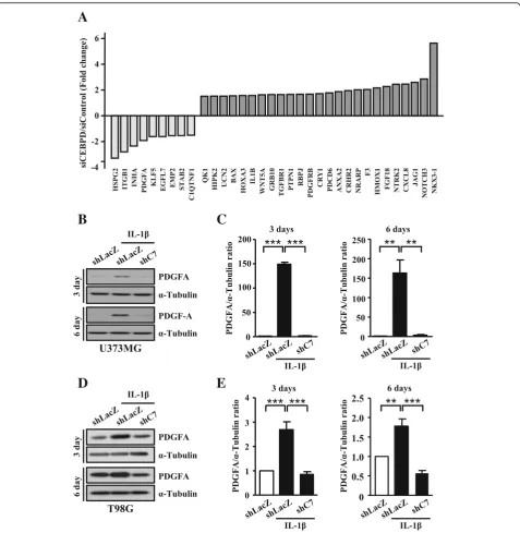
The identification of CEBPD-regulated genes in U373MG cells
To date, little was known about the relationship between CEBPD-regulated genes and stemness of GSCs. To identify the critical genes regulated by CEBPD in glioma, genome-wide profiling and comparisons were conducted using U373MG cells with and without the siRNA-mediated knockdown of CEBPD. As shown in Additional file 1: Table S1, we found that the expression of 342 genes were significantly reduced, and 510 genes were significantly in-creased in U373MG cells with CEBPD knockdown. Among these CEBPD-responsive genes, PDGFA (platelet-derived growth factor subunit A), downregulated in CEBPD-knockdown U373MG cells (Fig.3a), was of great interest in our study because of its critical role in tumor progression [21,22].
PDGFA gene is a direct target of CEBPD
Previous studies have shown that PDGFA, which binds and activates PDGF receptor tyrosine kinases, promotes cancer cell proliferation, invasion, and self-renewal [2,23]. However, the role of PDGFA in glioma in the inflamma-tory environment remains to be elucidated. We found that IL-1β-induced increase of PDGFA protein expression was remarkably attenuated in CEBPD-knockdown U373MG and T98G spheroid cells (Fig. 3b–e). PDGFA mRNA expression also showed a similar result in U373MG spher-oid cells with or without IL-1β treatment and CEBPD knockdown (Fig. 4a). Combined, these results suggest a strong link between PDGFA and CEBPD. Due to CEBPD’s function as a transcription factor, we then investigated if PDGFA was a downstream target of CEBPD. Using lu-ciferase reporter assay, we identified a potent CEBPD-responsive region in PDGFA promoter at −538/+ 70 bp (Fig.4b). In addition, in vivo DNA binding assay showed that the binding of CEBPD on thePDGFApromoter was observed in the presence of IL-1βin U373MG spheroid cells (Fig.4c). These data thus suggest that CEBPD regu-lates PDGFA transcription by binding to the PDGFA promoter region.
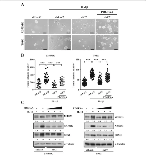
Recombinant protein PDGFAA rescues glioma stem cell properties in CEBPD-lacking spheroid cells
PDGFAA also partially reversed the effect of CEBPD knockdown-mediated downregulation of CD133, SOX2, and NANOG expressions (Fig. 5c). Combined, these ob-servations suggest that CEBPD regulates the GSCs stem-ness through PDGFA under inflammatory stimulation.
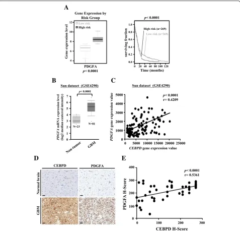
PDGFA expression is associated with poorer prognosis in GBM patients
We next examined whether PDGFA expression was as-sociated with the histopathology of glioma and patient
prognosis. The overall survival of patients with low PDGFA expression exhibited better prognosis than those with high PDGFA expression (Fig. 6a). Con-sistently, glioma samples expressed higherPDGFAthan non-tumor brain tissue (Fig. 6b). Similar results have been observed in the Bredel and Lee databases (Additional file 1: Figure S4a). We also found that the expression level of PDGFAsignificantly correlated with the expression level ofCEBPD in these glioma samples (Fig.6c and Additional file1: Figure S4b). Furthermore,
A
B
C
D
we examined the protein expression of PDGFA and CEBPD in tissue arrays of human glioma samples by immunohistochemical analyses. We found that the expression of PDGFA was significantly higher in glioma samples and was correlated with the CEBPD expression levels in these glioma samples (Fig.6d, e and Additional file1: Table S2). Taken together, these data suggest that the expression of PDGFA is increased in glioma and is correlated with CEBPD expression.
Discussion
The tumor microenvironment influences cancer ini-tiation, promotion, and malignancy [1, 3, 24]. Appro-ximately 30–50% of the cells in gliomas are microglia or macrophages, and the tumor-associated macrophages/ microglia (TAM) plays crucial roles in tumor growth, invasion, and metastasis [3]. TAM can secrete inflamma-tory factors and promote cancer stem cell formation. However, the mechanism of how inflammation induces
A
C
D
B
GSC spheroid formation still remains largely unclear. Our data demonstrate a novel function of CEBPD, which promotes glioma stem cell spheroid formation under the inflammatory environment. We show that CEBPD is responsive to IL-1β stimulation and enhances the
transcription and expression of PDGFA, which, in turn, promotes glioma stemness. IL-1β, IL-6, TGF-β, and EGF have been shown to promote glioma invasion from tumor-associated macrophage secretion [3]. Our pre-vious studies also show that these factors can also
A
B
C
D
E
induce CEBPD expression [9, 10,17]. However, whether inflammatory cytokines promote stemness is still un-clear. Herein, we provide evidences showing the upregu-lation of PDGFA expression and promotion of glioma stemness by CEBPD under inflammatory stimulation.
CEBPD plays a functional role in tumor formation and progression. Low expression of CEBPD in a variety of cancers has been observed, including hepatocellular car-cinoma and cervical cancer, and the hypermethylation in the CEBPD promoter mediated by polycomb group complex and DNA methyltransferase contributes to the silencing of CEBPD expression at this context [15, 25]. The overexpression of CEBPD in these cancers can in-duce cancer cell apoptosis and inhibit cell growth, thus implying CEBPD’s role as a tumor suppressor. On the
other hand, recent studies have demonstrated that CEBPD plays an oncogenic role when endogenous CEBPD expression is relatively higher or during indu-cible CEBPD expression in some cancer types, such as bladder cancer and lung cancer [17,26]. In these cancer cells, CEBPD can promote drug resistance, invasion, and lymphangiogenesis [17, 26]. However, the function of CEBPD in glioma stem cell spheroid formation remains less well understood. The GEO dataset shows that CEBPD is highly expressed in GBM compared to normal brain tissue. Additionally, we demonstrate that IL-1β can induce CEBPD transcription and expression in gli-oma cells, which, in turn, promotes gligli-oma stemness. Our observations thus favor the model of CEBPD func-tion promoting tumor growth in glioma.
A
C
B
A
B
C
A previous study suggests that constitutively active STAT3 confers cellular transformation, survival, prolifer-ation, anti-apoptosis, and chemosensitivity in several hu-man malignancies [17]. Moreover, STAT3 is known for its functions in regulating stem cell transcription factors and to maintain the stem cell phenotype and growth and proliferation of GSCs [2, 27]. In a bladder urothelial
carcinoma model, CEBPD expression is upregulated by STAT3 activation and promotes cancer drug resistance [17]. It is thus possible that STAT3 not only regulates the stemness transcription factors directly, but also in-directly regulates the stemness transcription factors through CEBPD expression. On the other hand, STAT3 activation can be stimulated through inflammatory factors
A
B
C
D
E
and cytokines, such as IL-6 and EGF [17,28]. Studies also show that IL-1βcan activate STAT3 in osteoblasts, Th17, and glioma [29–31]. According to bioinformatic datasets and previous studies, the glioma-associated microenviron-ment can increase expression levels of inflammatory factors and cytokines [1,3]. We thus propose a model of signaling pathways in the STAT3/CEBPD/PDGFA axis mediating inflammatory factor-induced stemness in glioblastoma. Further investigation of STAT3 function in glioblastoma could thus provide a better understanding of the detailed mechanism of inflammatory factor-induced stemness at this context.
PDGFA signaling maintains glioma stem cell growth and self-renewal [2, 23]. The stimulation of PDGFA signaling has been observed in GBMs and is associated with tumor initiation and malignant progression. Further-more, PDGFA signaling can also increase STAT3 activa-tion through autocrine or paracrine PDGFAA stimulaactiva-tion [2, 32]. PDGF can form various homo- or heterodimers, such as PDGFA/A, PDGFB/B, PDGFA/B, PDGFC/C, and PDGFD/D [21, 22]. Activation of two structurally related cell membrane tyrosine kinase receptors (PDGFR-A and PDGFR-B) can affect various physiological and patho-logical functions through PDGF binding [21,22]. Our data show that CEBPD affects PDGFRA expression through CEBPD knockdown array profile (transient and stable CEBPD-knockdown U373MG cells). In the future, we will continue investigating how CEBPD regulates PDGFRA expression in glioma.
Interestingly, we show thatIL-1β is upregulated when CEBPD expression is decreased in the siRNA experi-ment on U373 cells (Fig.3a). Our previous works show that CEBPD responsive miRNAs, including miR-4300 and miR-135a, are increased in CEBPD-overexpressing U373MG cells [33]. According to microRNA target pre-diction softwares (Targetscan, miRWalk, and miRanda), miR-4300 and miR-135a can target toIL-1β3′UTR and may further attenuate IL-1β expression. On the other hand, CEBPD knockdown has been found to enhance the expression of several IL-1β-induced inflammatory genes including interleukin-8, intracellular adhesion molecule-1, monocyte chemoattractant protein-1, and IL-1β in human brain pericytes [34]. IL-1β is a potent inflammatory stimulus, and its induction following CEBPD knockdown would appear to contradict its anti-inflammatory roles in astrocytes. Whether these changes induced by CEBPD knockdown actually lead to altered inflammatory events in vivo is still unclear, and further studies investigating the underlying mechanisms con-trolling this response are required.
Conclusions
In this study, we have demonstrated that IL-1β-induced CEBPD promotes glioma stemness in U373MG, T98G,
and PT#3-spheroid cells by elevating PDGFA expression. In addition, our data validate the suppression of CEBPD by knockdown system to inhibit the stemness effects through inhibition of PDGFA-induced stem cell markers (Additional file 1: Figure S5). This finding thus high-lights the importance of CEBPD and PDGFA regulation and implies both as potential targets for novel thera-peutic strategies against malignant glioma in inflamma-tory environments.
Additional file
Additional file 1:Table S1.CEBPD-responsive genes in U373MG cells. Table S2.H-score data shown each of the patient IHC images.Figure S1.CEBPD contributes to self-renewal in U373MG and T98G spheroid cells under IL-1βtreatment.Figure S2.Transient knockdown of CEBPD in U373MG cells reduces tumor sphere formation and attenuates stem cell transcription factor expression.Figure S3.Transient knockdown of CEBPD in PT#3 cells reduces tumor spheroid formation and attenuates stem cell transcription factor expression.Figure S4.PDGFA expression is clinically relevant in GBM patients.Figure S5.A schematic diagram illustrating the proposed model for the modulatory effect of CEBPD on glioma stem-like cell formation after IL-1βtreatment. (PPTX 6917 kb)
Abbreviations
CEBPD:CCAAT/enhancer-binding protein delta; GBM: Glioblastoma multiforme; GSCs: Glioma stem-like cells; IL-1β: Interleukin-1 beta; PDGFA: Platelet-derived growth factor subunit A; TAM: Tumor-associated macrophages/microglia; TNF-α: Tumor necrosis factor-alpha
Acknowledgements
We thank Yi-Chen Hsieh for the help in data analysis and Yi-Chao Lee for the technical assistance.
Authors’contributions
SM-W, HY-L, YL-C, and JY-C performed the experiments and analyzed the data. TI-H, TJ-K, and CY-K wrote the manuscript and organized all the figures. All authors read and approved the final manuscript.
Funding
This work was supported by the grants TMU105-AE1-B07 from Taipei Medical University and MOST 105-2320-B-038-068-MY2 and MOST 107-2320-B-038-002 from the Ministry of Science and Technology, Taiwan. This work was also supported by the“TMU Research Center of Cancer Translational Medicine” from The Featured Areas Research Center Program within the framework of the Higher Education Sprout Project by the Ministry of Education (MOE) in Taiwan.
Availability of data and materials
The datasets generated and analyzed during the current study are available from the corresponding authors on reasonable request.
Ethics approval and consent to participate
The use of human specimens was approved by the Institute Review Board (IRB)/Ethics Committee from the office of human research in Taipei Medical University (Taipei, Taiwan). The consent of the patient was obtained and approved by Taipei Medical University IRB protocols (N201006011).
Consent for publication Not applicable.
Competing interests
The authors declare that they have no competing interests.
Author details
Program for Neural Regenerative Medicine, College of Medical Science and Technology, Taipei Medical University and National Health Research Institutes, Taipei, Taiwan.3Department of Pathology, Cardinal Tien Hospital, School of Medicine College of Medicine, Fu Jen Catholic University, New Taipei City, Taiwan.4Research Center of Neuroscience, Taipei Medical University, Taipei, Taiwan.5TMU Research Center of Cancer Translational Medicine, Taipei, Taiwan.
Received: 16 January 2019 Accepted: 1 July 2019
References
1. Charles NA, Holland EC, Gilbertson R, Glass R, Kettenmann H. The brain tumor microenvironment. Glia. 2011;59(8):1169–80.
2. Cheng P, Wang J, Waghmare I, Sartini S, Coviello V, Zhang Z, et al. FOXD1-ALDH1A3 signaling is a determinant for the self-renewal and tumorigenicity of mesenchymal glioma stem cells. Cancer Res. 2016;76(24):7219–30. 3. Hambardzumyan D, Gutmann DH, Kettenmann H. The role of microglia
and macrophages in glioma maintenance and progression. Nat Neurosci. 2016;19(1):20–7.
4. Jackson M, Hassiotou F, Nowak A. Glioblastoma stem-like cells: at the root of tumor recurrence and a therapeutic target. Carcinogenesis. 2015;36(2):177–85. 5. Ko CY, Lin CH, Chuang JY, Chang WC, Hsu TI. MDM2 degrades deacetylated
nucleolin through ubiquitination to promote glioma stem-like cell enrichment for chemotherapeutic resistance. Mol Neurobiol. 2018;55(4):3211–23. 6. Barrett LE, Granot Z, Coker C, Iavarone A, Hambardzumyan D, Holland EC, et
al. Self-renewal does not predict tumor growth potential in mouse models of high-grade glioma. Cancer Cell. 2012;21(1):11–24.
7. Guan X, Hasan MN, Maniar S, Jia W, Sun D. Reactive astrocytes in glioblastoma multiforme. Mol Neurobiol. 2018;55(8):6927–38.
8. Garofalo S, Porzia A, Mainiero F, Di Angelantonio S, Cortese B, Basilico B, et al. Environmental stimuli shape microglial plasticity in glioma. Elife. 2017;6: e33415-42.
9. Ko CY, Chang LH, Lee YC, Sterneck E, Cheng CP, Chen SH, et al. CCAAT/ enhancer binding protein delta (CEBPD) elevating PTX3 expression inhibits macrophage-mediated phagocytosis of dying neuron cells. Neurobiol Aging. 2012;33(2):422 e11–25.
10. Wang SM, Hsu JC, Ko CY, Chiu NE, Kan WM, Lai MD, et al. Astrocytic CCAAT/enhancer-binding protein delta contributes to glial scar formation and impairs functional recovery after spinal cord injury. Mol Neurobiol. 2016;53(9):5912–27.
11. Wang SM, Lee YC, Ko CY, Lai MD, Lin DY, Pao PC, et al. Increase of zinc finger protein 179 in response to CCAAT/enhancer binding protein delta conferring an antiapoptotic effect in astrocytes of Alzheimer’s disease. Mol Neurobiol. 2015;51(1):370–82.
12. Lai HY, Hsu LW, Tsai HH, Lo YC, Yang SH, Liu PY, et al. CCAAT/enhancer-binding protein delta promotes intracellular lipid accumulation in M1 macrophages of vascular lesions. Cardiovasc Res. 2017;113(11):1376–88. 13. Wang SM, Lim SW, Wang YH, Lin HY, Lai MD, Ko CY, et al. Astrocytic CCAA
T/enhancer-binding protein delta contributes to reactive oxygen species formation in neuroinflammation. Redox Biol. 2018;16:104–12.
14. Chuang CH, Wang WJ, Li CF, Ko CY, Chou YH, Chuu CP, et al. The combination of the prodrugs perforin-CEBPD and perforin-granzyme B efficiently enhances the activation of caspase signaling and kills prostate cancer. Cell Death Dis. 2014;5:e1220.
15. Li CF, Tsai HH, Ko CY, Pan YC, Yen CJ, Lai HY, et al. HMDB and 5-AzadC combination reverses tumor suppressor CCAAT/enhancer-binding protein delta to strengthen the death of liver Cancer cells. Mol Cancer Ther. 2015;14(11):2623–33.
16. Wang YH, Wu WJ, Wang WJ, Huang HY, Li WM, Yeh BW, et al. CEBPD amplification and overexpression in urothelial carcinoma: a driver of tumor metastasis indicating adverse prognosis. Oncotarget. 2015;6(31):31069–84. 17. Wang WJ, Li CF, Chu YY, Wang YH, Hour TC, Yen CJ, et al. Inhibition of the EGFR/
STAT3/CEBPD Axis reverses cisplatin cross-resistance with paclitaxel in the urothelial carcinoma of the urinary bladder. Clin Cancer Res. 2017;23(2):503–13. 18. Bredel M, Bredel C, Juric D, Harsh GR, Vogel H, Recht LD, et al.
Functional network analysis reveals extended gliomagenesis pathway maps and three novel MYC-interacting genes in human gliomas. Cancer Res. 2005;65(19):8679–89.
19. Sun L, Hui AM, Su Q, Vortmeyer A, Kotliarov Y, Pastorino S, et al. Neuronal and glioma-derived stem cell factor induces angiogenesis within the brain. Cancer Cell. 2006;9(4):287–300.
20. Lee J, Kotliarova S, Kotliarov Y, Li A, Su Q, Donin NM, et al. Tumor stem cells derived from glioblastomas cultured in bFGF and EGF more closely mirror the phenotype and genotype of primary tumors than do serum-cultured cell lines. Cancer Cell. 2006;9(5):391–403.
21. Cao Y. Multifarious functions of PDGFs and PDGFRs in tumor growth and metastasis. Trends Mol Med. 2013;19(8):460–73.
22. Westermark B, Heldin CH, Nister M. Platelet-derived growth factor in human glioma. Glia. 1995;15(3):257–63.
23. Xu R, Ji J, Zhang X, Han M, Zhang C, Xu Y, et al. PDGFA/PDGFRalpha-regulated GOLM1 promotes human glioma progression through activation of AKT. J Exp Clin Cancer Res. 2017;36(1):193.
24. Sin WC, Aftab Q, Bechberger JF, Leung JH, Chen H, Naus CC. Astrocytes promote glioma invasion via the gap junction protein connexin43. Oncogene. 2016;35(12):1504–16.
25. Ko CY, Hsu HC, Shen MR, Chang WC, Wang JM. Epigenetic silencing of CCAAT/enhancer-binding protein delta activity by YY1/polycomb group/ DNA methyltransferase complex. J Biol Chem. 2008;283(45):30919–32. 26. Min Y, Ghose S, Boelte K, Li J, Yang L, Lin PC. C/EBP-delta regulates VEGF-C
autocrine signaling in lymphangiogenesis and metastasis of lung cancer through HIF-1alpha. Oncogene. 2011;30(49):4901–9.
27. Guryanova OA, Wu Q, Cheng L, Lathia JD, Huang Z, Yang J, et al. Nonreceptor tyrosine kinase BMX maintains self-renewal and tumorigenic potential of glioblastoma stem cells by activating STAT3. Cancer Cell. 2011;19(4):498–511.
28. Brooks GD, McLeod L, Alhayyani S, Miller A, Russell PA, Ferlin W, et al. IL6 trans-signaling promotes KRAS-driven lung carcinogenesis. Cancer Res. 2016;76(4):866–76.
29. Mori T, Miyamoto T, Yoshida H, Asakawa M, Kawasumi M, Kobayashi T, et al. IL-1beta and TNFalpha-initiated IL-6-STAT3 pathway is critical in mediating inflammatory cytokines and RANKL expression in inflammatory arthritis. Int Immunol. 2011;23(11):701–12.
30. Tanabe K, Nishimura K, Dohi S, Kozawa O. Mechanisms of interleukin-1beta-induced GDNF release from rat glioma cells. Brain Res. 2009;1274:11–20. 31. Whitley SK, Balasubramani A, Zindl CL, Sen R, Shibata Y, Crawford GE,
et al. IL-1R signaling promotes STAT3 and NF-kappaB factor recruitment to distal cis-regulatory elements that regulate Il17a/f transcription. J Biol Chem. 2018;293(41):15790–800.
32. Gong AH, Wei P, Zhang S, Yao J, Yuan Y, Zhou AD, et al. FoxM1 drives a feed-forward STAT3-activation signaling loop that promotes the self-renewal and tumorigenicity of glioblastoma stem-like cells. Cancer Res. 2015;75(11):2337–48.
33. Chu YY, Ko CY, Wang WJ, Wang SM, Gean PW, Kuo YM, et al. Astrocytic CCAAT/enhancer binding protein delta regulates neuronal viability and spatial learning ability via miR-135a. Mol Neurobiol. 2016;53(6):4173–88. 34. Rustenhoven J, Scotter EL, Jansson D, Kho DT, Oldfield RL, Bergin PS, et
al. An anti-inflammatory role for C/EBPdelta in human brain pericytes. Sci Rep. 2015;5:12132.
Publisher’s Note