R E S E A R C H
Open Access
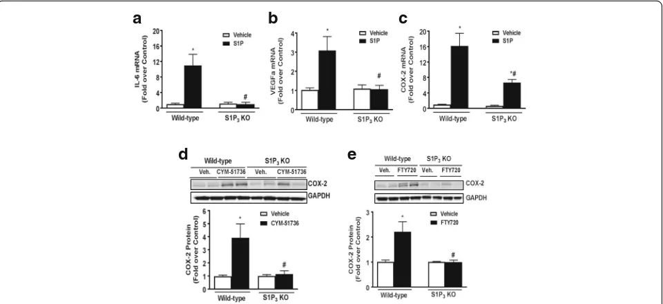
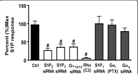
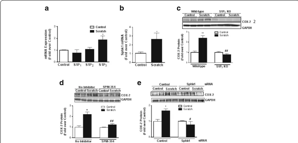
Sphingosine 1-phosphate receptor 3 and
RhoA signaling mediate inflammatory gene
expression in astrocytes
Stephanie S. Dusaban
1, Jerold Chun
2, Hugh Rosen
3, Nicole H. Purcell
1*and Joan Heller Brown
1*Abstract
Background:
Sphingosine 1-phosphate (S1P) signals through G protein-coupled receptors to elicit a wide range of
cellular responses. In CNS injury and disease, the blood-brain barrier is compromised, causing leakage of S1P from
blood into the brain. S1P can also be locally generated through the enzyme sphingosine kinase-1 (Sphk1). Our
previous studies demonstrated that S1P activates inflammation in murine astrocytes. The S1P
1receptor subtype has
been most associated with CNS disease, particularly multiple sclerosis. S1P
3is most highly expressed and upregulated
on astrocytes, however, thus we explored the involvement of this receptor in inflammatory astrocytic responses.
Methods:
Astrocytes isolated from wild-type (WT) or S1P
3knockout (KO) mice were treated with S1P
3selective drugs or
transfected with short interfering RNA to determine which receptor subtypes mediate S1P-stimulated inflammatory
responses. Interleukin-6 (IL-6), and vascular endothelial growth factor A (VEGFa) messenger RNA (mRNA) and
cyclooxygenase-2 (COX-2) mRNA and protein were assessed by q-PCR and Western blotting. Activation of RhoA was
measured using SRE.L luciferase and RhoA implicated in S1P signaling by knockdown of G
α
12/13proteins or by inhibiting
RhoA activation with C3 exoenzyme
.
Inflammation was simulated by in vitro scratch injury of cultured astrocytes.
Results:
S1P
3was highly expressed in astrocytes and further upregulated in response to simulated inflammation. Studies
using S1P
3knockdown and S1P
3KO astrocytes demonstrated that S1P
3mediates activation of RhoA and induction of
COX-2, IL-6, and VEGFa mRNA, with some contribution from S1P
2. S1P induces expression of all of these genes through
coupling to the G
α
12/13proteins which activate RhoA. Studies using S1P
3selective agonists/antagonists as well as
Fingolimod (FTY720) confirmed that stimulation of S1P
3induces COX-2 expression in astrocytes. Simulated inflammation
increased expression of Sphk1 and consequently activated S1P
3, demonstrating an autocrine pathway through which S1P
is formed and released from astrocytes to regulate COX-2 expression.
Conclusions:
S1P
3, through its ability to activate RhoA and its upregulation in astrocytes, plays a unique role in inducing
inflammatory responses and should be considered as a potentially important therapeutic target for CNS disease
progression.
Keywords:
Astrocytes, Central nervous system, Inflammation, RhoA, S1P, S1P
3* Correspondence:npurcell@ucsd.edu;jhbrown@ucsd.edu
1Department of Pharmacology, School of Medicine, University of California
San Diego, 9500 Gilman Drive, Biomedical Sciences Building Room 3024, La Jolla, CA 92093-0636, USA
Full list of author information is available at the end of the article
Background
Sphingosine 1-phosphate (S1P) is a bioactive lipid and G
protein-coupled receptor (GPCR) ligand formed within the
brain from sphingomyelin and is also present at high levels
in blood where it is bound to lipoproteins and stored in
erythrocytes [1
–
5]. There are five S1P receptor subtypes [6,
7] with S1P
1, S1P
2, S1P
3, and S1P
5(and in some reports
S1P
4) expressed in the CNS [8
–
13]. Astrocytes are activated
in response to CNS injury and diseases like multiple
scler-osis (MS) and undergo astrogliscler-osis characterized by
in-creases in proliferation, hypertrophy, and glial fibrillary
acidic protein (GFAP) expression [14
–
20]. S1P induces
astrogliosis when injected into the brain as evidenced by
in-creases in GFAP expression and astrocyte proliferation
[21
–
23]. The importance of S1P receptors in disease is
highlighted by the widespread acceptance of Fingolimod
(FTY720; Gilenya) as a first line oral drug to treat MS [24
–
27]. Phosphorylated fingolimod functions as an S1P
analogue that blocks lymphocyte egress through functional
inhibition of S1P
1signaling [28, 29]. Its efficacy in the EAE
mouse model of MS has also been linked to signaling
through S1P
1on astrocytes [30].
The predominant S1P receptor subtype detected by
quantitative-PCR (q-PCR) in cortical astrocytes is S1P
3,
al-though S1P
1is also expressed on astrocytes from rat and
mouse brain [8, 12, 31]. The potential importance of S1P
3signaling in astrocytes is suggested by the finding that this
receptor is upregulated in MS lesions and in response to
in-flammatory stimuli [32
–
34]. In a mouse model of Sandhoff
disease characterized by neuronal death and astrocyte
pro-liferation, deletion of S1P
3, along with the enzyme
sphingo-sine kinase (Sphk) which catalyzes the synthesis of S1P,
decreased astrogliosis and disease severity [35]. Importantly,
whereas S1P
1exclusively couples to the G protein G
α
i,
S1P
3couples promiscuously and its coupling to G
α
12/13ac-tivates the small G-protein RhoA [36
–
38]. Previous work
from our laboratory documented the importance of RhoA
activation in inducing astrocyte proliferation, gene
expres-sion, and inflammation in response to stimulation of
GPCRs for thrombin and S1P [39
–
44].
Here, we ask whether stimulation of the S1P
3recep-tor on astrocytes activates RhoA, is responsible for
in-flammatory gene expression, or can be locally engaged
by endogenously formed S1P in an in vitro model of
neuroinflammation. We demonstrate that S1P
3, and
not S1P
1, mediates induction of interleukin-6 (IL-6)
and vascular endothelial growth factor A (VEGFa)
mRNA, and cyclooxygenase-2 (COX-2) mRNA and
protein in mouse astrocytes and that this occurs
through S1P receptor coupling to G
α
12/13and RhoA.
We also demonstrate that simulated inflammation in
vitro leads to increases in expression of Sphk1 and S1P
3which could contribute to autocrine inflammatory
astrocyte signaling.
Methods
Agonists and inhibitors
Sources were as follows: S1P was obtained from Avanti
Polar Lipids. The S1P
3antagonist SPM-354 was synthesized
and characterized as described [45]. A cell permeable
botu-linum C3 toxin exoenzyme, which inhibits RhoA activation,
was obtained from cytoskeleton. Pertussis toxin, which
ribosylates and inactivates the alpha subunit of the G
ipro-tein, was used to block signaling through receptor coupling
to G
i(Tocris Bioscience). The S1P
3specific agonist
CYM-51736 was provided by the Rosen laboratory, and the
func-tional S1P
1antagonist and MS therapeutic, FTY720
(Fingo-limod),
and S1P
2antagonist JTE-013
was from Cayman
Chemicals. S1P was used at 0.5
μ
M. SPM-354 was used at
5
μ
M. C3 exoenzyme was used at 0.5
μ
g/mL. FTY720 was
used at 100 nM. CYM-51736 was used at 10
μ
M,
JTE-013
at 1
μ
M, and pertussis toxin at 100 ng/ml.
Animals
All procedures were performed in accordance with NIH
Guide and Care and Use of Laboratory Animals and
ap-proved by the Institutional Animal Care and Use
Commit-tee at the University of California San Diego. C57BL/6
wild-type and homozygous S1P
3KO mice which do not
ex-hibit any gross phenotypic abnormalities [36, 46] were used
for astrocyte isolation.
Primary culture of astrocytes
Astrocytes were isolated from P1-P3 postnatal WT and
S1P
3KO mice [44]. Purity of astrocytes was determined to
be ~95% based on GFAP staining. In all experiments, WT
and S1P
3KO astrocytes were used at passage 2. Astrocytes
were cultured in six-well plates, maintained in high-glucose
DMEM supplemented with 10% FBS/2 mM glutamine/
100 units/ml penicillin/100
μ
g/ml streptomycin (Invitrogen,
Carlsbad, CA) at 37 °C in a humidified 5% CO
2-incubator.
Cells used for experiments were at 80% confluency and
serum-starved for 18
–
24 h prior to agonist treatment.
siRNA transfections
Pre-designed mouse short interfering RNA (siRNA)
smart-pools for S1P
1, S1P
2, S1P
3, and control siRNA were
pur-chased from Bioneer. Pre-designed mouse siRNA for G
α
12,
G
α
13, G
α
q, and sphingosine kinase 1 were purchased from
fresh media. Following overnight incubation, media
con-taining siRNA was removed, and cells were washed.
Astro-cytes were serum-starved for 18
–
24 h prior to treatment.
The table below lists the predesigned or three smartpool
siRNA sequences used in these studies.
SRE.L luciferase assay
Astrocytes were cultured on 12-well plates and
trans-fected with 500 ng of SRE.L and 50 ng of Renilla as an
internal control using DharmaFECT-3 as described
above. Following overnight incubation, cells were
serum-starved for 18
–
24 h prior to S1P treatment for 8 h. Cells
were lysed, and luciferase activities were measured using
the Dual-Luciferase Reporter Assay System (Promega).
In vitro scratch
injury
model
WT or S1P
3KO astrocytes were cultured on six-well
plates and grown to confluence followed by serum
star-vation for 18
–
24 h. To stimulate astrogliosis and
inflam-mation, plates were scratched with a 200-
μ
L pipette tip
six times (three vertical and three horizontal) across the
dish [47]. Cells were harvested and lysed after 1 h
scratch for mRNA analysis or after 8 h for Western
blot-ting as described below.
q-PCR
For gene expression analyses, RNA was extracted from
as-trocytes using an RNeasy kit (Invitrogen) [40].
Complemen-tary DNA (cDNA) was synthesized with High-Capacity
cDNA Reverse Transcription Kit (Applied Biosystems ABI)
and real-time q-PCR performed with TaqMan Universal
Master Mix II, with UNG (Applied Biosystems ABI). To
analyze gene expression in mouse astrocytes treated with
S1P or scratch wounding, gene-specific primers for COX-2,
IL-6, VEGFa, S1P
1, S1P
2, S1P
3, Sphk1, and GAPDH (as an
internal control) were used (Integrated DNA Technologies).
S1P
3KO astrocytes were analyzed for the levels of S1P
1and
S1P
2and were found to have no significant compensatory
changes (data not shown). Data were normalized to internal
GAPDH, and fold change was determined according to a
published protocol [48]. Values for comparison for a single
gene across multiple samples was determined using cycle
threshold (Ct) data fitted to a standard curve. For
compari-son of multiple transcripts in a single sample, then the 2
−ΔΔCtmethod was applied to the Ct value [48].
Western blotting
Astrocytes were lysed in RIPA buffer (20 mm Tris, 250 mm
NaCl, 3 mm EDTA, 3 mm EGTA, and 20 mm
β
-glycerophosphate) supplemented with sodium vanadate,
leupeptin, aprotinin,
p
-nitrophenyl phosphate, and
phenyl-methylsulfonyl fluoride. BCA analysis was performed using
the Micro BCA Protein Assay Kit (ThermoFisher Scientific)
to determine protein concentration. Equal amounts of
pro-tein (10
μ
g) were loaded onto 4
–
12% 10-well or 15-well
SDS-PAGE gels (Invitrogen NuPage System). Gels were
transferred to PVDF membranes (Millipore), and the
result-ing blot was probed with specific antibodies. The COX-2
antibody (Cayman #160126) was used at 1:500 dilution, and
the band running at 72 kDa band was quantitated. The
GAPDH antibody (Cell Signaling Technology #2118) was
used at 1:1000 dilution, and a band at 37 kDa was
quanti-tated. Rabbit secondary antibody was used at 1:4000
dilu-tion. Fold changes were determined by densitometry and
normalized to accompanying GAPDH blots.
Statistical analysis
Statistical differences were determined using Tukey
’
s
multi-comparison analysis following one-way ANOVA with Prism
software (GraphPad).
p
< 0.05 was considered significant.
Results
S1P
3is highly expressed in astrocytes and mediates
increases in COX-2 protein expression
We used q-PCR to compare directly the levels of mRNA
expression for S1P
1-3in the cultured mouse astrocytes
used in the studies presented here. Relative expression
levels for S1P
3(4.7) > S1P
1(1.6) > S1P
2(0.7) mRNA were
established using absolute quantitative-PCR (Fig. 1a). To
examine S1P receptor subtype involvement in induction
of the inflammatory gene COX-2, all three receptor
sub-types were knocked down prior to treatment with S1P.
Knockdown with S1P
1siRNA (78% decrease in S1P
1mRNA) had no effect on COX-2 protein expression in
cells stimulated for 6 h with S1P. Knockdown of S1P
3(80%) significantly attenuated
S
1P-stimulated COX-2
ex-pression, and the combination of S1P
2and S1P
3knock-down was most effective (Fig. 1b). Knockknock-down of S1P
2(83%) diminished but did not significantly decrease
S1P-induced COX-2 protein expression nor did pretreatment
with the selective S1P
2antagonist JTE-013 (Fig. 1c). In
contrast, SPM-354, a bitopic antagonist that has a
siRNA Sense Sequence Antisense SequenceS1P1 GAUAUCAUAGUCCGGCAUU
CCGGAGCUUUGAUUUUGCA CGGACCUAUUAGCAGGCGU
AAUGCCGGACUAUGAUAUC UGCAAAAUCAAAGCUCCGG ACGCCUGCUAAUAGGUCCG
S1P2 CUGUACGUCCGAAUCUACU
CACUUCUGGAGUGCCAGUA CCUCGGUCUUUAGCCUCCU
AGUAGAUUCGGACGUACAG UACUGGCACUCCAGAAGUG AGGAGGCUAAAGACCGAGG
S1P3 UCUUGGUCACCUGUAGCUU
UGUACAGGAUGUAUACGAU AGACAUCGGGUGCAUCCAA
AAGCUACAGGUGACCAAGA AUCGUAUACAUCCUGUACA UUGGAUGCACCCGAUGUCU
Gα12 UGACUUCGUUAUAAAGAAATT UUUCUUUAUAACGAAGUCATG
Gα13 CCAUAAUCCUCUUCUUAAATT UUUAAGAAGAGGAUUAUGGAG
Gαq GGUGGAUAGUAUUAUCCUATT UAGGAUAAUACUAUCCACCAG
Sphingosine kinase 1
significantly higher affinity for S1P
3than for S1P
2(1840-fold) or S1P
1(30-fold) [45], decreased S1P-induced
COX-2 expression by more than 70% (Fig. 1d) further
confirming the predominant role of S1P
3activation in
COX-2 protein expression. The S1P
2and S1P
3receptor
subtypes can serve redundant functions in regulation of
RhoA and other downstream responses in some cell
sys-tems [36, 49] but our data indicates that S1P
3mRNA is
most highly expressed in mouse astrocytes and plays the
predominant role in mediating COX-2 protein
expres-sion in response to S1P.
S1P
3is required for induction of inflammatory genes in
astrocytes
To provide further evidence that S1P
3is the receptor on
astrocytes that is primarily responsible for the induction
of COX 2 and other inflammatory genes, astrocytes were
isolated from WT and S1P
3KO mice. The ability of S1P
to increase inflammatory gene mRNA was examined at
1 h treatment since it was demonstrated that mRNA for
IL-6 and COX-2 were significantly increased by S1P at
this early time [40]. Deletion of S1P
3fully prevented
S1P-mediated increases in IL-6 (Fig. 2a) and VEGFa
(Fig. 2b) mRNA. Induction of COX-2 mRNA (Fig. 2c)
was markedly but not fully attenuated paralleling the
changes in COX-2 protein expression shown in Fig. 1b
which suggests some redundancy in S1P
2and S1P
3sig-naling to COX2 expression. Next, we used a recently
generated S1P
3receptor allosteric agonist, CYM-51736,
which is more specific than the previous S1P
3agonists
[50, 51]. CYM-51736 increased COX-2 protein in WT
but not in S1P
3KO astrocytes (Fig. 2d), consistent with
its specificity and the ability of S1P
3activation to
regu-late COX-2 mRNA expression. Finally, we tested
FTY720 (fingolimod), which acts as an agonist at both
S1P
1and S1P
3. FTY720 treatment increased COX-2
pro-tein expression in WT but not in S1P
3KO astrocytes
(Fig. 2e), suggesting that its agonist actions at S1P
3in-duce astrocyte inflammatory genes.
S1P
3signals through G
α
12/13and RhoA to induce gene
expression
The G
α
12/13proteins are the G-protein family members
that most effectively couple GPCRs to RhoA exchange
COX-2 GAPDH Veh. Veh. S1P S1P No Inhibitor JTE-013
COX-2
GAPDH Veh. Veh. S1P S1P No Inhibitor SPM-354
a
b
c
d
Fig. 1S1P3is highly expressed in astrocytes and mediates COX-2 expression. In WT astrocytes,aS1P1, S1P2, and S1P3mRNA expression was
mea-sured by absolute PCR.bQuantification and Western blot of COX-2 protein expression after S1P treatment (0.5μM, 6 h) following knockdown of S1P1, S1P2, S1P3, and S1P2/3with siRNA (2μM). Data shown are mean ± SEM of values from three independent experiments run in triplicate.
COX-2 was normalized to GAPDH and expressed relative to control siRNA vehicle treated. *p< 0.05 and **p< 0.01 between vehicle and S1P-treated groups and #p< 0.05 and ##p< 0.01 between control siRNA S1P-treated and S1P receptor siRNA S1P-treated groups.cQuantification and Western blot of COX-2 expression after pretreatment with the S1P2antagonist JTE-013 for 30 min (1μM) followed by S1P treatment (0.5μM, 6 h). Data
shown are mean ± SEM of values from three independent samples. COX-2 was normalized to GAPDH and expressed relative to vehicle control. *p< 0.05 between vehicle and S1P-treated groups.dQuantification and Western blot of COX-2 expression after pretreatment with the S1P3
factors and thus to activation of RhoA. To demonstrate
that S1P
3activates inflammatory gene expression by
sig-naling through G
α
12/13, we used siRNAs to achieve
com-bined knockdown of G
α
12and G
α
13(92% decrease in
G
α
12mRNA and 90% decrease in G
α
13mRNA). In
addition, RhoA was functionally inhibited by
pretreat-ment of cells with C3 exoenzyme. Both interventions
significantly decreased S1P-mediated increases in
COX-2 protein (Fig. 3a, b). We further demonstrated that
in-duction of COX-2, IL-6, and VEGFa mRNAs by S1P
were attenuated by knockdown of G
α
12and G
α
13(Fig. 3c
–
e).
The SRE.L luciferase reporter gene contains a
trun-cated TCF-independent binding site for serum response
factor (SRF) and is widely used as a readout for activated
RhoA, which regulates genes through SRF and its
tran-scriptional co-activator MRTF-A [52, 53]. S1P markedly
increased SRE.L luciferase activity (16 to 50-fold,
de-pending on the experiment). The S1P response (shown
as 100% in the averaged experiments in Fig. 4) was
at-tenuated by functional blockade of Rho signaling with
C3 treatment and by knockdown of S1P
2, S1P
3, and
G
α
12/13, but not by knockdown of S1P
1or G
α
q.
Inhib-ition of G
α
ifunction by pretreatment with pertussis toxin
(PTX) was also without effect on S1P-stimulated SRE.L
activation. These data demonstrate S1P
3and S1P
2coupling to G
α
12/13to activate RhoA, which in turn
regu-lates COX-2 and other inflammatory genes in astrocytes.
S1P
3and Sphk1 are upregulated in response to in vitro
wounding and mediate COX-2 expression
To determine whether the signaling pathway delineated
above could be activated under pathophysiological
con-ditions, we used an in vitro scratch injury model to
simulate localized inflammation of cultured astrocyte
[47]. Within an hour after cells were scratched, S1P
3(but not S1P
1or S1P
2) mRNA was increased relative to
control unscratched cells (Fig. 5a) as was the mRNA
level for sphingosine kinase 1 (Sphk1), the enzyme that
catalyzes the synthesis of S1P (Fig. 5b) [54]. Our
previ-ous studies showed that injuring astrocytes increases
COX-2 expression and that the media from scratched
cells contains substances that contribute to this response
[40]. To determine whether this localized inflammatory
response could be mediated by the actions of S1P,
formed from the elevated Sphk1 and acting on astrocyte
S1P
3, we repeated the scratch injury studies using S1P
3KO astrocytes. The increase in COX-2 protein
expres-sion was lost, indicating that the ability of scratch to
elicit this response requires S1P
3(Fig. 5c). We confirmed
this further by comparing wild-type cells that were
sub-ject to scratch injury in the presence or absence of the
a
d
e
b
c
Fig. 2S1P3is required for induction of inflammation in astrocytes. WT and S1P3KO astrocytes were treated with vehicle or S1P (0.5μM) for 1 h, and IL-6
(a), VEGFa (b), and COX-2 (c) mRNA expression was measured by q-PCR. IL-6, VEGFa, and COX-2 were normalized to GAPDH, and fold increase expressed relative to the WT or KO vehicle treated. Data shown are mean ± SEM of three independent experiments run in triplicate. *p< 0.01 between vehicle and S1P-treated groups and #p< 0.01 between WT and KO S1P-treated groups.dQuantification and Western blot of COX-2 expression in WT and S1P3KO
astrocytes treated with CYM-51736 (10μM, 6 h). Data shown are mean ± SEM of values from three independent experiments run in triplicate. COX-2 was normalized to GAPDH and expressed relative to the WT or KO vehicle treated.eQuantification and Western blot of COX-2 expression in FTY720-treated (100 nM, 6 h) WT and S1P3KO astrocytes. Data shown are mean ± SEM of values from three independent experiments run in triplicate.
S1P
3inhibitor SPM-354 used in Fig. 1d. Pharmacological
blockade of S1P
3, like genetic deletion of the receptor,
prevented scratch-induced COX-2 expression (Fig. 5d).
Finally, to test the importance of localized formation of
S1P, we knocked down Sphk1 using siRNA (90%
reduc-tion in Sphk1). Scratch injury failed to increase COX-2
protein expression when Sphk1 was downregulated
(Fig. 5e) implicating this enzyme, its product, and S1P
3activation in localized astrocyte COX-2 induction.
Discussion
Neuroinflammation, which underlies many
neurodegen-erative processes including those involved in Alzheimer
’
s
disease, Parkinson
’
s disease, and MS, is increasingly
rec-ognized as a hallmark of CNS pathology [19, 20, 55
–
58].
Astrocytes were once considered as structural elements
in the brain but subsequently emerged as functionally
important for neuronal guidance, maintenance of the
BBB, and structural and metabolic support of neurons
[15, 17, 18]. In addition, astrocytes, like microglia, are
now known to contribute to neuroinflammation [14, 17,
19, 56, 59]. The lysophospholipid S1P regulates
astro-gliosis and inflammatory responses in the CNS; however,
the role of the individual S1P receptor subtypes in these
processes has not been clearly delineated [23, 30, 31, 33,
34, 60, 61].
Astrocytes contribute to neuroinflammation by
upreg-ulating proinflammatory mediators such us IL-6,
MCP-1, TNF-
α
, iNOS, and COX-2 [15, 17, 40, 62]. Induction
of COX-2 in astrocytes increases generation of reactive
oxygen species (ROS), as well as formation of
prosta-noids that play a prominent role in inflammation, and
thus further contribute to neuronal cell death and
de-myelination in diseases such as MS [20, 56]. Moreover,
Fig. 3S1P signals through Gα12/13and RhoA to induce COX-2 expression.aCOX-2 protein expression was measured in WT cells pretreated with C3
exoenzyme (0.5μg/mL) for 4 h prior to vehicle or S1P (0.5μM) treatment for 6 h. COX-2 was normalized to GAPDH and expressed relative to the averaged ± inhibitor controls. Representative Western blot and data shown are mean ± SEM from three independent experiments run in triplicate. The
blotrepresents a single gel where unnecessary lanes have been removed. **p< 0.01 between vehicle and S1P-treated groups and ##p< 0.01 between S1P-treated ± inhibitor groups.bQuantification and Western blot of COX-2 protein levels after knockdown of Gα12/13with siRNA (2μM) followed by
S1P treatment (5μM, 6 h). The blot represents a single gel where unnecessary lanes have been removed. Data shown are mean ± SEM of values from four independent experiments run in triplicate. COX-2 was normalized to GAPDH and expressed relative to the siRNA control. mRNA expression levels of COX-2 (c), IL-6 (d), and VEGFa (e) were measured by q-PCR following knockdown of Gα12/13with siRNA (2μM) and S1P treatment (0.5μM, 1 h). Data
shown are mean ± SEM from three independent experiments run in triplicate. COX-2, IL-6, and VEGFa were normalized to GAPDH and fold increase expressed relative to the vehicle-treated control siRNA. *p< 0.05 and **p< 0.01 between vehicle and S1P-treated groups and #p< 0.05 and ##p< 0.01 between control and Gα12/13S1P-treated groups
Fig. 4S1P2, S1P3, and Gα12/13are required for Rho activation in
astrocytes. WT astrocytes were transfected with an SRE.L luciferase reporter construct to assess Rho activation following knockdown with control siRNA or siRNA (2μM) targeting S1P1, S1P2, S1P3, Gα12/
13, or Gαq. Cells were also treated with pertussis toxin (PTX, 100 ng/
mL, 24 h) to inhibit Gαior with the C3 exoenzyme (0.5μg/mL, 4 h)
astrocytes produce VEGF which plays a role in the
break-down of the blood-brain barrier, a step critical to the entry
of pathogenic lymphocytes into the brain [63
–
67]. Our
data demonstrate that an important mechanism for
induc-tion of inflammatory cytokines and cytotoxic mediators
such as IL-6, COX-2, and VEGFa in astrocytes is through
their exposure to S1P and activation of S1P
3.
Both S1P
1and S1P
3are expressed on astrocytes [8, 12]
and are upregulated on reactive astrocytes that
contrib-ute to inflammation associated with CNS disease [32, 33,
35, 61, 68]. In response to inflammatory stimuli or in
CNS pathologies, Sphk1, an enzyme that generates S1P,
is also increased in astroglial cells [23, 34, 35, 69, 70].
Our findings using siRNA and S1P
3KO astrocytes
dem-onstrate mechanistically that agonist binding to S1P
3sig-nals to inflammatory responses through S1P
3coupling
to G
α
12/13and activation of RhoA. We also show here,
using an astrocyte scratch injury assay, that S1P
3and
Sphk1 expression are increased by simulated
inflam-mation and demonstrate by their knockout and
down-regulation, respectively, that they are involved in an
autocrine signaling loop to increase COX-2 expression.
While S1P
2could also signal through G
α
12/13and RhoA
to contribute to COX-2 expression ([37, 71
–
73] and
Fig. 4) and appears to serve this role when S1P
3is
downregulated (Fig. 1b), the relatively low expression of
this receptor subtype and its lack of upregulation in
re-sponse to wounding suggests limited involvement in
astrocyte inflammatory responses (Fig. 1c). Thus, it
ap-pears that S1P
3, and its autocrine activation by S1P
gen-erated through Sphk1, are poised to mediate astrocytic
inflammatory responses that could contribute to the
progression of CNS neuropathology.
S1P signaling in the CNS has important
pathophysio-logical consequences [21, 28
–
30, 33
–
35, 40, 61, 74].
Much research has focused on S1P
1as the primary
target for the MS drug FTY720 (fingolimod). While a
well-recognized effect of fingolimod is to functionally
antagonize S1P
1receptors on lymphocytes and thereby
prevent their egress into the blood and access to the
brain, S1P
1localized to astrocytes contributes
signifi-cantly to the effects of this drug in an experimental
model of MS [75]. The basis for also considering S1P
3signaling in MS is that this receptor subtype is
upregu-lated in astrocytes during MS and in EAE and that it is a
target for fingolimod [33, 61]. Notably, fingolimod
causes transient bradycardia that appears, at least in the
mouse, to be due to its agonist actions on S1P
3[24, 25,
76
–
78]. While it is clear that fingolimod downregulates
S1P
1, and thus acts as a functional antagonist, its ability
2
a
d
e
b
c
Fig. 5S1P3and Sphk1 are upregulated in response to in vitro scratch injury and mediate COX-2 expression. In WT astrocytes,aS1P1, S1P2, S1P3
andbSphK1 mRNA expression were measured 1 h after scratch injury on culture plates. Data shown are mean ± SEM from three independent experiments run in triplicate. Fold increase is relative to the scratch control for each receptor subtype or Spkh1. *p< 0.05 between control and scratch-treated groups. COX-2 protein levels were measured after 8 h of scratch incS1P3KO astrocytes ordWT astrocytes pretreated with
to similarly downregulate and thus act as a functional
antagonist of S1P
3is controversial [61, 77, 79, 80]
.
A
re-cent study demonstrated that continuous treatment with
FTY20, initiated at the onset of disease in an EAE
model, reduced S1P
3expression at day 22 [61]. While
this indicates that S1P
3is downregulated by FTY720
treatment, this could reflect reversal of the disease
process/inflammation (and its accompanying increases
in S1P
3gene expression) rather than downregulation at
the receptor level. Our data with FTY720 (like that
examining bradycardia) demonstrate that FTY720 acts
as an agonist, eliciting COX-2 induction, over a period
of at least 6 h. Our data further establish that it is S1P
3-mediated RhoA signaling, not effects of S1P
1and G
α
i,
that lead to maladaptive astrocyte inflammation. Thus,
agonism at astrocyte S1P
3by fingolimod or other drugs
could contribute to neuroinflammation and worsen disease
progression, particularly when S1P
3are upregulated and
S1P availability increased through activation of sphingosine
kinase. Further studies using S1P
1/3double knockout mice
are ongoing and should indicate whether blocking S1P
3, in
addition to S1P
1, would have additional therapeutic benefit.
The importance of S1P
3and RhoA signaling in CNS
disease could be logically extended to consideration of
any of the myriad GPCRs found on astrocytes [32] that
couple to RhoA signaling. We and others have shown
that PAR1, the receptor for thrombin, couples through
RhoA to mediate proliferation and inflammatory
re-sponses in astrocytes [39, 40]. Thrombin is also
in-creased in the injured brain [81, 82], and an antagonist
of protease activated receptor 1 (PAR1) reduces clinical
symptoms in EAE mice [83]. Thus, the evidence that
S1P
3and other GPCRs that stimulate RhoA can
contrib-ute to sustained inflammatory responses suggests this
pathway as a critical target for blocking
neuroinflamma-tion in MS and other CNS diseases.
Conclusions
Our findings demonstrate that S1P
3and Sphk1 are
me-diators of inflammatory signaling and are upregulated in
astrocytes in response to injury. S1P
3couples to G
α
12/13and activated RhoA to induce COX-2, IL-6, and VEGFa
mRNA as well as COX-2 protein expression in
astro-cytes. The data suggest that blocking S1P
3, as well as the
clinically relevant S1P
1, could have therapeutic benefit
for limiting CNS inflammatory disease progression.
Abbreviations
BBB:Blood-brain barrier; CNS: Central nervous system;
COX-2: Cyclooxygenase 2; EAE: Experimental Autoimmune Encephalomyelitis; GFAP: Glial fibrillary acidic protein; GPCR: G protein-coupled receptor; IL-6: Interleukin 6; KO: Knockout; MS: Multiple Sclerosis; NF-κB: Nuclear factor kappa B; PAR1: Protease activated receptor 1; PTX: pertussis toxin; q-PCR: Quantitative-PCR; S1P: Sphingosine 1-phosphate, S1P1, Sphingosine
1-phosphate receptor 1; S1P2: Sphingosine 1-phosphate receptor 2;
S1P3: Sphingosine 1-phosphate receptor 3; S1P4: Sphingosine 1-phosphate
receptor 4; S1P5: Sphingosine 1-phosphate receptor 5; Sphk1: Sphingosine
kinase-1; VEGFa: Vascular Endothelial Growth Factor A; WT: Wild-type
Acknowledgements
We thank Melissa S. Barlow for her capable assistance with the animal breeding and genotyping and Jeffery M. Smith for the technical assistance.
Funding
This work was supported by National Institutes of Health Grants
T32GM007752 (to S.S.D.), HL114949 (to N.H.P.), MH084512 (to H.R.), DA019674 (to J.C.), and HL028143 and GM36927 (to J.H.B.).
Availability of data and materials Not applicable
Authors’contributions
SSD, NHP, and JHB designed of the study, carried out the experiments, and participated in the data analysis and manuscript preparation. JC and HR contributed to the data analysis and interpretation of the results. All authors reviewed the results and approved the final version of the manuscript.
Competing interests
The authors declare that they have no competing interests.
Consent for publication Not applicable
Ethics approval
All procedures were performed in accordance with NIH Guide and Care and Use of Laboratory Animals and approved by the Institutional Animal Care and Use Committee at the University of California San Diego.
Publisher
’
s Note
Springer Nature remains neutral with regard to jurisdictional claims in published maps and institutional affiliations.
Author details
1Department of Pharmacology, School of Medicine, University of California
San Diego, 9500 Gilman Drive, Biomedical Sciences Building Room 3024, La Jolla, CA 92093-0636, USA.2Sanford Burnham Prebys Medical Discovery Institute, La Jolla, CA 92037, USA.3Department of Chemical Physiology, The Scripps Research Institute, La Jolla, CA 92037, USA.
Received: 1 September 2016 Accepted: 17 May 2017
References
1. Camerer E, Regard JB, Cornelissen I, Srinivasan Y, Duong DN, Palmer D, Pham TH, Wong JS, Pappu R, Coughlin SR. Sphingosine-1-phosphate in the plasma compartment regulates basal and inflammation-induced vascular leak in mice. J Clin Invest. 2009;119:1871–9.
2. Anelli V, Bassi R, Tettamanti G, Viani P, Riboni L. Extracellular release of newly synthesized sphingosine-1-phosphate by cerebellar granule cells and astrocytes. J Neurochem. 2005;92:1204–15.
3. Pappu R, Schwab SR, Cornelissen I, Pereira JP, Regard JB, Xu Y, Camerer E, Zheng YW, Huang Y, Cyster JG, Coughlin SR. Promotion of lymphocyte egress into blood and lymph by distinct sources of sphingosine-1-phosphate. Science. 2007;316:295–8.
4. Yatomi Y, Ohmori T, Rile G, Kazama F, Okamoto H, Sano T, Satoh K, Kume S, Tigyi G, Igarashi Y, Ozaki Y. Sphingosine 1-phosphate as a major bioactive lysophospholipid that is released from platelets and interacts with endothelial cells. Blood. 2000;96:3431–8.
5. Yatomi Y, Igarashi Y, Yang L, Hisano N, Qi R, Asazuma N, Satoh K, Ozaki Y, Kume S. Sphingosine 1-phosphate, a bioactive sphingolipid abundantly stored in platelets, is a normal constituent of human plasma and serum. J Biochem. 1997;121:969–73.
6. Ishii I, Fukushima N, Ye X, Chun J. Lysophospholipid receptors: signaling and biology. Annu Rev Biochem. 2004;73:321–54.
8. Rao TS, Lariosa-Willingham KD, Lin FF, Palfreyman EL, Yu N, Chun J, Webb M. Pharmacological characterization of lysophospholipid receptor signal transduction pathways in rat cerebrocortical astrocytes. Brain Res. 2003;990:182–94.
9. Tham CS, Lin FF, Rao TS, Yu N, Webb M. Microglial activation state and lysophospholipid acid receptor expression. Int J Dev Neurosci. 2003;21: 431–43.
10. Toman RE, Payne SG, Watterson KR, Maceyka M, Lee NH, Milstien S, Bigbee JW, Spiegel S. Differential transactivation of sphingosine-1-phosphate receptors modulates NGF-induced neurite extension. J Cell Biol. 2004;166:381–92.
11. Yu N, Lariosa-Willingham KD, Lin FF, Webb M, Rao TS. Characterization of lysophosphatidic acid and sphingosine-1-phosphate-mediated signal transduction in rat cortical oligodendrocytes. Glia. 2004;45:17–27. 12. Zhang Y, Chen K, Sloan SA, Bennett ML, Scholze AR, O'Keeffe S, Phatnani
HP, Guarnieri P, Caneda C, Ruderisch N, Deng S, Liddelow SA, Zhang C, Daneman R, Maniatis T, Barres BA, Wu JQ. An RNA-sequencing transcriptome and splicing database of glia, neurons, and vascular cells of the cerebral cortex. J Neurosci. 2014;34:11929–47.
13. Choi JW, Chun J. Lysophospholipids and their receptors in the central nervous system. Biochim Biophys Acta. 2013;1:20–32.
14. Hamby ME, Sofroniew MV. Reactive astrocytes as therapeutic targets for CNS disorders. Neurotherapeutics. 2010;7:494–506.
15. Ridet JL, Malhotra SK, Privat A, Gage FH. Reactive astrocytes: cellular and molecular cues to biological function. Trends Neurosci. 1997;20:570–7. 16. Sofroniew MV. Molecular dissection of reactive astrogliosis and glial scar
formation. Trends Neurosci. 2009;32:638–47.
17. Sofroniew MV. Multiple roles for astrocytes as effectors of cytokines and inflammatory mediators. Neuroscientist. 2014;20:160–72.
18. Sofroniew MV, Vinters HV. Astrocytes: biology and pathology. Acta Neuropathol. 2010;119:7–35.
19. Miljkovic D, Timotijevic G, Mostarica Stojkovic M. Astrocytes in the tempest of multiple sclerosis. FEBS Lett. 2011;585:3781–8.
20. Williams A, Piaton G, Lubetzki C. Astrocytes—friends or foes in multiple sclerosis? Glia. 2007;55:1300–12.
21. Sorensen SD, Nicole O, Peavy RD, Montoya LM, Lee CJ, Murphy TJ, Traynelis SF, Hepler JR. Common signaling pathways link activation of murine PAR-1, LPA, and S1P receptors to proliferation of astrocytes. Mol Pharmacol. 2003; 64:1199–209.
22. Pebay A, Toutant M, Premont J, Calvo CF, Venance L, Cordier J, Glowinski J, Tence M. Sphingosine-1-phosphate induces proliferation of astrocytes: regulation by intracellular signalling cascades. Eur J Neurosci. 2001;13:2067–76.
23. Moon E, Han JE, Jeon S, Ryu JH, Choi JW, Chun J. Exogenous S1P exposure potentiates ischemic stroke damage that is reduced possibly by inhibiting S1P receptor signaling. Mediators Inflamm. 2015;2015:492659.
24. Cohen JA, Barkhof F, Comi G, Hartung HP, Khatri BO, Montalban X, Pelletier J, Capra R, Gallo P, Izquierdo G, Tiel-Wilck K, de Vera A, Jin J, Stites T, Wu S, Aradhye S, Kappos L. Oral fingolimod or intramuscular interferon for relapsing multiple sclerosis. N Engl J Med. 2010;362:402–15.
25. Kappos L, Radue EW, O'Connor P, Polman C, Hohlfeld R, Calabresi P, Selmaj K, Agoropoulou C, Leyk M, Zhang-Auberson L, Burtin P. A placebo-controlled trial of oral fingolimod in relapsing multiple sclerosis. N Engl J Med. 2010;362:387–401.
26. Cohen JA, Chun J. Mechanisms of fingolimod's efficacy and adverse effects in multiple sclerosis. Ann Neurol. 2011;69:759–77.
27. Kihara Y, Mizuno H, Chun J. Lysophospholipid receptors in drug discovery. Exp Cell Res. 2015;333:171–7.
28. Brinkmann V, Lynch KR. FTY720: targeting G-protein-coupled receptors for sphingosine 1-phosphate in transplantation and autoimmunity. Curr Opin Immunol. 2002;14:569–75.
29. Mandala S, Hajdu R, Bergstrom J, Quackenbush E, Xie J, Milligan J, Thornton R, Shei GJ, Card D, Keohane C, Rosenbach M, Hale J, Lynch CL, Rupprecht K, Parsons W, Rosen H. Alteration of lymphocyte trafficking by sphingosine-1-phosphate receptor agonists. Science. 2002;296:346–9.
30. Choi JW, Gardell SE, Herr DR, Rivera R, Lee CW, Noguchi K, Teo ST, Yung YC, Lu M, Kennedy G, Chun J. FTY720 (fingolimod) efficacy in an animal model of multiple sclerosis requires astrocyte sphingosine 1-phosphate receptor 1 (S1P1) modulation. Proc Natl Acad Sci U S A. 2011;108:751–6.
31. Mullershausen F, Craveiro LM, Shin Y, Cortes-Cros M, Bassilana F, Osinde M, Wishart WL, Guerini D, Thallmair M, Schwab ME, Sivasankaran R, Seuwen K,
Dev KK. Phosphorylated FTY720 promotes astrocyte migration through sphingosine-1-phosphate receptors. J Neurochem. 2007;102:1151–61. 32. Hamby ME, Coppola G, Ao Y, Geschwind DH, Khakh BS, Sofroniew MV.
Inflammatory mediators alter the astrocyte transcriptome and calcium signaling elicited by multiple G-protein-coupled receptors. J Neurosci. 2012; 32:14489–510.
33. Van Doorn R, Van Horssen J, Verzijl D, Witte M, Ronken E, Van Het Hof B, Lakeman K, Dijkstra CD, Van Der Valk P, Reijerkerk A, Alewijnse AE, Peters SL, De Vries HE. Sphingosine 1-phosphate receptor 1 and 3 are upregulated in multiple sclerosis lesions. Glia. 2010;58:1465–76.
34. Fischer I, Alliod C, Martinier N, Newcombe J, Brana C, Pouly S. Sphingosine kinase 1 and sphingosine 1-phosphate receptor 3 are functionally upregulated on astrocytes under pro-inflammatory conditions. PLoS One. 2011;6:e23905. 35. Wu YP, Mizugishi K, Bektas M, Sandhoff R, Proia RL. Sphingosine kinase 1/ S1P receptor signaling axis controls glial proliferation in mice with Sandhoff disease. Hum Mol Genet. 2008;17:2257–64.
36. Ishii I, Ye X, Friedman B, Kawamura S, Contos JJ, Kingsbury MA, Yang AH, Zhang G, Brown JH, Chun J. Marked perinatal lethality and cellular signaling deficits in mice null for the two sphingosine 1-phosphate (S1P) receptors, S1P(2)/LP(B2)/EDG-5 and S1P(3)/LP(B3)/EDG-3. J Biol Chem. 2002;277:25152–9.
37. Siehler S, Manning DR. Pathways of transduction engaged by sphingosine 1-phosphate through G protein-coupled receptors. Biochim Biophys Acta. 2002;1582:94–9.
38. Mutoh T, Rivera R, Chun J. Insights into the pharmacological relevance of lysophospholipid receptors. Br J Pharmacol. 2012;165:829–44.
39. Dusaban SS, Kunkel MT, Smrcka AV, Brown JH. Thrombin promotes sustained signaling and inflammatory gene expression through the CDC25 and Ras-associating domains of phospholipase C-epsilon. J Biol Chem. 2015;44:26776–83.
40. Dusaban SS, Purcell NH, Rockenstein E, Masliah E, Cho MK, Smrcka AV, Brown JH. Phospholipase C epsilon links G protein-coupled receptor activation to inflammatory astrocytic responses. Proc Natl Acad Sci U S A. 2013;110:3609–14.
41. Post GR, Collins LR, Kennedy ED, Moskowitz SA, Aragay AM, Goldstein D, Brown JH. Coupling of the thrombin receptor to G12 may account for selective effects of thrombin on gene expression and DNA synthesis in 1321N1 astrocytoma cells. Mol Biol Cell. 1996;7:1679–90.
42. Walsh CT, Radeff-Huang J, Matteo R, Hsiao A, Subramaniam S, Stupack D, Brown JH. Thrombin receptor and RhoA mediate cell proliferation through integrins and cysteine-rich protein 61. FASEB J. 2008;22:4011–21. 43. Yu OM, Miyamoto S, Brown JH. Myocardin-related transcription factor a and
yes-associated protein exert dual control in G protein-coupled receptor-and RhoA-mediated transcriptional regulation receptor-and cell proliferation. Mol Cell Biol. 2015;36:39–49.
44. Citro S, Malik S, Oestreich EA, Radeff-Huang J, Kelley GG, Smrcka AV, Brown JH. Phospholipase Cepsilon is a nexus for Rho and Rap-mediated G protein-coupled receptor-induced astrocyte proliferation. Proc Natl Acad Sci U S A. 2007;104:15543–8.
45. Sanna MG, Vincent KP, Repetto E, Nguyen N, Brown SJ, Abgaryan L, Riley SW, Leaf NB, Cahalan SM, Kiosses WB, Kohno Y, Brown JH, McCulloch AD, Rosen H, Gonzalez-Cabrera PJ. Bitopic sphingosine 1-phosphate receptor 3 (S1P3) antagonist rescue from complete heart block: pharmacological and genetic evidence for direct S1P3 regulation of mouse cardiac conduction. Mol Pharmacol. 2016;89:176–86.
46. Ishii I, Friedman B, Ye X, Kawamura S, McGiffert C, Contos JJ, Kingsbury MA, Zhang G, Brown JH, Chun J. Selective loss of sphingosine 1-phosphate signaling with no obvious phenotypic abnormality in mice lacking its G protein-coupled receptor, LP(B3)/EDG-3. J Biol Chem. 2001;276:33697–704. 47. Pan H, Wang H, Wang X, Zhu L, Mao L. The absence of Nrf2 enhances NF-kappaB-dependent inflammation following scratch injury in mouse primary cultured astrocytes. Mediators Inflamm. 2012;2012:217580.
48. Schmittgen TD, Livak KJ. Analyzing real-time PCR data by the comparative C(T) method. Nat Protoc. 2008;3:1101–8.
49. Means CK, Xiao CY, Li Z, Zhang T, Omens JH, Ishii I, Chun J, Brown JH. Sphingosine 1-phosphate S1P2 and S1P3 receptor-mediated Akt activation protects against in vivo myocardial ischemia-reperfusion injury. Am J Physiol Heart Circ Physiol. 2007;292:H2944–51.
51. Guerrero M, Poddutoori R, Urbano M, Peng X, Spicer TP, Chase PS, Hodder PS, Schaeffer MT, Brown S, Rosen H, Roberts E. Discovery, design and synthesis of a selective S1P(3) receptor allosteric agonist. Bioorg Med Chem Lett. 2013;23:6346–9.
52. Cen B, Selvaraj A, Burgess RC, Hitzler JK, Ma Z, Morris SW, Prywes R. Megakaryoblastic leukemia 1, a potent transcriptional coactivator for serum response factor (SRF), is required for serum induction of SRF target genes. Mol Cell Biol. 2003;23:6597–608.
53. Miralles F, Posern G, Zaromytidou AI, Treisman R. Actin dynamics control SRF activity by regulation of its coactivator MAL. Cell. 2003;113:329–42. 54. Kohama T, Olivera A, Edsall L, Nagiec MM, Dickson R, Spiegel S. Molecular
cloning and functional characterization of murine sphingosine kinase. J Biol Chem. 1998;273:23722–8.
55. Ellwardt E, Zipp F. Molecular mechanisms linking neuroinflammation and neurodegeneration in MS. Exp Neurol. 2014;262 Pt A:8–17.
56. Glass CK, Saijo K, Winner B, Marchetto MC, Gage FH. Mechanisms underlying inflammation in neurodegeneration. Cell. 2010;140:918–34.
57. Heneka MT, O'Banion MK, Terwel D, Kummer MP. Neuroinflammatory processes in Alzheimer's disease. J Neural Transm. 2010;117:919–47. 58. Minghetti L. Role of COX-2 in inflammatory and degenerative brain
diseases. Subcell Biochem. 2007;42:127–41.
59. Sofroniew MV. Astrocyte barriers to neurotoxic inflammation. Nat Rev Neurosci. 2015;16:249–63.
60. Hoffmann FS, Hofereiter J, Rubsamen H, Melms J, Schwarz S, Faber H, Weber P, Putz B, Loleit V, Weber F, Hohlfeld R, Meinl E, Krumbholz M. Fingolimod induces neuroprotective factors in human astrocytes. J Neuroinflammation. 2015;12:184.
61. Colombo E, Di Dario M, Capitolo E, Chaabane L, Newcombe J, Martino G, Farina C. Fingolimod may support neuroprotection via blockade of astrocyte nitric oxide. Ann Neurol. 2014;76:325–37.
62. Burgos M, Fradejas N, Calvo S, Kang SU, Tranque P, Lubec G. A proteomic analysis of PKCepsilon targets in astrocytes: implications for astrogliosis. Amino Acids. 2011;40:641–51.
63. Schachtrup C, Ryu JK, Helmrick MJ, Vagena E, Galanakis DK, Degen JL, Margolis RU, Akassoglou K. Fibrinogen triggers astrocyte scar formation by promoting the availability of active TGF-beta after vascular damage. J Neurosci. 2010;30:5843–54.
64. Argaw AT, Asp L, Zhang J, Navrazhina K, Pham T, Mariani JN, Mahase S, Dutta DJ, Seto J, Kramer EG, Ferrara N, Sofroniew MV, John GR. Astrocyte-derived VEGF-A drives blood-brain barrier disruption in CNS inflammatory disease. J Clin Invest. 2012;7:2454–68.
65. Haseloff RF, Blasig IE, Bauer HC, Bauer H. In search of the astrocytic factor(s) modulating blood-brain barrier functions in brain capillary endothelial cells in vitro. Cell Mol Neurobiol. 2005;25:25–39.
66. Abbott NJ, Ronnback L, Hansson E. Astrocyte-endothelial interactions at the blood-brain barrier. Nat Rev Neurosci. 2006;7:41–53.
67. Baeten KM, Akassoglou K. Extracellular matrix and matrix receptors in blood-brain barrier formation and stroke. Dev Neurobiol. 2011;71:1018–39. 68. Garris CS, Wu L, Acharya S, Arac A, Blaho VA, Huang Y, Moon BS, Axtell RC,
Ho PP, Steinberg GK, Lewis DB, Sobel RA, Han DK, Steinman L, Snyder MP, Hla T, Han MH. Defective sphingosine 1-phosphate receptor 1 (S1P1) phosphorylation exacerbates TH17-mediated autoimmune neuroinflammation. Nat Immunol. 2013;14:1166–72.
69. Paugh BS, Bryan L, Paugh SW, Wilczynska KM, Alvarez SM, Singh SK, Kapitonov D, Rokita H, Wright S, Griswold-Prenner I, Milstien S, Spiegel S, Kordula T. Interleukin-1 regulates the expression of sphingosine kinase 1 in glioblastoma cells. J Biol Chem. 2009;284:3408–17.
70. Nayak D, Huo Y, Kwang WX, Pushparaj PN, Kumar SD, Ling EA, Dheen ST. Sphingosine kinase 1 regulates the expression of proinflammatory cytokines and nitric oxide in activated microglia. Neuroscience. 2010;166:132–44. 71. Volzke A, Koch A, Meyer Zu Heringdorf D, Huwiler A, Pfeilschifter J.
Sphingosine 1-phosphate (S1P) induces COX-2 expression and PGE2 formation via S1P receptor 2 in renal mesangial cells. Biochim Biophys Acta. 1841;2014:11–21.
72. Sobel K, Monnier L, Menyhart K, Bolinger M, Studer R, Nayler O, Gatfield J. FTY720 phosphate activates sphingosine-1-phosphate receptor 2 and selectively couples to Galpha12/13/Rho/ROCK to induce myofibroblast contraction. Mol Pharmacol. 2015;87:916–27.
73. Skoura A, Sanchez T, Claffey K, Mandala SM, Proia RL, Hla T. Essential role of sphingosine 1-phosphate receptor 2 in pathological angiogenesis of the mouse retina. J Clin Invest. 2007;117:2506–16.
74. Bryan L, Kordula T, Spiegel S, Milstien S. Regulation and functions of sphingosine kinases in the brain. Biochim Biophys Acta. 2008;1781:459–66. 75. Groves A, Kihara Y, Chun J. Fingolimod: direct CNS effects of sphingosine
1-phosphate (S1P) receptor modulation and implications in multiple sclerosis therapy. J Neurol Sci. 2013;328:9–18.
76. Forrest M, Sun SY, Hajdu R, Bergstrom J, Card D, Doherty G, Hale J, Keohane C, Meyers C, Milligan J, Mills S, Nomura N, Rosen H, Rosenbach M, Shei GJ, Singer II, Tian M, West S, White V, Xie J, Proia RL, Mandala S. Immune cell regulation and cardiovascular effects of sphingosine 1-phosphate receptor agonists in rodents are mediated via distinct receptor subtypes. J Pharmacol Exp Ther. 2004;309:758–68.
77. Brinkmann V, Billich A, Baumruker T, Heining P, Schmouder R, Francis G, Aradhye S, Burtin P. Fingolimod (FTY720): discovery and development of an oral drug to treat multiple sclerosis. Nat Rev Drug Discov. 2010;9:883–97. 78. Sanna MG, Liao J, Jo E, Alfonso C, Ahn MY, Peterson MS, Webb B, Lefebvre
S, Chun J, Gray N, Rosen H. Sphingosine 1-phosphate (S1P) receptor subtypes S1P1 and S1P3, respectively, regulate lymphocyte recirculation and heart rate. J Biol Chem. 2004;279:13839–48.
79. Sensken SC, Staubert C, Keul P, Levkau B, Schoneberg T, Graler MH. Selective activation of G alpha i mediated signalling of S1P3 by FTY720-phosphate. Cell Signal. 2008;20:1125–33.
80. Chun J, Brinkmann V. A mechanistically novel, first oral therapy for multiple sclerosis: the development of fingolimod (FTY720, Gilenya). Discov Med. 2011;12:213–28.
81. Nicole O, Goldshmidt A, Hamill CE, Sorensen SD, Sastre A, Lyuboslavsky P, Hepler JR, McKeon RJ, Traynelis SF. Activation of protease-activated receptor-1 triggers astrogliosis after brain injury. J Neurosci. 2005;25:4319–29.
82. Davalos D, Baeten KM, Whitney MA, Mullins ES, Friedman B, Olson ES, Ryu JK, Smirnoff DS, Petersen MA, Bedard C, Degen JL, Tsien RY, Akassoglou K. Early detection of thrombin activity in neuroinflammatory disease. Ann Neurol. 2014;75:303–8.
83. Kim HN, Kim YR, Ahn SM, Lee SK, Shin HK, Choi BT. Protease activated receptor-1 antagonist ameliorates the clinical symptoms of experimental autoimmune encephalomyelitis via inhibiting breakdown of blood-brain barrier. J Neurochem. 2015;135:577–88.
• We accept pre-submission inquiries
• Our selector tool helps you to find the most relevant journal • We provide round the clock customer support
• Convenient online submission • Thorough peer review
• Inclusion in PubMed and all major indexing services • Maximum visibility for your research
Submit your manuscript at www.biomedcentral.com/submit