Journal of Experimental Biology and Agricultural Sciences
http://www.jebas.org ISSN No. 2320 – 8694 Journal of Experimental Biology and Agricultural Sciences, April - 2019; Volume – 7(2) page 186 – 193
ISOLATION AND CHARACTERIZATION OF SOIL MENDING
Azotobacter
spp.
FROM RHIZOSPHERE OF CHILLI (
Capsicum annum
L.)
Swapna
1*, K. Tamil Vendan
1, Mahadevaswamy
1, D. S. Aswathanarayana
2, R. C. Gundappagol
11
Department of Agricultural Microbiology, College of Agriculture, University of Agricultural Sciences, Raichur - 584104. 2
Department of Plant Pathology, College of Agriculture, University of Agricultural Sciences, Raichur - 584104.
Received – September 23, 2018; Revision – October 26, 2018; Accepted – March 29, 2019 Available Online – April 10, 2019
DOI: http://dx.doi.org/10.18006/2019.7(2).186.193
ABSTRACT
Seventy five Azotobacter sp were isolated from rhizospheric soils of chilli grown in Raichur, Koppal, Bellary, Yadagiri and Kalaburagi region (parts of Hyderabad Karnataka) and characterized by using both morphological and biochemical tests. Compound Microscopic observation showed that all the isolates were rod shaped and Gram-negative. These isolates were capable of producing mildly heat and stress resistant cyst under nutrient deprivation and physical stress conditions. Interestingly, light brown to dark brown pigmentation varying with isolate are observed after 7 to 14 days after incubation at 37 0C. These isolates were capable of utilizing Protein (gelatin), starch, tryptophan whereas, production of ammonia is observed at the end of protein degradation. Most of the isolates were capable of reducing nitrate, a positive sign for the fixation of free nitrogen.
* Corresponding author KEYWORDS
Azotobacter
Brown pigment
Gelatin
Starch
Ammonia
Aerobe
E-mail: [email protected] (Swapna)
Peer review under responsibility of Journal of Experimental Biology and Agricultural Sciences.
All the article published by Journal of Experimental Biology and Agricultural Sciences is licensed under a Creative Commons Attribution-NonCommercial 4.0 International License Based on a work at www.jebas.org.
Production and Hosting by Horizon Publisher India [HPI] (http://www.horizonpublisherindia.in/).
Journal of Experimental Biology and Agricultural Sciences http://www.jebas.org
Isolation and Characterization of soil mending Azotobacter isolates isolated from Rhizosphere of Chilli 187
1 Introduction
Azotobacter is a Gram-negative bacterium belongs to family
Azotobacteracea. Most of the members ofthis family belongs to gamma subclass of Proteobacteria and have capability of free nitrogen fixation by utilizing nitrogenase enzyme (Komagata et al., 2014). Member of the genus Azotobacter are pleomorphic (bacillary to spherical shape) and their size ranges from 1.0 to 3.8 μm. Many of the Azotobacter sp produces exaggerated amounts of pigments varying from black to brown (A. chroococcum), pinkish (A. macrocytogenes), greenish fluorescent (A. vinelandii, A. agilis,
and A. paspali), grayish blue (A. insignis) and yellow (A. beijerinckii) (Jiménez et al., 2011). This pigmentation protects these bacteria from UV, desiccation, Superoxide radicals, and drought. In addition, under adverse conditions member of genus
Azotobacter produces cysts, which are resistant to mild heat and desiccation (Orville et al., 1961) and provides a comparative
advantage to compete in rhizosphere with pathogens. During the process of encystment respiration rate is comparatively decreased (Socolofsky & Orville, 1962) however, they can metabolize the carbon (Orville et al., 1961).
Azotobacter sp. commonly dispersed in neutral to alkaline soils (Aquilanti et al., 2004); widely grow at optimum temperature range of 28- 32 °C. Roots are usually teeming with menagerie of microbes; root colonization is one of the most important steps in the interaction of bacteria and host plants (Narula et al., 2007). These
bacteria have proved to be beneficial for the plants by directly nitrogen fixation, phosphate solubilization, antibiosis, inducing systemic resistance, phytohormone production, lowering ethylene concentration. In PGPR bacteria, Azotobacter have specific place and frequently used to enhance the growth and yield of plants.
Chilli (Capsicumannum L.) is an important vegetable crop, and its socio-cultural role is remarkable worldwide and it is an indispensable condiment of Indian cuisine because of its
pungency, color, flavor and aroma. Continuous use of chemical fertilizers disrupts soil ecology and environment; degrades soil fertility, which leads to harmful effects on human and animal
health (Ayala & Rao, 2002); and contaminates groundwater resources (Joshi et al., 2006). Further, the use of chemical fertilizers and pesticides to control plant diseases and pathogens has decreased fertility status of the soil and created unhealthy
environment. In response to this, researchers from worldwide have focused their effort to develop alternative measures to
synthetic chemicals for promoting plant growth and yield. This study aims to isolate and to characterize the Azotobacter isolates from rhizospheric soils of chilli grown in HK region (Hyderabad Karnataka regions of India). Characterization of isolated isolates
carried out by morphological and biochemical tests, which gives a brief idea regarding prevalence of Azotobacter sp in dry land
regions of Karnataka. It is imperative to understand the situation
before proceeding to solve the problem hence we tried to explore the presence of beneficial organisms near rhizosphere as much as possible.
2 Material and methods
2.1 Collection of soil samples
A total of one fifty (150) chilli rhizospheric soil samples were collected from the different districts of HK regions (Hyderabad Karnataka) including Raichur (16.18’23.3’N 77̊ 9 ’20’E), Koppal
(15.35̊ N 76.15̊ E), Bellary (15̊ 06’N76 ̊ 55’E), Yadagiri (16.77 ̊ N 77.13 ̊ E) and Kalaburagi (17.12 ̊ N 76.31 ̊ E). For soil sample collection, top 15 cm of soil carefully collected by using sterile soil scoop and packed immediately by using sterile polythene
cover. Samples placed in the deep freezer (below -15 0C) and processed next day for isolation and characterization of
Azotobacter species.
2.2 Isolation of Azotobactersp
Azotobacter sp were isolated from rhizospheric soils of chilli by serial dilution plate technique. Soil samples serially diluted up to
10-4 as per the standard protocol, from 10-4 dilution an aliquot of 0.1 ml of uniformly mixed soil suspension aseptically spread on Waksman No.77 N-free agar medium (Mannitol: 10.0 g , CaCO3:
5.0 g, K2HPO4: 0.5 g, MgSO4: 0.2 g, NaCl: 0.01 g, Agar: 20 g,
Distilled water: 1000 ml and pH: 7.0) (Allen, 1953). Thereafter the inoculated plates incubated at 30 ⁰C for 4-6 days in standard BOD incubator.
2.3 Morphological characterization of Azotobacter isolates All the isolates of Azotobacter were examined for their cell morphology, colony morphology and gram reaction as per the standard procedures given by Cappuccino & Sherman (1992).
2.4 Cyst formation
The Azotobacter isolates cultured for 7 days on Waksman No. 77 (Nitrogen free) agar medium. Individual culture was picked aseptically on to the clean grease and oil free slide and stained with Violamine stain for 45 seconds followed by washing and counter staining with crystal violet to observe the presence of cysts (Winogradsky, 1949).
2.5 Biochemical characterization of Azotobacter isolates
Biochemical characterizations of Azotobacter sp were carried out by employing the standard procedures given by Cappuccino & Sherman (1992). Different biochemical tests performed for
Journal of Experimental Biology and Agricultural Sciences http://www.jebas.org
188 Swapna et al.
2.5.1 Catalase test
Azotobacter isolates aseptically inoculated on labeled nutrient agar slants by streak and incubated at 30 0C for 24 h. After incubation, the slants were flooded with 3 -4 drops of 3 per cent (%) hydrogen peroxide and examined for the production of bubbles or foam (Cappuccino & Sherman, 1992).
2.5.2 Starch hydrolysis
Organisms capable of hydrolyzing starch to maltose possess the enzyme amylase. By this test, the presence or absence of this enzyme in the organisms was ascertained. Azotobacter isolates streaked on the starch agar plates and incubated at 37⁰C for 24 h, then iodine solution was flooded on plates for 5 to 10 min. Plates showing halo clear zone near colonies are considered as positive whereas plates without halo zone are considered as negative for the test (Cappuccino & Sherman, 1992).
2.5.3 Indole production test
Ability to hydrolyze tryptophan with the production of indole is not a characteristic of all microorganisms and therefore serves as biochemical marker. For this test SIM agar supplemented with tryptophan is inoculated with individual bacterial test culture in respectively labeled tubes thereafter tubes incubated at 37 ⁰C for 48 h. Presence of indole is detected by adding 1 ml of Kovac’s reagent to each tube, formation of cherry red color ring on the top of the tube is positive test.
2.5.4 Gelatin liquefaction
Pre-sterilized nutrient gelatin deep tubes inoculated with
Azotobacter isolates and incubated at 28⁰ C for 24 h. After incubation, the tubes were kept in a refrigerator at 4⁰C for 30 min. The tubes with Azotobacter isolates remained liquefied are
taken as positive for the test (Cappuccino & Sherman, 1992).
2.5.5 Nitrate reduction
Azotobacter isolates inoculated in to the pre-sterilized trypticase nitrate semisolid medium (supplemented with 1 % KNO3) and kept it for incubation at 37 ⁰C for 48 h. Addition
of three drops of α-napthylamine and one drop of sulphanilic acid results in development of red color indicates positive for the test whereas no color change for negative for the test (Cappuccino & Sherman, 1992).
2.5.6 Ammonification
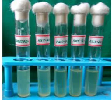
Pure culture of Azotobacter isolates were inoculated into tubes containing 10 ml peptone water and incubated at 30⁰ C for 48 h. After incubation, Nessler′s reagent (0.5 ml) was added to each tube (Cappuccino & Sherman, 1992) and the tubes were observed for the development of yellow color.
3 Results and Discussion
3.1 Isolation of Azotobacter isolates
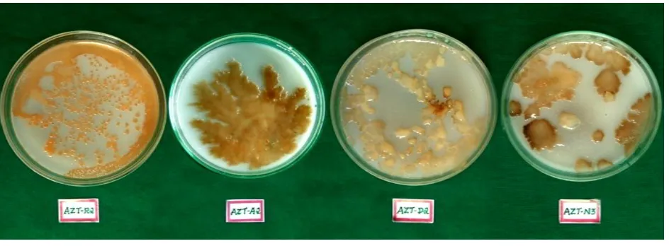
From the collected 150 rhizospheric soil samples, total of 75 overtly grown Azotobacter isolates were isolated from rhizospheric soil of chilli after 4-6 days of incubation. All the 75 Azotobacter isolates showed small/medium, milky white, round, raised colonies on Waksman No. 77 plates (Figure: 1) during early growth. Colonies have slow growth up to 2 days later on growth was very fast (Wu et al., 2006).
3.2 Morphological characterization of Azotobacter isolates
All the collected 75 Azotobacter isolates were taken for the morphological characterization. Result of morphological identification revealed Gram negative, rod shaped, forming white
Journal of Experimental Biology and Agricultural Sciences http://www.jebas.org
Isolation and Characterization of soil mending Azotobacter isolates isolated from Rhizosphere of Chilli 189
raised, wrinkled edge colony on Waksman No. 77 medium.
Pigment production differs with individual isolate as dark brown and light brown (Table 1; Figure 1) (Jacobson, 2000).These
observations are in agreement with the characteristics of
Azotobacter described in Bergey’s manual of determinative bacteriology (Brenner et al., 2005). The formation of brown insoluble
Table 1 Morphological and biochemical characteristics of Azotobacter isolates isolated from Chilli Rhizosphere soil of Hyderabad Karnataka region
Sl. No. Isolate code Cell shape Motility Colony color Gram reaction CT SH GL NR A IT
1 AZT-J1 Rod Motile Light brown Negative + + + + + +
2 AZT-J2 Rod Motile Light brown Negative + + + - + +
3 AZT-J3 Rod Motile Light brown Negative + + + - + +
4 AZT-J4 Rod Motile Brown Negative + + + + + +
5 AZT-J5 Rod Motile Dark brown Negative + + + - + +
6 AZT-J6 Rod Motile Dark brown Negative + + + + + +
7 AZT-J7 Rod Motile Dark brown Negative + + + - + +
8 AZT-J8 Rod Motile Dark brown Negative + + + - + +
9 AZT-J9 Rod Motile Light brown Negative + + + - + +
10 AZT-J10 Rod Motile Light brown Negative + + + + + +
11 AZT-J11 Rod Motile Light brown Negative + + + - + +
12 AZT-J12 Rod Motile Dark brown Negative + + + - + +
13 AZT-J13 Rod Motile Dark brown Negative + + + - + +
14 AZT-J14 Rod Motile Dark brown Negative + + + + + +
15 AZT-J15 Rod Motile Light brown Negative + + + - + +
16 AZT-Y1 Rod Motile Light brown Negative + + + - + +
17 AZT-Y2 Rod Motile Dark Brown Negative + + + - + +
18 AZT-Y3 Rod Motile Dark Brown Negative + + + - + +
19 AZT-Y4 Rod Motile Light brown Negative + + + + + +
20 AZT-Y5 Rod Motile Light brown Negative + + + - + +
21 AZT-Y6 Rod Motile Dark Brown Negative + + + + + +
22 AZT-Y7 Rod Motile Brown Negative + + + + + +
23 AZT-Y8 Rod Motile Brown Negative + + + + + +
24 AZT-Y9 Rod Motile Light brown Negative + + + + + +
25 AZT-Y10 Rod Motile Light brown Negative + + + - + +
26 AZT-Y11 Rod Motile Brown Negative + + + - + +
27 AZT-Y12 Rod Motile Light brown Negative + + + - + +
28 AZT-Y13 Rod Motile Light brown Negative + + + + + +
29 AZT-Y14 Rod Motile Dark Brown Negative + + + + + +
30 AZT-Y15 Rod Motile Dark Brown Negative + + + - + +
31 AZT-R1 Rod Motile Brown Negative + + + - + +
32 AZT-R2 Rod Motile Brown Negative + + + + + +
33 AZT-R3 Rod Motile Light brown Negative + + + - + +
34 AZT-R4 Rod Motile Light brown Negative + + + - + +
35 AZT-R5 Rod Motile Brown Negative + + + - + +
Journal of Experimental Biology and Agricultural Sciences http://www.jebas.org
190 Swapna et al.
Sl. No. Isolate code Cell shape Motility Colony color Gram reaction CT SH GL NR A IT
37 AZT-R7 Rod Motile Light brown Negative + + + - + +
38 AZT-R8 Rod Motile Dark brown Negative + + + - + +
39 AZT-R9 Rod Motile Dark brown Negative + + + + + +
40 AZT-R10 Rod Motile Dark brown Negative + + + - + +
41 AZT-R11 Rod Motile Light brown Negative + + + - + +
42 AZT-R12 Rod Motile Light brown Negative + + + - + +
43 AZT-R13 Rod Motile Dark brown Negative + + + + + +
44 AZT-R14 Rod Motile Light brown Negative + + + - + +
45 AZT-R15 Rod Motile Light brown Negative + + + - + +
46 AZT-G1 Rod Motile Dark Brown Negative + + + + + +
47 AZT-G2 Rod Motile Brown Negative + + + + + +
48 AZT-G3 Rod Motile Light brown Negative + + + + + +
49 AZT-G4 Rod Motile Light brown Negative + + + + + +
50 AZT-G5 Rod Motile Dark brown Negative + + + - + +
51 AZT-G6 Rod Motile Dark brown Negative + + + - + +
52 AZT-G7 Rod Motile Dark brown Negative + + + - + +
53 AZT-G8 Rod Motile Dark brown Negative + + + - + +
54 AZT-G9 Rod Motile Dark brown Negative + + + - + +
55 AZT-G10 Rod Motile Dark brown Negative + + + + + +
56 AZT-G11 Rod Motile Light brown Negative + + + + + +
57 AZT-G12 Rod Motile Light brown Negative + + + - + +
58 AZT-G13 Rod Motile Dark brown Negative + + + - + +
59 AZT-G14 Rod Motile Light brown Negative + + + + + +
60 AZT-G15 Rod Motile Dark Brown Negative + + + - + +
61 AZT-B1 Rod Motile Brown Negative + + + - + +
62 AZT-B2 Rod Motile Light brown Negative + + + + + +
63 AZT-B3 Rod Motile Light brown Negative + + + - + +
64 AZT-B4 Rod Motile Light brown Negative + + + - + +
65 AZT-B5 Rod Motile Dark Brown Negative + + + - + +
66 AZT-B6 Rod Motile Brown Negative + + + - + +
67 AZT-B7 Rod Motile Light brown Negative + + + + + +
68 AZT-B8 Rod Motile Light brown Negative + + + + + +
69 AZT-B9 Rod Motile Light brown Negative + + + - + +
70 AZT-B10 Rod Motile Dark Brown Negative + + + + + +
71 AZT-B11 Rod Motile Brown Negative + + + + + +
72 AZT-B12 Rod Motile Light brown Negative + + + - + +
73 AZT-B13 Rod Motile Light brown Negative + + + + + +
74 AZT-B14 Rod Motile Light brown Negative + + + - + +
75 AZT-B15 Rod Motile Dark Brown Negative + + + + + +
Journal of Experimental Biology and Agricultural Sciences http://www.jebas.org
Isolation and Characterization of soil mending Azotobacter isolates isolated from Rhizosphere of Chilli 191
pigments is characteristic of species A. chroococcum (Gibbs & Shapton, 1968). All the isolates formed cysts under nutrient and physical stress conditions. Earlier reports shows that Azotobacter
can survive in the form of cysts up to 24 years in dry soil conditions and the formation of cyst is the unique identification
characters of genus Azotobacter (Hitchins & Sadoff, 1973; Page & Shivprasad, 1991; Moreno et al.,1999).
3.3 Biochemical characterization of efficient Azotobacter isolates
All the Azotobacter isolates showed positive results for catalase test, starch hydrolysis gelatin liquefaction, ammonification, and
indole test. Few isolates were negative for nitrate reduction. In this manner result of present study are in agreement with the findings of previous researchers (Juarez et al., 2005; Murumkar et
al,2012; Jenifer et al., 2013; Kumar et al., 2014) (Table 1 and Figure 2,3,4 and 5).
Conclusion
Our study aimed in the isolation and characterization of isolates
from northern districts of Karnataka. Usually these districts fall under dry lands or rain fed areas, which is a major deterrent for the growth of crops, which require plethora of water and fertilizer. Promotion of water conservation and limited fertilizer application
is impeccably practical. Most of the Azotobacter isolates can fix nitrogen and conserves the moisture by producing excess
exopolysaccharides. In conjunction with this, these Azotobacter sps., also promote plant growth by several other mechanisms like phosphate solubilization, antagonism, production of phytohormone, and degradation of toxic compounds. In the
present investigation, some isolates were able to reduce the nitrate under anaerobic conditions, which may help the plant by fixing
Figure 5 Yellow coloration in peptone broth upon addition of Nessler’s reagent inoculated with efficient Azotobacter isolates
indicating positive for ammonification process Figure 2 Presence of bubbling/ foaming after addition of
hydrogen peroxide in the nutrient agar slants inoculated with Azotobacter isolates indicating positive for the catalase test
Figure 3 Motility test positive for Azotobacter isolates
Figure 4 No formation of red color in nitrate broth inoculated with efficient Azotobacter isolates up on addition of nitrate test
Journal of Experimental Biology and Agricultural Sciences http://www.jebas.org
192 Swapna et al.
more nitrogen, or reduce the available nitrogen for the plant.
Ammonification by all the isolates was favorably enhancing the plant growth. All isolates are producing cysts after the exponential phase of growth or under adverse conditions due to the lack of
nutrients. Many of the isolates shown the production of copious amounts of brown to black color pigmentation, there is an inclined pigmentation was also observed as the maximum temperature
increases with location. Pigmentation serves as protective agent against extreme heat and cold, UV, act as a cellular scavenger against free radicals, Reactive Oxygen Species (ROS), drugs, oxidents, xenobiotics and moreover melanized cells are resistant
to certain antifungal too.
Azotobacter species maintains the natural habitat of the soil and increases crop yield by 20-30 %. It replaces chemical nitrogen and phosphorus by 25 % in addition to stimulating the plant growth. Finally, it can provide protection against drought and some soil
borne diseases.
Conflict of interest
No potential conflict of interest was reported by the authors.
References
Allen ON (1953) Experiments in Soil Bacteriology. Burgess publication company, Minneapology Minnesota, U.S.A. Pp: 69-70.
Aquilanti L, Favilli F, Cleminti F (2004) Comparison of different strategies for isolation and preliminary identification of
Azotobacter from soil samples. Soil Biology and Biochemistry36: 1475-1483.
Ayala S, Rao EVSP (2002) Perspective of soil fertility management with a focus on fertilizer use for crop productivity. Current Science82: 797-807.
Brenner DJ, Krieg NR, Staley JT (2005) Bergy’s Manual of Systematic Bacteriology. In: The Proteobacteria part B, The Gammaproteobacteria Second Edition, Springer Science and Business Media, Inc., New York, USA.
Cappuccino JC, Sherman N (1992) Microbiology: A Laboratory Manual (3rded), Benjamin cummings Publishing Company, New York, Pp. 125-179.
Gibbs BM, Shapton DA (1968) Identification methods for
microbiologists. Society of Applied Microbiology, No.2 pp.xi+212 pp.
Hitchins VM, Sadoff HL (1973) Morphogenesis of cysts in
Azotobactervinelandii. Journal of Bacteriology104:492-498.
Jacobson E (2000) Pathogenic Roles for Fungal Melanins.
Clinical Microbiology Reviews 13 : 708 - 717.
Jenifer AMR, Reena A, Aysha OS, Valli S, Nirmala P, Vinothkumar P (2013) Isolation of siderophore producing bacteria from rhizosphere soil and their antagonistic activity against
selected fungal plant pathogens. International Journal of Current Microbiology and Applied Science2 : 59-65.
Jiménez DJ, Montaña JS, Martínez MM (2011) Characterization of free Nitrogen Fixing Bacteria of the genus Azotobacter in Organic Vegetable Grown Colombian Soils. Brazilian Journal of Microbiology 42: DOI: 10.1590/S1517-83822011000300003.
Joshi KK, Kumar V, Dubey RC, Maheshwari DK (2006) Effect of chemical fertilizer adaptive variants, Pseudomonas aeruginosa
GRC2 and Azotobacter chroococcum AC1 on Macrophomina phaseolina causing charcoal rot of Brassica juncea. Korean Journal of Environmental Agriculture 1: 228-235. DOI: 10.5338/KJEA.2006.25.3.228.
Juarez B, Martinez-Toledo MV, Gonzalez-Lopez J (2005) Growth of Azotobacter chroococcum in chemically defined media containing p-hydroxybenzoic acid and protocatechuic acid. Chemosphere59 :1361-1365.
Komagata K, Iino T, Yamada Y (2014) The Family
Acetobacteraceae. Rosenberg E, DeLong EF, Lory S, Stackebrandt E, Thompson F (Eds.) The Prokaryotes. Springer, Berlin, Heidelberg, Pp. 3-78. https://doi.org/10.1007/978-3-642-30197-1_396.
Kumar A, Singh R, Giri DD, Singh PK, Pandey KD (2014) Effect of Azotobacter chroococcum CL13 inoculation on growth and curcumin content of Turmeric (Curcuma longa L.). International Journal of Current Microbiology and Applied Science3 : 275-283.
Moreno J, Vargas-Garcla C, Lopez MJ, Sanchez-serrano G (1999) Growth and exopolysaccharide production by Azotobacter vinelandii in media containing phenolic acids. Journal of Applied Microbiology 86: 430-445.
Murumkar DR, Borkar SG, Chimote VP (2012) Diversity of cell
morphology, nitrogenase activity and DNA profile of Azotobacter
isolates from soils of Maharastra. Bioinfolet9 : 851-858.
Journal of Experimental Biology and Agricultural Sciences http://www.jebas.org
Isolation and Characterization of soil mending Azotobacter isolates isolated from Rhizosphere of Chilli 193
Orville W, Marilyn G, Neumann, Socolofsky MD (1961)
Development and germination of the Azotobacter cyst. The Journal of Biophysical and Biochemical Cytology10: 555-565.
Page WJ, Shivprasad S (1991) Azotobacter salinestris sp. nov., a
Sodium-Dependent, Microaerophilic, and Aeroadaptive
Nitrogen-Fixing Bacterium. International Journal of Systematic Bacteriology41: 369-376. doi: 10.1099/00207713-41-3-369.
Socolofsky MD, Orville W (1962) Resistance of the Azotobacter
cyst. Journal of Bacteriology 84:119-124.
Winogradsky SN (1949) Microbiologie du soil problems et methods (e.f.). The Azotobacteraceae. Bacteriological Review 18: 199.