Technology (IJRASET)
An Approach to
Contrast Enhancement Using
Local Features Method
M.J.Preethi1, Mrs.R.Mekala2
¹P.G Scholar, Department of Computer Science and Engineering, Anna University, India ²Professor, Department of Computer Science and Engineering, Anna University, India
Abstract: Digital Image forensic analysis is utilized to identify the integrity and authenticity of the images. This paper provides the technique for the detection of contrast enhancement through Principle component Analysis by establishing co-occurrence matrix for the feature deduction. PCA is to standardize the data in image. Real-world data sets usually exhibit relationships among their variables. These relationships are often linear, or at least approximately so, making them amenable to common analysis techniques. One such technique is principal component analysis ("PCA"), which rotates the original data to new coordinates, making the data as "flat" as possible. The features extracted are passed through the PCA data mining for better classification.
Keywords: Contrast Enhancement, image manipulation, Histogram modification, peak/gap bins, PCA, co-occurrence matrix, detection of image.
I. INTRODUCTION
Images are the most common and convenient means of conveying or transmitting the information. An image is significance of thousand terms. Pictures in brief convey information on positions, sizes and inter-relationships among objects. They describe spatial information that we can recognize as objects. Human beings are superior in deriving information from such images, because of our native visual and mental abilities. About 75% of the information received by human is in pictorial form.
The image enhancement is one of the significant techniques in digital image processing. It has an important role in many fields such as medical image analysis, remote sensing, high description television, hyper spectral image processing, microscopic imaging etc. The contrast is the difference in visual properties that distinguish an object from other object and from the background. In other words, it is the difference between the darker and the lighter pixels of image.
Enhanced image can also be described as if certain of fog have been removed from the image. There are a number of reasons for an image to have poor disparity: ·
The device used for imaging is of poor quality.
The undesirable outside conditions at the time of acquisition
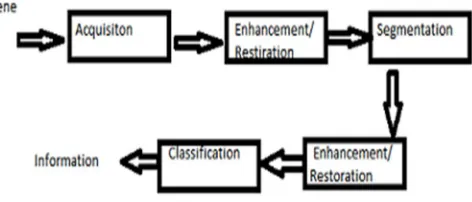
[image:2.612.200.436.566.667.2]Contrast enhancement is an important factor for image enhancement. In this technique, contrast of an image is improved to make the image better for human vision. Image enhancement methods based on redistributing the probability densities are indirect methods of contrast enhancement. In these methods, the image intensities can be redistributed within the dynamic range without defining a specific contrast term. Histogram modification techniques such as histogram equalization (HE) is one of the most frequently used technique. The fundamental principle of Histogram equalization is to make the histogram of the enhanced image to have approximately uniform distribution so that the dynamic range of the image can be fully exploited.
Figure 1: Generic model for Enhancement
Co-Technology (IJRASET)
Occurrence Matrix which uses a matrix representation for the pixels in the input images. Using this techniques better classification of the images is possible.
II. RELATEDWORKS
Arici et al (2009) [1] introduced a contrast enhancement technique as an optimization problem that minimizes a cost function. This method is called as Global Contrast Enhancement using Histogram Modification (GCEHM).Here, the level of enhancement is adjusted by penalty terms. It incorporates noise robustness, black/white stretching and mean brightness preservation in the optimization module. This method improved only the visual quality of the image without considering the features in the image. Hence, it is suitable only in display applications.
Asadi Amiri et al (2010) [2] explains the technique known as Adaptive gamma correction using weighting distribution (AGCWD) was presented that modify histograms and enhance contrast in digital images. A hybrid HM (histogram modification) method was proposed by combining TGC (Transform based gamma correction) and THE (Traditional histogram equalization) methods. In this method cumulative distribution function (CDF) is utilized directly and normalized gamma function is applied to modify the transformation curve. In adaptive gamma correction (AGC) method compensated CDF is used as an adapted parameter. The AGC method increases low intensity and avoids significant decrement of high intensity. In Weighting distribution the input histogram or probability distribution function (PDF) is modified in such way that the infrequent gray levels are given relatively more probabilities (or weights) than the frequent gray levels. Results of this proposed technique have shown that this method produced enhanced images of comparable or higher quality than those produced using previous methods.
Bedi et al (2013) [3] proposed that Image Fusion is one of the major research fields in image processing. Image fusion process can be defined as the integration of information from a number of registered images without the introduction of distortion. It is often not possible to get an image that contains all relevant objects in focus. One way to overcome this problem is image fusion, in which one can acquire a series of pictures with different focus settings and fuse them to produce an image with extended depth of field which helps in clinical diagnosis. Image fusion techniques can improve the quality and increase the application of these data. The proposed paper uses multi-image Contrast enhancement for PCA fusion of medical images. The objective of this paper is to propose a technique for fusion of human brain MRI images based on Principal Component Analysis and to improve the visibility of medical images by applying contrast enhancement existing techniques. The PCA fusion technique adopted here improve resolution of the images. The PCA algorithm builds a fused image of several input images as a weighted superposition of all input images. The resulting images contains enhanced information as compared to individual images and also apply Contrast Enhancement technique to improve visibility of medical image details without introducing unrealistic visual appearances and/or unwanted artifacts. It also gives the quality comparison study of original medical images before fusion, after applying PCA and various existing techniques for contrast enhancement for those medical images.
Chalekar et al (2014) [4] Contrast Stretching to expand the range of brightness values in an image the contrast enhancement techniques are used, so that the image can be efficiently displayed in a manner desired by the analyst. The level of contrast in an image may vary due to poor illumination or improper setting in the acquisition sensor device. Therefore, there is a need to manipulate the contrast of an image in order to compensate for difficulties in image acquisition. The idea behind contrast stretching is to increase the dynamic range of the gray levels in the image being processed. The idea is to modify the dynamic range of the grey-levels in the image. Linear Contrast Stretch is the simplest contrast stretch algorithm that stretches the pixel values of a low-contrast image or high-low-contrast image by extending the dynamic range across the whole image spectrum from 0 – (L-1).
Shefali Gupta et al (2014) [5] proposed that images having low contrast are usually captured in dark or bright environments. So pre-processing of such images becomes necessary to make the images suitable for other image pre-processing applications. Image enhancement is a common problem. The histogram equalization (HE) technique is widely used for this purpose because it is simple and effective. However, it produces undesirable visual artifacts in the output image because the mean brightness of the image is changed. This paper presents a review of different techniques that can be used for contrast enhancement. The ultimate aim of these techniques is to preserve the input mean brightness so that the image looks natural in appearance.
Technology (IJRASET)
procedure is used to improve the image quality from first stage to second stage with edge preservation. The wavelet Thresholding methods used for removing random noise has been researched extensively due to its effectiveness and simplicity. However, not much has been done to make the threshold values adaptive to the spatially changing statistics of images. Such adaptively can improve the wavelet Thresholding performance because it allows additional local information of the image (such as the identification of smooth or edge regions) to be incorporated into the algorithm.
III. PROPOSED MODEL
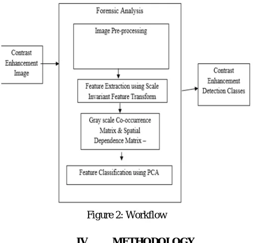
[image:4.612.179.434.297.541.2]The model describes the detection of contrast enhancement in the input images using PCA. The input image is first pre-processed where the noises in the image are removed. Then the whole image is splitted into blocks. The features are extracted using scale Invariant Feature Transform where the local features in the images are detected. An object is recognized in a new image by individually comparing each feature from the new image to this database and finding candidate matching features based on Euclidean distance of their feature vectors. From the full set of matches, subsets of key points that agree on the object and its location, scale, and orientation in the new image are identified to filter out good matches. Then the similarity is measured using Gray scale Co-occurrence Matrix (GLCM) and Spatial Dependence Matrix computation. The GLCM is a method which produce a tabulation of how often different combinations of pixel brightness values (grey levels) occur in an image.
Figure 2: Workflow
IV. METHODOLOGY
The enhancements in the images are done using the PCA methodology via GLCM method. At first the images are processed via GLCM technique where matrix format is used for representing the pixel values in the images. The features in the images are extracted using PCA method as mentioned below.
A. Modelling Gray level -Co- occurrence Matrix
A statistical method of examining texture that considers the spatial relationship of pixels is the gray-level co-occurrence matrix (GLCM), also known as the gray-level spatial dependence matrix. The GLCM functions characterize the texture of an image by calculating how often pairs of pixel with specific values and in a specified spatial relationship occur in an image, creating a GLCM, and then extracting statistical measures from this matrix. (The texture filter functions, described in Texture Analysis cannot provide information about shape, i.e., the spatial relationships of pixels in an image.)
B. Feature classification using PCA
Technology (IJRASET)
Then the block wise separation is done and the RGB channels are separated. Apply PCA for each block and find the maximum brightness of the image. Finally merge the whole block and proceed bulk PCA. The following steps takes place in PCA:-
1) Calculate the mean and standard deviation of the features in the image.
2) Subtract the sample mean from each observation, then dividing by the sample standard deviation. This centres and scales the
data.
3) Calculating the coefficients of the principal components and their respective variances is done by finding the Eigen functions of
the sample covariance matrix.
4) The matrix contains the coefficients for the principal components. The diagonal elements store the variance of the respective
principal components. We can extract the diagonal.
5) The maximum variance in data results in maximum information content which is required for better classification.
V. RESULTS ANDDISCUSSIONS
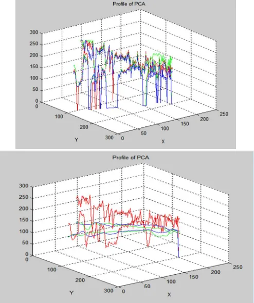
[image:5.612.183.427.277.381.2]The input image is processed using the GLCM method and the overall contrast enhancement is achieved in the given image. The PCA method when applied to the input image the differences in their pixel values due to contrast enhancement is identified easily when compared to the Histogram using Peak/Gap bins.
Figure 3: Original and Forged image using PCA
In the above image the contrast is increased and the resulting enhanced image and the original one is shown. The chart depicting the differences in the image is done and it is shown below.
[image:5.612.184.431.416.711.2]Technology (IJRASET)
From the above analyses, it is clear that the identification of the forged images are identified easily using the Principal Component Analysis method and the error rate is minimized subsequently.
Figure 5: Comparison result
VI. CONCLUSION
In this paper, the forged mechanism for the detection of enhanced digital images has been done. The analysis report shows that the
PCA technique will be the better one for the detection of the changes in the images better than other techniques such as Histogram
methods. The differences between the images can also be self-learned from the two images. (i.e. From original to that of enhanced one)
REFERENCES
[1] T. Arici, S. Dikbas, and Y. Altunbasak, “A histogram modificationframework and its application for image contrast enhancement,” IEEE Trans. Image Process., vol. 18, no. 9, pp. 1921–1935, Sep. 2009.
[2] S. Asadi Amiri, H. Hassanpour, A.K. Pouyan, “Texture Based Image Enhancement Using Gamma Correction”, Middle-East Journal of Scientific Research, 2010, Vol. 6, pp. 569-574.
[3] Dr. S. S. Bedi, Rati Khandelwal, “CONTRAST ENHANCEMENT FOR PCA FUSION OF MEDICAL IMAGES,” Journal of Global Research in Computer ScienceVolume 4, No. 3, March 2013.
[4] Ms. K.T.Chalekar, Prof T. Yengantiwar, “REVIEW PAPER ON IMAGE CONTRAST ENHANCEMENT TECHNIQUES,” International Journal of Advanced Research in ComputerEngineering & Technology Volume 3 Issue 3, March 2014.
[5] Shefali Gupta, Yadwinder Kaur, “Review of Different Histogram Equalization Based Contrast Enhancement Techniques,” International Journal of Advanced Research in Computer and Communication Engineering Vol. 3, Issue 7, July 2014.
[6] Vikas D Patil, Sachin D. Ruikar, “PCA Based Image Enhancement in Wavelet Domain” International Journal of Engineering Trends and Technology- Volume3Issue1- 2012.
[7] H. Farid, “Image forgery detection,” IEEE Signal Process. Mag., vol. 26, no. 2, pp. 16–25, Mar. 2009.
[8] J. Fan, H. Cao, and A. C. Kot, “Estimating EXIF parameters based on noise features for image manipulation detection,” IEEE Trans. Inf. Forensics Security, vol. 8, no. 4, pp. 608–618, Apr. 2013.
[9] Swaminathan, M. Wu, and K. J. R. Liu, “Digital image forensics via intrinsic fingerprints,” IEEE Trans. Inf. Forensics Security, vol. 3, no. 1, pp. 101–117, Mar. 2008.
[10] .C. Popescu and H. Farid, “Exposing digital forgeries by detecting traces of resampling,” IEEE Trans. Signal Process., vol. 53, no. 2, pp. 758–767, Feb. 2005. [11] G. Cao, Y. Zhao, R. Ni, and A. C. Kot, “Unsharp masking sharpening detection via overshoot artifacts analysis,” IEEE Signal Process. Lett., vol. 18, no. 10, pp.
603–606, Oct. 2011.
[12] M. C. Stamm and K. J. R. Liu, “Forensic estimation and reconstruction of a contrast enhancement mapping,” in Proc. IEEE Int. Conf. Acoust., Speech Signal, Dallas, TX, USA, Mar. 2010, pp. 1698–1701.
[13] S. Remya, “Digital Image Forgery Detection by Contrast Enhancement”, Volume 16, Issue 5, Ver. IX , PP 01-07, Sep. 2014
[14] J. O’Brien and H. Farid, “Exposing photo manipulation with inconsistent reflections,” ACM Trans. Graph., vol. 31, no. 1, pp. 1–11, 2012.
[15] M. Stamm and K. Liu, “Blind forensics of contrast enhancement in digital images,” in 15th IEEE Int. Conference on Image Processing, 2008. ICIP 2008, Oct. 2008, pp. 3112–3115.
Parameters Histogram Method
PCA
PSNR 53.4025 18.8311
MSE 0.2970 851.072
Maximum Error Rate
0.9994 0.99