Copyright © 2002, American Society for Microbiology. All Rights Reserved.
Genotyping of Hepatitis C Virus Types 1, 2, 3, and 4 by a One-Step
LightCycler Method Using Three Different Pairs of
Hybridization Probes
Matthias Schröter,
1Bernhard Zöllner,
1Peter Schäfer,
1Olfert Landt,
2Ulrich Lass,
2Rainer Laufs,
1and Heinz-Hubert Feucht
1*
Institut für Medizinische Mikrobiologie und Immunologie, Universitätsklinikum Hamburg-Eppendorf,
20246 Hamburg,
1and TIB MOLBIOL, 10829 Berlin,
2Germany
Received 10 July 2001/Returned for modification 24 November 2001/Accepted 20 January 2002
Determination of hepatitis C virus (HCV) genotypes has become increasingly important during the last years
for prediction of the clinical course and the outcome of antiviral therapy. Therefore, numerous different
methods have been developed to enable HCV genotyping. However, many of them are very laborious and
expensive, leading to limited usage in daily routine diagnostics. We have established a method which combines
the speed of the new LightCycler technology with the use of amplification products generated for diagnostic
quantitative HCV RNA determination. Differentiation of HCV genotypes is performed with these amplicons in
a single step by using fluorophore-labeled hybridization probes. Although currently only two different acceptor
fluorophores are available for the LightCycler, types 1, 2, 3, and 4, which are by far the prevailing HCV
genotypes in Europe and the United States, can be distinguished. Genotypes of specimens from 190 chronically
HCV-infected patients were determined by the LightCycler method and compared with the results of nucleotide
sequencing. Concordant results were obtained for all samples. This new method offers a fast and convenient
possibility to determine the quantitative HCV RNA load and the genotype in large-scale settings within about
4 h.
Hepatitis C Virus (HCV) is the main causative agent of
chronic posttransfusion hepatitis and poses an elevated risk for
development of liver cirrhosis and hepatocellular carcinoma
(2, 6, 28). Much effort has been undertaken to establish
ther-apeutic strategies against HCV infection. Both duration of and
sustained response to the current standard therapy regimens
are strongly associated with the HCV genotype (3, 10, 12, 30).
For this reason, besides quantitative PCR, genotyping has
be-come increasingly important in laboratory routine diagnostics.
The most-effective way to determine these two parameters
would be the usage of the same amplicons for diagnostic HCV
RNA detection and for genotyping (19).
The demand for quantitative assays to monitor the viral load
during antiviral therapy (8, 12) has led to the development of
a variety of amplification procedures. Recently, we have
de-veloped a LightCycler assay which fulfils the need for fast
real-time quantitative detection of HCV RNA (21). To meet
the increasing need for fast and easily performable genotyping
assays, we have developed a genotyping method using
Light-Cycler technology. Amplicons of the 5
⬘
noncoding region,
which are generated during the routine determination of the
quantitative viral load, are used for genotyping by
fluorophore-labeled oligonucleotides. The results obtained by this new
as-say were compared to those obtained by nucleotide
sequenc-ing, which is currently regarded as the “gold standard” (23).
MATERIALS AND METHODS
Serum samples.Sera from 190 chronically HCV infected patients were in-cluded in the retrospective study. The samples were selected for their genotype, which had been determined prior to the study by nucleotide sequencing accord-ing to the system proposed by Simmonds and coworkers (23). The enrolled collection represents the distribution of the main HCV genotypes in the United
States and Europe (18, 20) and consisted of 1a isolates (n⫽35), 1b isolates (n
⫽52), 2a isolates (n⫽13), 2b isolates (n⫽17), 2c isolates (n⫽3), 3a isolates
(n⫽62), 4a isolates (n⫽15), and one 4c isolate.
Samples derived from 30 healthy blood donors served as negative controls.
LightCycler PCR.LightCycler PCR was performed as recently described (21) with slight modifications. Nucleic acids were extracted automatically by MagNA Pure (Roche Molecular Biochemicals, Indianapolis, Ind.). Reverse transcription
and PCR were performed in a single step usingTth(LightCycler RNA Master
SYBR Green I Kit) containing 3.25 mM Mn acetate by using 5 pmol each of
primers 27 (5⬘-TCCACCATGAATCACTCCC-3⬘; positions 27 to 43 according
to numbering of nucleotide sequences as previously described [2]) and KY81as
(5⬘-CGGAACCGGTGAGTACACC-3⬘; positions 169 to 150). PCR was
per-formed in 45 cycles with 1 s at 95°C (denaturation), 3 s at 55°C (annealing), and 8 s at 72°C (extension), and fluorescence detection was performed by melting
point (Tm) curve analysis. Results were expressed as international units per
milliliter according to different standards as described previously (21). After
completing PCR, 2l was diluted in 198l of H2O, and 2l of this dilution was
used for genotyping.
LightCycler typing.PCR was performed using 2l of LightCycler-DNA Mas-ter Hybridization Probes (LightCycler-DNA MasMas-ter Hybridization Probes Kit;
Roche Diagnostics GmbH, Mannheim, Germany) with 3 mM MgCl2by using 5
pmol each of primers 27 and KY81as. For genotype detection 3 pmol of each hybridization probe was added to the reaction mixture. The position and se-quences of the hybridization probes are shown in Fig. 1. Each probe pair consists of two oligonucleotides, which are labeled with different fluorophores. One
probe, called the detection probe, is labeled at the 5⬘-end with a LightCycler-Red
fluorophore (LC Red 640 or LC Red 705). The other probe, called the anchor
probe, is labeled at the 3⬘end with fluorescein. Only after hybridization do the
two probes come in close proximity, resulting in fluorescence resonance energy transfer between the two fluorophores. During fluorescence resonance energy transfer, fluorescein, the donor fluorophore, is excited by the light source of the
* Corresponding author: Mailing address: Institut für Medizinische
Mikrobiologie und Immunologie, Universitätsklinikum
Hamburg-Ep-pendorf, Martinistrasse 52, 20246 Hamburg, Germany. Phone: 49
(0)40 42803 3159. Fax: 49 (0)40 42803 4062. E-mail: [email protected]
-hamburg.de.
2046
on May 15, 2020 by guest
http://jcm.asm.org/
LightCycler instrument, and part of the excitation energy is transferred to Light-Cycler Red, the acceptor fluorophore. The emitted fluorescence of the acceptor fluorophore is measured.
PCR was performed in 40 cycles with 5 s at 95°C (denaturation), 15 s at 52°C
(annealing), and 15 s at 72°C (extension). After the final cycle,Tmanalysis of all
samples and controls was performed within the range from 37 to 77°C. The fluorescence signals of the LC Red 640-labeled probes were first measured in channel 2 (F2), and afterwards the signals of the LC Red 705 probe were
measured using F3. The four genotypes were discriminated by the differentTms
of the amplicons (Table 1).
Nucleotide sequencing.Determination of genotypes and subtypes by nucleo-tide sequencing had been performed as previously described (5). Sequencing was performed using an ABI Prism 310 Genetic Analyzer (PE Applied Biosystems, Weiterstadt, Germany). The sequencing reaction was performed as recom-mended by the manufacturer (PE Applied Biosystems). Genotypes were deter-mined by comparison with published reference sequences according to the clas-sification proposed by Simmonds and coworkers (23).
Statistical analysis.The mean values and confidence intervals were calculated using the WinSTAT program package (Springer Electronic Media, Heidelberg, Germany). For statistical analysis of the differences between the respective HCV
genotypes, Student’sttest was used. To estimate the reproducibility of the assay,
three repeated measures of different samples were compared statistically using the Friedman test.
RESULTS
HCV genotypes of samples from 190 chronically infected
patients were determined by nucleotide sequencing according
to the system proposed by Simmonds and coworkers (23).
Among the genotypes determined, 44% (
n
⫽
87) belonged
to genotype 1, 17% (
n
⫽
33) belonged to genotype 2, 31% (
n
⫽
62) belonged to genotype 3, and 8% (
n
⫽
16) belonged to
genotype 4. All of these samples were tested by the new
Light-Cycler typing method. Concordant results were obtained with
all 190 samples.
Quantitative viral titers in these samples ranged from 10
3to
9
⫻
10
6IU/ml, representing the dynamic range described
ear-lier for the LightCycler HCV PCR (21). No differences
regard-ing the titers were observed between the respective genotypes.
A detection limit of 10
3IU/ml was identical for all genotypes
by LightCycler PCR and LightCycler typing assay.
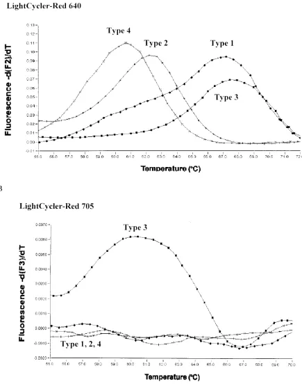
T
manalysis was performed, and the fluorescence signals
were first measured in F2 for LC Red 640 emission. Genotypes
1 and 3 displayed similar
T
ms, but genotype 2 and genotype 4
isolates could easily be determined by their different
T
ms (Fig.
2A). For better discrimination of type 1 and 3 isolates,
T
ms
were additionally determined using measurement of LC Red
705 emission in F3. Signals were exclusively obtained with
genotype 3 isolates as shown in Fig. 2B. Samples with
T
ms in
the typical range for type 1 and 3 isolates in F2 and no signal
in F3 belonged to genotype 1.
The mean
T
ms and the 95% confidence intervals of the
respective genotypes are shown in Table 1. In no case was an
overlap of
T
ms between the respective genotypes observed.
Statistical analysis using Student’s
t
test revealed that the
T
ms
of each of the respective genotypes 1, 2, 3, and 4 differ
signif-icantly (
P
⬍
0.001) when measured in F2.
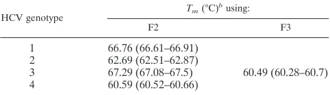
[image:2.587.46.531.75.299.2]Likewise, reproducibility of the LightCycler typing method
FIG. 1. Comparison of nucleotide sequences of the region used for LightCycler typing. Numbering of nucleotide sequences is as previously
described (2). Dots indicate identical nucleotides between the different subtypes. In the boxes at the top of the figure, the sequences and positions
of the hybridization probes are indicated. In parentheses is indicated which HCV genotype is detectable by the respective oligonucleotide.
TABLE 1.
T
ms observed for the respective genotypes in this study
aHCV genotype Tm(°C)
busing:
F2 F3
1
66.76 (66.61–66.91)
2
62.69 (62.51–62.87)
3
67.29 (67.08–67.5)
60.49 (60.28–60.7)
4
60.59 (60.52–60.66)
aUsing F2, genotypes 2 and 4 can be identified unequivocally. In contrast,
genotypes 1 and 3 have nearly identicalTms in this analysis but can be
distin-guished using F3. Only the hybridization probes designed for type 3 isolates emit measurable fluorescence when F3 is used.
bValues are means. Values in parentheses are 95% confidence intervals.
on May 15, 2020 by guest
http://jcm.asm.org/
[image:2.587.43.284.616.686.2]FIG. 2. The respective HCV genotypes are distinguished by their different
T
ms. (A)
T
manalysis was performed, and the fluorescence signals
were first measured in F2 for LC Red 640 emission. Genotypes 1 and 3 display similar
T
ms, but genotype 2 and genotype 4 isolates can easily be
determined by different
T
mvalues. (B) For discrimination of type 1 and 3 isolates,
T
ms are determined using measurement of LC Red 705 emission
in F3. Signals are exclusively obtained with genotype 3 isolates as shown.
on May 15, 2020 by guest
http://jcm.asm.org/
was very high. To analyze the variance of repeated runs,
iso-lates of all four genotypes were tested three times on different
days. Using the Friedman test no statistically significant
differ-ence could be observed among the results of each day. Every
sample could be typed clearly, and no differences were noted
between the results of the respective days.
Thirty samples from healthy blood donors served as negative
controls. None of these showed a positive result.
DISCUSSION
A new rapid PCR-based HCV genotyping method was
de-veloped using the LightCycler technology. Recently, a method
using LightCycler technology for the detection and
quantifica-tion of HCV RNA in serum samples was described by our
group (21). As there is evidence that the HCV genotype is one
of the main factors determining the outcome of antiviral
ther-apy (3, 10, 12, 30). there is an increasing demand for fast and
highly reproducible genotyping assays. Nucleotide sequencing
is currently regarded as the reference method for genotyping
of HCV isolates. However, this procedure is not considered
suitable for routine laboratory settings because it is very
labo-rious and time-consuming (7, 20, 22). Therefore, several
dif-ferent tests like PCR with genotype-specific primers,
restric-tion fragment length polymorphism assay, cleavase fragment
length polymorphism assay, hybridization techniques (line
probe assay), and serological assays have been developed for
HCV genotype determination (1, 13–17, 20, 25).
A further strategy to reduce expenditure and requirement
for time is the use of only one system for both quantitative
HCV PCR and genotyping as recently suggested (19). A main
problem of this approach is the need for highly conserved
regions for routine PCR testing on the one hand and regions
with sufficient variability for genotype discrimination on the
other hand. The currently accepted typing system is based on
nucleotide sequences of the NS-5B region of HCV (23). Due
to its variability this region is not suitable for routine PCR
testing. When using other genomic regions for genotyping,
misclassifications may arise due to inconstancies in the viral
genome. However, a consistent feature of studies evaluating
typing methods using different regions of HCV is that
se-quence relationships between subgenomic regions always
re-flect those of the complete genome (24, 26).
For quantitative HCV RNA determination, the 5
⬘
noncod-ing region is the preferred area, because it is highly conserved.
This region was chosen for the development of the LightCycler
typing assay because it has been described as a useful target for
both quantitative LightCycler PCR (21) and HCV genotyping
by in-house tests and commercially available assays like the
INNO-LiPA HCV II LineProbe assay (9, 19, 27). By using
three pairs of hybridization probes, differentiation of the
ge-notypes is possible in a single reaction. This significantly
de-creases time expenditure and costs and thus enables
high-throughput examinations in routine settings (22). Comparison
of the results obtained by LightCycler typing and NS-5B
nu-cleotide sequencing revealed identical genotypes in all
sam-ples. The HCV subtype, although not identifiable by the new
assay, did not influence the LightCycler typing result. Among
the genotype 1 isolates, 35 classified as subtype 1a isolates were
detected equally as well as 52 subtype 1b isolates. Likewise, all
of the genotype 2 subtypes—among them 13 subtype 2a, 17
subtype 2b, and 3 subtype 2c isolates—were classified with
equal efficiency. The type 3 isolates belonged exclusively to
subtype 3a, and among the type 4 isolates there was no
differ-ence between 1 4c and 15 4a isolates.
Using the LightCycler, detection of amplification products
can either be performed using SYBR Green or hybridization
probes. While SYBR Green has been proven useful for
quan-titative HCV PCR (21), differentiation of HCV genotypes can
be accomplished using different hybridization probes. Until
now, only two different acceptor fluorophores have been
de-veloped to be used with the LightCycler. Therefore, the
num-ber of genotypes identifiable by this assay is restricted, and no
subtypes can be distinguished so far. However, by far the
pre-vailing HCV genotypes of Europe and the United States (18,
20) could be included. The prevalence of other genotypes is
very low in our patient population. In our laboratory we found
only two genotype 5 isolates and one genotype 6 isolate among
specimens from more than 3,000 HCV infected patients (data
not shown). From the clinical point of view, the knowledge of
the precise HCV subtype seems to be unnecessary for
thera-peutic decisions. In this context it is most important to
deter-mine the genotype and to discriminate between genotype 1 and
non-1 types (11, 19). Patients infected with genotype 1 isolates
seem to have a less favorable response to antiviral treatment
than those infected with genotype 2 or 3 (4, 29) and need a
different therapy regimen (30).
Our assay revealed a wide dynamic range that extended over
a 4-log range of HCV input.
One striking advantage of the LightCycler typing method is
the speed with which amplification and detection of amplicons
are achieved. Within about 4 h, including RNA extraction,
both the quantitative PCR and genotyping can be performed.
REFERENCES
1.Andonov, A., and R. K. Chaudhary.1995. Subtyping of hepatitis C virus
isolates by a line probe assay using hybridization. J. Clin. Microbiol.33:254–
256.
2.Choo, Q. L., K. H. Richman, J. H. Han, K. Berger, C. Lee, C. Dong, C. Gallegos, D. Coit, R. Medina Selby, P. J. Barr, A. J. Weiner, D. W. Bradley, G. Kuo, and M. Houghton.1991. Genetic organization and diversity of
hepatitis C virus. Proc. Natl. Acad. Sci. USA88:2451–2455.
3.Davis, G. L., and J. N. Lau.1997. Factors predictive of a beneficial response
to therapy of hepatitis C. Hepatology26(Suppl. 3):122.S-127.S.
4.European Association for the Study of Liver. 1999. EASL international
consensus conference on hepatitis C. Consensus statement. J. Hepatol.30:
956–961.
5.Feucht, H. H., M. Schröter, B. Zöllner, S. Polywka, H. Nolte, and R. Laufs.
1997. The influence of age on the prevalence of hepatitis C virus subtypes 1a
and 1b. J. Infect. Dis.175:685–688.
6.Feucht, H. H., B. Zöllner, M. Schröter, S. Polywka, P. Buggisch, H. Nolte, and R. Laufs.1999. High rate of chronicity in HCV infection determined by antibody confirmatory assay and PCR in 4110 patients during long-term
follow-up. J. Clin. Virol.13:43–51.
7.Forns, X., and J. Bukh.1998. Methods for determining hepatitis C virus
genotype. Viral Hep. Rev.4:1–19.
8.Hagiwara, H., N. Hayashi, E. Mita, T. Takehara, A. Kasahara, H. Fusamoto, and T. Kamada.1993. Quantitative analysis of hepatitis C virus RNA in
serum during interferon alpha therapy. Gastroenterology104:877–883.
9.Halfon, P., P. Trimoulet, M. Bourliere, H. Khiri, V. de Lédinghen, P. Couzi-gou, J. M. Feryn, P. Alcaraz, C. Renou, H. J. A. Fleury, and D. Ouzan.2001.
Hepatitis C virus genotyping based on 5⬘noncoding sequence analysis
(Tru-gene). J. Clin. Microbiol.39:1771–1773
10.Heathcote, E. J., M. L. Shiffman, W. G. E. Cooksley, G. M. Dusheiko, S. S. Lee, L. Balart, R. Reindollar, R. K. Reddy, T. L. Wright, A. Lin, J. Hoffman, and J. De Pamphilis.2000. Peginterferon alfa-2a in patients with chronic
hepatitis C and cirrhosis. N. Engl. J. Med.343:1673–1680.
11.Krekulova, L., V. Rehak, A. E. Wakil, E. Harris, and L. W. Riley.2001. Nested restriction site-specific PCR to detect and type hepatitis C virus
on May 15, 2020 by guest
http://jcm.asm.org/
(HCV): a rapid method to distinguish HCV subtype 1b from other
geno-types. J. Clin. Microbiol.39:1774–1780.
12.Martinot-Peignoux, M., P. Marcellin, M. Pouteau, C. Castelnau, N. Boyer, M. Poliquin, C. Degott, I. Descombes, V. Le Breton, and V. Milotova.1995. Pretreatment hepatitis C virus RNA levels and hepatitis C virus genotype are main and independent prognostic factors of sustained response to interferon
alpha therapy in chronic hepatitis. Hepatology22:1050–1056.
13.Okamoto, H., K. Kurai, S. Okada, K. Yamamoto, H. Lizuka, T. Tanaka, S. Fukuda, F. Tsuda, and S. Mishiro.1992. Full-length sequence of a hepatitis C virus genome having poor homology to reported isolates: comparative
study of four distinct genotypes. Virology188:331–341.
14.Okamoto, H., Y. Sugiyama, S. Okada, K. Kurai, Y. Akahane, Y. Sugai, T. Tanaka, K. Sata, F. Tsuda, and M. Miyakawa.1992. Typing hepatitis C virus by polymerase chain reaction with type specific primers: application to
clin-ical surveys and tracing infectious sources. J. Gen. Virol.73:673–679.
15.Okamoto, H., H. Tokita, M. Sakamoto, M. Horikita, M. Kojima, H. Iizuka, and S. Mishiro.1993. Characterization of the genomic sequence of type V (or 3a) hepatitis C virus isolates and PCR primers for specific detection.
J. Gen. Virol.74:2385–2390.
16.Pawlotsky, J. M., L. Prescott, P. Simmonds, C. Pellet, P. Laurent-Puig, C. Labonne, F. Darthuy, J. Remire, J. Duval, C. Buffet, J. P. Etienne, D. Dhumeaux, and E. Dussaix.1997. Serological determination of hepatitis C virus genotype: comparison with a standardized genotyping assay. J. Clin.
Microbiol.35:1734–1739.
17.Prescott, L. E., A. Berger, J. M. Pawlotsky, P. Conjeevaram, I. Pike, and P. Simmonds.1997. Sequence analysis of hepatitis C virus variants producing
discrepant results with two different genotyping assays. J. Med. Virol.53:
237–244.
18.Ross, R. S., S. Viazov, K. Renzing-Kohler, and M. Roggendorf. 2000. Changes in the epidemiology of hepatitis C infection in Germany: shift in the
predominance of hepatitis C subtypes. J. Med. Virol.60:122–125.
19.Ross, R. S., S. O. Viazov, C. D. Holtzer, A. Beyou, A. Monnet, C. Mazaure, and M. Roggendorf.2000. Genotyping of hepatitis C virus isolates using
CLIP sequencing. J. Clin. Microbiol.38:3581–3584.
20.Schröter, M., H. H. Feucht, P. Schäfer, B. Zöllner, and R. Laufs.1999. Serological determination of HCV subtypes 1a, 1b, 2a, 2b, 3a, and 4a by a
recombinant immunoblot assay. J. Clin. Microbiol.37:2576–2580.
21.Schröter, M., B. Zöllner, P. Schäfer, R. Laufs, and H. H. Feucht.2001.
Quantitative detection of hepatitis C virus RNA by Light Cycler PCR and
comparison with two different PCR assays. J. Clin. Microbiol.39:765–768.
22.Schröter, M., B. Zöllner, P. Schäfer, R. Laufs, and H. H. Feucht.2001. Comparison of three HCV genotyping assays: a serological method as a reliable and inexpensive alternative to PCR based assays. J. Clin. Virol.
23:57–63.
23.Simmonds, P., E. C. Holmes, T. A. Cha, S.-W. Chan, F. McOmish, B. Irvine, E. Beall, P. L. Yap, J. Kolberg, and M. S. Urdea.1993. Classification of hepatitis C virus into six major genotypes and a series of subtypes by
phy-logenetic analysis of the NS-5 region. J. Gen. Virol.74:2391–2399.
24.Simmonds, P., D. B. Smith, F. McOmish, P. L. Yap, J. Kolberg, M. S. Urdea, and E. C. Holmes.1994. Identification of genotypes of hepatitis C virus by sequence comparisons in the core, E1 and NS-5 regions. J. Gen. Virol.
75:1053–1061.
25.Sreevatsan, S., J. B. Bookout, F. M. Ringpis, M. R. Pottathil, D. J. Marshall, M. De Arruda, C. Murvine, L. Fors, R. M. Pottathil, and R. R. Barathur.
1998. Algorithmic approach to high-throughput molecular screening for alpha interferon-resistant genotypes in hepatitis C patients. J. Clin.
Micro-biol.36:1895–1901.
26.Stuyver, L., W. Vanarnhem, A. Wyseur, F. Hernandez, E. Delaporte, and G. Maertens.1994. Classification of hepatitis C viruses based on phylogenetic analysis of the envelope 1 and nonstructural 5b regions and identification of
five additional subtypes. Proc. Natl. Acad. Sci. USA91:10134–10138.
27.Stuyver, L., A. Wyseur, W. van Arnhem, F. Hernandez, and G. Maertens.
1996. Second-generation LineProbe assay for hepatitis C virus genotyping.
J. Clin. Microbiol.34:2259–2266.
28.Takada, A., M. Tsutsumi, S. C. Zhang, T. Okanoue, T. Matsushima, S. Fujiyama, and M. Komatsu.1996. Relationship between hepatocellular car-cinoma and subtypes of hepatitis C virus: a nationwide analysis. J.
Gastro-enterol. Hepatol.11:166–169.
29.Zein, N. N., J. Rakela, E. L. Krawitt, K. R. Reddy, T. Tominaga, D. H. Persing, and the Collaborative Group.1996. Hepatitis C virus genotypes in the United States: epidemiology, pathogenicity and response to interferon
therapy. Ann. Intern. Med.125:634–639.
30.Zeuzem, S., S. V. Feinmann, J. Rasenack, E. J. Heathcote, M. Y. Lai, E. Gane, J. O’Grady, J. Reichen, M. Diago, A. Lin, J. Hoffman, and M. J. Brunda.2000. Peginterferon alfa-2a in patients with chronic hepatitis C. N.
Engl. J. Med.343:1723–1724.