Copyright © 2000, American Society for Microbiology. All Rights Reserved.
MINIREVIEW
Quantitative Molecular Analysis of Virus Expression
and Replication
MASSIMO CLEMENTI*
Department of Biomedical Sciences, University of Trieste, Trieste, Italy
Analysis of virus expression in vitro and in vivo using the highly sensitive quantitative methods developed during the last 10 years is at present an absolute requirement for addressing the pathogenic mechanisms of viral infections and the virus-host interactions at the molecular level. In medical virology, the availability of methods and strategies able to address in vivo the relationship between virus expression and disease out-come is playing a crucial role in pathogenic research. These studies have documented that virus load in blood or in tissues is an important correlate of disease outcome, as documented in infections with human immunodeficiency virus type 1 (HIV-1) (3, 5, 17, 35, 63, 70), hepatitis B virus (HBV) (10, 31), hepatitis C virus (HCV) (25, 34, 54–56), human cytomegalovi-rus (HCMV) (87), Epstein-Barr vicytomegalovi-rus (50, 88), human papil-lomaviruses (HPVs) (89), and human T-lymphotropic virus type 1 (42). Moreover, while the rationale for the development of new antiviral compounds is a direct consequence of a pre-cise understanding of virus life cycle, identification of the vi-rologic correlates of disease progression in vivo using quanti-tative methods has had a major role in the planning of effective treatments in viral infections of humans. Basic science ap-proaches have also extensively employed quantitative molecu-lar procedures. In virology, these approaches have shown that a number of events in the life cycle of many viruses (as well as those driving virus-host interactions) are more complex than originally defined. For instance, the characterization of the viral transcriptional profile and its dynamics using quantitative methods has uncovered, in some cases, complex processes or novel dynamic features. Importantly, together with new data, the application of quantitative methods to basic virologic re-search has generated new working hypotheses. Overall, the potential of virologic investigations has increased dramatically following the development of reliable quantitative techniques for viral nucleic acids, and from this point of view, quantitative molecular technology represents an important hallmark of the virology of the 1990s.
It has recently been observed that the new technologies (including those allowing absolute quantitation of viral nucleic acids) are driving the research agenda (9). However, despite the intense effort of the research community, several questions concerning the technical development and the methodology of specific applications and the role of quantitative parameters in basic and medical virology remain unanswered. Firstly, it is important to verify whether or not an ideal molecular method for the quantitative analysis of viral nucleic acids is currently available. Secondly, although a preliminary diagnosis in clinical
virology does not require quantitation, it should be clarified whether direct quantitative molecular methods are likely to provide, in the near future, a real alternative to classic culture techniques or immunological assays in the laboratory evalua-tion of most (all) viral infecevalua-tions. Thirdly, the real prognostic-diagnostic role of the different quantitative molecular param-eters analyzed in vivo (cell-free viral genome molecules in plasma or in different compartments, analysis of different classes of viral transcripts in infected cells, and provirus copy numbers in infected cells in retroviral infections) should be evaluated in most viral infections. Fourthly, it should be clar-ified whether quantitative methods are invariably necessary and/or sufficient for monitoring specific antiviral treatments. These general questions and other aspects concerning the bi-ology of specific viral agents and the relevant features of the virus-host interplay highlight the central role of the current research in this field. Due to the general implications of quan-titative methods, the correct answers to these outstanding questions are expected to contribute significantly to the iden-tification of future objectives for molecular research in virology and to the development of effective diagnostic strategies for viral infections.
QUANTITATIVE TECHNIQUES FOR VIRAL NUCLEIC ACIDS
Although the present report aims at addressing the present and future impact of quantitative molecular methods in virol-ogy and not at providing technical guidelines, a brief critical comment on available procedures is necessary for a clear un-derstanding of the current research trends. Different quantita-tive techniques and methodologies for nucleic acid species have been developed in the last 10 years; most of them have first been optimized in virologic applications and later applied to other biological and biomedical fields. Thus, virologic ap-plications may be regarded as an “icebreaker” for quantitative methods aimed at determining the copy numbers of nucleic acids present at low concentrations in biological samples.
Ideally, a quantitative assay for viral nucleic acids should be endowed with (i) high sensitivity (in several conditions, the detection of very low levels of viral nucleic acids is required), (ii) flexibility (viral nucleic acids of different natures and present at highly different concentrations in biological samples should be quantified with identical efficiency), and (iii) repro-ducibility (comparative evaluation is necessary in most cases). The assay should also (iv) allow absolute (not relative) quan-titation of nucleic acid copy numbers and (v) be suitable for widespread routine application (fast and safe and requiring limited handling). Unfortunately, available methods do not meet all these requirements.
Conventional PCR amplification (80, 81) currently provides * Mailing address: Department of Biomedical Sciences, Section of
Microbiology, University of Trieste, Via Alexander Fleming, 22, I-34100 Trieste, Italy. Phone: 39 040 6767186. Fax: 39 040 577435. E-mail: [email protected].
2030
on May 15, 2020 by guest
http://jcm.asm.org/
high sensitivity and specificity for the purpose of detecting specific nucleic acid sequences present in low amounts in bio-logical samples. Furthermore, PCR has demonstrated very high flexibility; other enzymatic amplification techniques, such as ligase chain reaction (8) and isothermal amplification meth-ods (68), have not yet proved to be equally versatile. However, PCR is not per se a quantitative technique, and a commonly experienced feature of PCR amplifications is the low repro-ducibility level of the amount of product yield, even under the most stringent assay conditions. Among the methods proposed to overcome this problem, only competitive PCR (cPCR) (33) has proved to be sufficiently reliable for the absolute quanti-tation of DNA and RNA species (18, 20). A large number of virologic applications of cPCR have clearly shown its flexibility and reliability (3, 5, 19, 30, 44, 51, 62, 75–77, 84, 88, 92). Although theoretical considerations and practical data indicate that cPCR may be regarded as the reference method for the quantitative analysis of nucleic acid species (21), the relatively high technical complexity of cPCR applications and the need for experienced operators unfortunately represent important obstacles to the widespread routine use of this procedure.
An alternative method for the direct quantitative analysis of nucleic acids based on signal amplification after hybridization is designated branched DNA (66). Although in its early ver-sions this technique displayed lower sensitivity than PCR-based procedures, the changes made in the method in the last few years have increased the signal-to-noise ratio, significantly improving sensitivity. This method exhibits several positive characteristics that could allow its widespread application as a diagnostic tool. These characteristics include simpler and faster sample preparation for branched DNA than for other molecular methods and better tolerance of target sequence variation (71); the latter feature may be important when se-quences of viruses exhibiting inter- or intrasubject variability are to be quantified.
More recently, a new fluorogenic probe-based PCR method-ology (designated TaqMan; Roche Molecular Systems, Somer-ville, N.J.) has been developed and used in virologic applica-tions. This technique is a real-time sequence detection system which employs a dual-labeled fluorogenic probe. The probe contains a fluorescent reporter at the 5⬘end and a quencher at the 3⬘ end. The use of this probe, combined with the 5⬘-3⬘
nuclease activity ofTaqpolymerase, allows direct quantitation of the PCR product by the detection of the fluorescent re-porter released during the exponential phase of PCR amplifi-cation. This technique is very simple and fast (it does not require a postamplification step), able to quantify efficiently both RNA and DNA nucleic acid species, potentially appro-priate for routine application, and at least as sensitive as other PCR-based applications (36, 37, 43, 48, 58, 64). Major draw-backs of real-time amplification are presently the time-con-suming and largely empiric work necessary for the optimiza-tion of new applicaoptimiza-tions and the inability to quantify variable sequences.
Overall, a wide range of molecular techniques are currently available to the research community. Although most of them exhibit interesting features for specific applications, theoretical considerations and technical data suggest that none is the ideal quantitative method appropriate for universal use in molecular virology, sufficiently flexible and reliable for both routine diagnostic applications and investigation of the pathogenic mechanisms of viral diseases in vivo and in vitro. In the light of this evidence, further methodological research in this impor-tant area is still of great importance.
MOLECULAR CORRELATES AND DYNAMICS OF VIRAL ACTIVITY
Natural history and pathogenicity studies of viral diseases have largely employed quantitative molecular methods to as-sess viral nucleic acids. These studies have supplied a profile of viral activity during the different phases of acute and persistent viral infections, contributed to a better understanding of virus-host interactions, allowed the application of mathematical mod-els to evaluate the intrahost viral dynamics, and, finally, pro-vided a theoretical basis for therapeutic antiviral intervention. This process and the application of quantitative molecular methods to in vitro studies have revolutionized research strat-egies in basic and medical virology and have greatly influenced the diagnostic methodology of human viral infections. For this principal reason and in view of a more widespread use of quan-titative molecular methods in virology in the next few years, a more accurate understanding of the biological and pathogenic correlates of the different quantitative indices obtained in the study of viral infections may be of crucial importance.
Strategies to address the dynamics of systemic viral activity
in vivo.In vivo, systemic viral activity is a formal entity that
consists of a sum of dynamic processes, including productive infection of target cells, release of virions outside the infected cell and eventually in the blood compartment, and de novo infection of permissive cells. The virus variables influencing the level of systemic viral activity and cell-free virus dynamics in-clude degree of viral expression and host cell range (14); host variables include the specific (humoral and cytotoxic) immune response and (as documented in HIV-1 infection) polymor-phism of genes coding for cell receptors of viruses.
The vast majority of quantitative in vivo studies have high-lighted the role of cell-free viremia as a reliable index of mean viral activity in several infections. Indeed, viremia-based stud-ies have provided clear evidence that changes in virus load during the different phases of persistent infections (including HIV-1, HCV, and HCMV infections) can be efficiently evalu-ated by measuring cell-free virus in plasma samples (5, 51, 73, 87) and that substantial increases in viral load parallel (and, in some cases, even predict) the progression of viral disease (16, 39, 60, 61, 88, 95). These findings have greatly contributed in the last few years to a clearer understanding of the virologic correlates of disease progression, to driving new attempts at understanding the pathogenic potential of viruses, and to de-signing effective antiviral strategies. Although recent research has pointed out the potential of other quantitative parameters (including viral transcription pattern and, in retrovirus infec-tion, provirus copy numbers) and although in some cases virus compartmentalization may influence the exact correspondence between cell-free plasma viremia and systemic viral activity (discussed below), the analysis of viral genome molecules in plasma samples is still a major molecular correlate of systemic viral activity at the level of the whole body in many human viral infections.
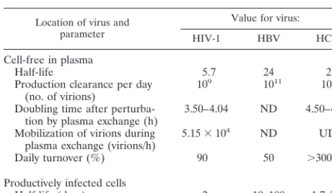
The evaluation of patients undergoing potent antiviral treat-ments has allowed the dynamics of cell-free virus in plasma to be addressed in vivo for HIV-1, HCV, and HBV infections (39, 45, 65, 67, 73, 95). Importantly, these approaches have docu-mented the dynamics of cell-free virions in plasma (half-lives being approximately 5.7, 24, and 2.7 hours in HIV-1, HBV, and HCV infection, respectively) and the turnover of infected cells (Table 1). These values, which reflect the different biologies and pathogenic potentials of these viruses (HBV is believed to be noncytopathic, whereas HIV-1 can kill productively infected cells within a few days), unequivocally document the high viral turnover that characterizes these infections in vivo. The
on May 15, 2020 by guest
http://jcm.asm.org/
standing of these features is expected to allow effective treat-ment and (eventually) eradication strategies to be designed and developed in the light of the specific dynamics of each infection. Although cell-free viremia is currently regarded as a mirror of systemic viral activity in many infections in vivo, viral turn-over is generally at its maximum where target cells principally are localized (i.e., in a specific organ or body fluid). Thus, in many viral infections, the amount of viral genome molecules that can be measured in blood samples (the net balance among the amount of virions released by producing cells, their sequestration in extravascular body fluids or other compart-ments, and their clearance from circulation) does not reflect exactly (depending on the type of infection, the range of infected cells, and the level of circulation in that organ) the actual viral activity taking place in target tissues or organs. A recent attempt to evaluate the relationship between the number of HIV-1-producing cells in lymph nodes and plasma viral load has documented a significant correlation between these two parameters, despite highly divergent viremia levels in the subjects under study (40). On the other hand, no correla-tion has been observed between HCV load in the liver (eval-uated either as HCV RNA molecules or as specific HCV antigens in liver cells) and plasma HCV RNA (7). Further-more, although the measurement of HCMV DNA in blood is a reliable index of the degree of HCMV dissemination, quan-titation of plasma virus often does not permit identification of the organ localization of this virus, which requires detection and quantitation of virus in samples taken locally (11, 32).
The correct understanding of the factors influencing the correlation between viral activity at the level of target cells and the number of cell-free genome molecules in plasma in the different viral infections is clearly necessary to interpret the data supplied by an increasing body of viremia-based studies carried out in vivo. To address this issue, my laboratory has recently analyzed the dynamic features of cell-free viremia in two persistent human infections (HIV-1 and HCV infections) after perturbation by plasma exchange (2, 53). The data doc-umented substantial differences in the dynamic features of the two viruses. In fact, although in both cases the dramatic reduc-tion in genome copy numbers determined by plasma exchange was rapidly followed by restoration of previous levels (with a doubling time ranging from 3.50 to 4.04 h for HIV-1 and from 4.50 to 4.60 h for HCV), in HIV-1 infection, but not in HCV, mobilization of extravascular cell-free virions also occurred
during the 2-h plasma exchange procedure (on average, 5.15⫻
104viral genome molecules per h). Taken together, these re-sults point to the existence in HIV-1 infection of an extravas-cular compartment of cell-free virus (probably the fluid of the lymphoid circulation which is [or tends to be] in balance with the vascular compartment), while in HCV infection, the res-toration of plasma viremia levels within a few hours of plasma exchange is principally due to newly produced virions. These data, obtained by a direct approach (subtraction of cell-free virus from plasma), are in substantial agreement with those documenting high turnover of cell-free virus in HCV (65) and HIV-1 (73) infections in subjects under treatment with antivi-rals.
Overall, these results highlight a substantially different sce-nario from that imagined before the introduction of quantita-tive molecular methods. Very high viral turnover has been observed during the symptomless phases of important, persis-tent human infections. In this context, all pathogenic events and the virus-host relationships should be analyzed in vivo in the light of the data from viral dynamics.
Quantitative analysis of viral transcription in vitro and in
vivo.The sensitivity and specificity performances of most
quan-titative methods have provided in the last few years a simple approach to the evaluation of gene transcription in vivo and in vitro. A precise understanding of the dynamics of virus tran-scription has allowed the direct evaluation of the latency-ac-tivity of herpesviruses. Recent studies of latent HCMV and herpes simplex virus type 1 and 2 infections have provided new insights into the dynamic pattern of virus expression, with potential implications for diagnosis and treatment (79, 83, 86, 94). In addition, the close correlation observed for HCMV infection between expression of the late HCMV transcripts in peripheral blood mononuclear cells (PBMCs) and levels of viral DNA molecules in plasma (12) has suggested a potential diagnostic use for quantitative analysis of HCMV mRNAs in nonblood samples such as bronchoalveolar cells (13). In other DNA viruses, such as HPVs, the potential pathogenic role of high levels of HPV type 16 expression is the subject of a recent investigation (41).
A typical example of the role of strategies aimed at revealing the pattern of viral transcripts in different phases of the infec-tion comes from the comparative evaluainfec-tion of infecinfec-tion activ-ity in samples from patients with diverging disease progression. In HIV-1 infection, consistent evidence indicated that progres-sion of disease is driven by an increase in viral load evaluated as cell-free plasma virus; it was unclear, however, to what extent this increase stems from the dysregulation of the mo-lecular mechanisms governing virus gene expression at the transcriptional or posttranscriptional levels. To address this issue, several quantitative virologic parameters (including pro-virus transcriptional activity and splicing pattern) have been analyzed for subjects with nonprogressive HIV infection and compared with those of matching groups of progressor pa-tients. It was observed not only that high levels of unspliced (US) and multiply spliced (MS) viral transcripts in PBMCs correlate with the decrease in CD4⫹T cells (1, 6, 23, 27, 82),
following the general trend of systemic HIV-1 activity, but also that MS mRNA levels in PBMCs are closely associated with the number of productively infected cells (6), since the half-life of this class of transcripts after administration of a potent protease inhibitor is very consistent with that of productively infected cells (39, 95). The transcriptional pattern observed during in vitro infections of T-cell lines, primary PBMCs, and monocytes/macrophages supports these findings.
[image:3.612.53.293.91.231.2]Quantitative molecular analysis has simplified the evaluation of the dynamic pattern of viral mRNAs in different target cells. TABLE 1. Dynamic features of HIV-1, HBV, and
HCV infections in vivoa
Location of virus and parameter
Value for virus:
HIV-1 HBV HCV
Cell-free in plasma
Half-life 5.7 24 2.7
Production clearance per day
(no. of virions) 10
9 1011 1012
Doubling time after
perturba-tion by plasma exchange (h) 3.50–4.04 ND 4.50–4.60 Mobilization of virions during
plasma exchange (virions/h) 5.15⫻10
4 ND UD
Daily turnover (%) 90 50 ⬎300
Productively infected cells
Half-life (days) 2 10–100 1.7–70 aThe data for HIV-1 are derived from the work of Wei et al. (95), Ho et al.
(39), Perelson et al. (73), and Bagnarelli et al. (2). The data for HBV are derived from the work of Nowak et al. (67). The data for HCV are derived from the work of Neumann et al. (65) and Manzin et al. (53). ND, not done; UD, undetectable.
on May 15, 2020 by guest
http://jcm.asm.org/
This allows the relative contribution of different cell subsets to a given infection to be calculated, as demonstrated in HIV-1 infection (6). In this infection, the molecular data for virus expression in cultured macrophages (S. Aquaro, P. Bagnarelli, M. Clementi, T. Guenci, R. Calio, and C.-F. Perno, Dynamics of HIV replication in primary macrophages and modulation by antiviral drugs, presented at the 6th Conference on Retrovi-ruses and Opportunistic Infections, Chicago, Ill., 31 January to 4 February 1999) have confirmed and extended previous anal-yses of HIV-1 infectivity (91), lending strong support to the hypothesis of a role for these cells as an effective long-term in vivo reservoir in HIV-1 infection (96). In this context, the accuracy of studies aimed at defining the cell tropism of a virus in vivo and the role of virus reservoirs in disease progression can be improved by evaluating, besides other indices of ongo-ing infection, the pattern of viral mRNAs and the dynamics of virus gene expression.
Virus compartmentalization and tropism in vivo.An
inter-esting aspect of viral dynamics studies, i.e., the presence of distinct compartments for viral infections in vivo, has been addressed using either biological or molecular approaches, in-cluding quantitative techniques for viral nucleic acids. The availability of methods to investigate this aspect has opened new prospects for the understanding of the pathogenesis of viral disease and of the mechanisms of virus transmission.
In HIV-1 infection, early data have shown that HIV-1 iso-lates from semen samples are frequently biologically unrelated to plasma isolates (93). More recently, remarkable sequence heterogeneity of viral quasispecies from plasma and genital secretions has been observed (101), together with the absence of a correlation between cell-free HIV-1 loads in plasma and those in semen (49). Taken together, these results suggest that plasma and semen are separate compartments and that local factors (including inflammation and other infections) may have significant effects on HIV-1 concentration in semen (and, con-sequently, on infectivity).
Furthermore, several reports in the last few years have in-dicated that HCV is capable of infecting cells other than hepa-tocytes (15, 28, 52, 100, 102); this finding has suggested that accurate analysis of HCV tropism in vivo could be a useful strategy toward a greater understanding of the HCV patho-genic potential and the development of effective antiviral strat-egies. Although conflicting results have been obtained to date for the role of HCV in several human lymphoproliferative diseases (22, 24, 69), this example documents the potential of pathogenic research in medical virology by the application of quantitative methods.
QUANTITATIVE METHODS FOR VIRAL NUCLEIC ACIDS AND ANTIVIRAL TREATMENTS
The introduction of new antiviral agents into preclinical and clinical use will greatly expand in the near future the treatment options available for acute and persistent viral infections. A major consequence of this new scenario will be the acute need for reliable parameters to evaluate the efficacy of therapies in real time and to monitor them (in some cases for months or years). Theoretical studies (57, 97) and early experimental evidence (4, 7, 11, 32, 39, 45, 59, 67, 95) have indicated unam-biguously that most quantitative molecular methods are able to provide information on changes in systemic viral activity and that they are thus suitable for following up infected patients treated with antivirals. While there is no doubt of the useful-ness of these methods in evaluating the efficacy of any antiviral treatment in vivo, several new questions have been raised. Among these, it seems important to verify whether (i) a single
quantitative parameter (i.e., cell-free genome copy numbers in plasma) is sufficient to monitor viral infections during treat-ment or, alternatively, whether other indices (in addition to cell-free virus, viral transcripts in infected cells, viral load in different compartments, and proviral copy numbers in retrovi-ral infections) are necessary to evaluate exhaustively the effi-cacy of antiviral compounds over time, and (ii) virologic indices other than those documenting the level of systemic viral activ-ity are necessary to assess specific antiviral treatments. In other words, it is important to evaluate whether, in different infec-tions, the plasma viral load may constitute a reliable index of selection of drug-resistant variants or whether more-specific quantitative assays are required.
Although potent antiretroviral therapy can at present con-trol HIV-1 infection, suggesting that virus eradication might be at hand (72), a long-lived reservoir of infectious virus persists in CD4⫹T cells. Furthermore, it has been shown that very high
concentrations of protease inhibitors are necessary to suppress HIV-1 production in infected macrophages (74). Thus, even in patients under effective therapy and showing suppression of plasma RNA, HIV-1 DNA is easily recovered from PBMCs (98), and it has recently been shown that the dynamics of proviral HIV-1 DNA copy numbers in PBMCs from patients under effective antiretroviral therapy document the crucial role of latently infected cells (which are insensitive to current an-tiviral treatments) in HIV-1 persistence (29). More recently, decay of proviral HIV-1 DNA copy numbers and specific viral transcripts (US and MS) has been observed for PBMCs from patients with sustained response to the anti-HIV-1 treatment (26); this decay occurs in two phases, but the ratio between US and MS HIV-1 transcripts tends subsequently to remain stable for months, indicating that current therapies are unable to eradicate the infection, at least within a few decades. These data also suggest that measurements of different viral nucleic acid species are crucial to the accurate monitoring of antiviral therapies in HIV-1 infection.
In HCV infection, the involvement of a direct cytopathic effect or of an immune-mediated mechanism in the progres-sion of the hepatic damage observed in chronic hepatitis C is still a matter of controversy. Similarly, conflicting results have been obtained for the pathogenic role of high HCV RNA levels in persistently infected subjects and for the capability of cell-free virus in plasma of documenting sustained response to interferon treatment (46, 47, 98). Recently, it has been served that an accurate profile of viral replication can be ob-tained only by monthly testing (since longer intervals could miss viremia fluctuations, frequent in these patients) and that HCV RNA levels are more stable in asymptomatic HCV car-riers than in patients with the biochemical activity of liver disease (78). Although early reports addressing the role of HCV viremia levels in subjects under treatment with inter-feron (or with combinations of ribavirin and interinter-feron) have highlighted the potential usefulness of this parameter (90), further insights into this particular aspect will probably be obtained when the new antiviral compounds interfering with specific steps of the viral life cycle (such as the function of the protease-helicase HCV gene product) reach the phase of clin-ical evaluation. Thus, specific antiviral therapy and its moni-toring could effectively contribute to the understanding of HCV disease pathogenesis.
In the routine diagnosis of HCMV infection, molecular niques have largely replaced traditional culture-based tech-niques. In this infection, a high systemic viral load generally correlates with HCMV disease (11); this correlation is strong in the HIV-1-infected population and in organ transplantation recipients but less clear in allogeneic bone marrow
on May 15, 2020 by guest
http://jcm.asm.org/
tation recipients. A reduction in systemic HCMV load also correlates with response to the specific antiviral treatment (77), but (due to the scarce data currently available) further research is needed to evaluate the role of HCMV load as a surrogate marker for drug resistance in different clinical con-ditions.
Finally, considerable effort is currently being directed at the development of new antiviral chemotherapeutic agents. The introduction of potent viral inhibitors in monotherapy or com-bination therapy regimens has resulted in a marked improve-ment in clinical response in a small number of viral infections. However, selection of drug-resistant variants during long-term antiviral treatments is an outstanding clinical problem during treatment of persistent infections. In this context, monitoring of these therapies implies not only analysis of viral load and of other indices of viral expression but also the introduction of widespread drug sensitivity testing. Indeed, early experience in HIV-1 infection has documented that the routine use of reli-able, real-time methods to test the sensitivity of replicative viral strains could drive a more effective therapeutic interven-tion in HIV-1 infecinterven-tion (38, 85). Since only incomplete data are available at present on the role of genotypic and phenotypic drug resistance testing in human infections other than those with HIV-1, thorough research into this specific aspect will be absolutely necessary when new compounds are proposed for routine use in medical virology.
In conclusion, considerable improvements in the laboratory monitoring of antiviral therapies have been achieved by the introduction of quantitative molecular techniques as routine diagnostic methods. However, the assessment of viremia levels alone does not appear sufficient to provide complete data for real-time information on treatment efficacy. The evaluation of other molecular parameters is necessary in some cases; more-over, the frequent selection of drug-resistant viral mutants requires the introduction of additional molecular assays for the early detection of the genotypic and phenotypic features of replicative viral strains.
CONCLUDING REMARKS
The data obtained in the last 10 years have unambiguously indicated that absolute quantitation of viral nucleic acid spe-cies is a crucial prerequisite for future developments in virol-ogy. The different features of the existing techniques for the assessment of viral nucleic acid copy numbers have allowed the widespread application of quantitative studies to themes of basic and medical virology. An important new area of research in virology has been developed which is directly dependent on the widespread application of highly sensitive and reliable quantitative methodologies. The availability of these methods has significantly contributed to the study of the natural history and pathogenesis of viral infections and virus-host relation-ships and to addressing the efficacy of antiviral therapies in real time. However, we need to consider that (i) further technical improvements are necessary since the available quantitative techniques are affected by important limitations, (ii) more than one quantitative index of viral activity is required in specific in vivo situations for a reliable evaluation of viral activity, and (iii) quantitative methods, albeit necessary, are not sufficient to address all the aspects relevant for a complete diagnosis in the monitoring of antiviral therapies, including virus resistance to inhibitory compounds. All this indicates that further research in this area is needed.
ACKNOWLEDGMENTS
This study and the research activity of my group in the present field have been supported by grants from Istituto Superiore di Sanita` (I.S.S.) (Progetto di Ricerca sull’AIDS e Progetto Epatite Virale), Consiglio Nazionale delle Ricerche (C.N.R.) (Progetto Biotecnologie), and Ministero dell’Universita` e della Ricerca Scientifica e Tecnologica (MURST).
REFERENCES
1.Bagnarelli, P., C. Balotta, A. Valenza, F. Mazzola, M. C. Colombo, M. Violin, M. Galli, and M. Clementi.1997. Patterns of HIV-1 transcripts in peripheral blood lymphocytes from long-term nonprogressors and typical progressor patients. J. Acquir. Immune Defic. Syndr.15(Suppl. I):S69–S71. 2.Bagnarelli, P., M. Candela, A. Valenza, A. Manzin, L. Solforosi, F. Maz-zola, L. Butini, M. Montroni, A. Gabrielli, P. E. Varaldo, and M. Clementi.
1996. Dynamic features of human immunodeficiency virus type 1 (HIV-1) viremia: kinetics of cell-free HIV RNA after therapeutic plasma exchange. J. Infect. Dis.176:801–804.
3.Bagnarelli, P., S. Menzo, A. Valenza, A. Manzin, M. Giacca, F. Ancarani, G. Scalise, P. E. Varaldo, and M. Clementi.1992. Molecular profile of human immunodeficiency virus type 1 infection in symptomless patients and in patients with AIDS. J. Virol.66:7328–7335.
4.Bagnarelli, P., S. Menzo, A. Valenza, S. Paolucci, S. Petroni, G. Scalise, R. Sampaolesi, A. Manzin, P. E. Varaldo, and M. Clementi.1995. Quantitative molecular monitoring of human immunodeficiency virus type 1 activity during therapy with specific antiretroviral compounds. J. Clin. Microbiol.
33:16–23.
5.Bagnarelli, P., A. Valenza, S. Menzo, A. Manzin, G. Scalise, P. E. Varaldo, and M. Clementi.1994. Dynamics of molecular parameters of human im-munodeficiency virus type 1 activity in vivo. J. Virol.68:2495–2502. 6.Bagnarelli, P., A. Valenza, S. Menzo, R. Sampaolesi, P. E. Varaldo, L.
Butini, M. Montroni, C.-F. Perno, S. Aquaro, D. Mathez, J. Leibowitch, C. Balotta, and M. Clementi.1996. Dynamics and modulation of human im-munodeficiency virus type 1 transcripts in vitro and in vivo. J. Virol.70:
7603–7613.
7.Ballardini, G., A. Manzin, F. Giostra, R. Francesconi, P. Groff, A. Grassi, L. Solforosi, S. Ghetti, D. Zauli, M. Clementi, and F. B. Bianchi.1997. Quantitative liver parameters of HCV infection: relation to HCV geno-types, viremia, and response to interferon treatment. J. Hepatol.26:779– 786.
8.Barany, F.1991. Genetic disease detection and DNA amplification using cloned thermostable ligase. Proc. Natl. Acad. Sci. USA88:189–193. 9.Bell, J. I.1999. Clinical research is dead; long live clinical research. Nat.
Med.5:477–478.
10.Berger, A., J. Braner, H. W. Doerr, and B. Weber.1998. Quantification of viral load: clinical relevance for human immunodeficiency virus, hepatitis B virus and hepatitis C virus infection. Intervirology41:24–34.
11.Boeckh, M., and G. Boivin.1998. Quantitation of cytomegalovirus: meth-odologic aspects and clinical applications. Clin. Microbiol. Rev.11:533–554. 12.Boivin, G., J. Handfield, E. Toma, R. Lalonde, and M. G. Bergeron.1999. Expression of the late cytomegalovirus (CMV) pp150 transcript in leuko-cytes of AIDS patients is associated with high viral DNA load in leukoleuko-cytes and presence of CMV DNA in plasma. J. Infect. Dis.179:1101–1107. 13.Boivin, G., C. A. Olson, M. R. Quirk, B. Kringstad, M. I. Hertz, and M. C.
Jordan.1996. Quantitation of cytomegalovirus DNA and characterization of viral gene expression in bronchoalveolar cells of infected patients with or without pneumonitis. J. Infect. Dis.173:1304–1312.
14.Bonhoeffer, S., R. M. May, G. M. Shaw, and M. A. Nowak.1997. Virus dynamics and drug therapy. Proc. Natl. Acad. Sci. USA94:6971–6976. 15.Bouffard, P., P. H. Hayashi, R. Acevedo, M. Levy, and J. B. Zeldis.1992.
Hepatitis C virus infection is detected in a monocyte/macrophage subpopu-lation of peripheral blood mononuclear cells of infected patients. J. Infect. Dis.166:1276–1280.
16.Bowen, E. F., C. A. Sabin, P. Wilson, P. D. Griffiths, C. C. Davey, M. A. Johnson, and V. C. Emery.1997. Cytomegalovirus (CMV) viraemia de-tected by polymerase chain reaction identifies a group of HIV-positive patients at high risk of CMV disease. AIDS11:889–893.
17.Cao, Y., L. Qin, L. Zhang, J. Safrit, and D. D. Ho.1995. Virologic and immunologic characterization of long-term survivors of human immunode-ficiency virus type 1 infection. N. Engl. J. Med.332:201–208.
18.Clementi, M., P. Bagnarelli, S. Menzo, A. Valenza, A. Manzin, and P. E. Varaldo.1993. Clearance of HIV viremia after seroconversion. Lancet
341:315–316.
19.Clementi, M., P. Bagnarelli, A. Manzin, and S. Menzo.1994. Competitive polymerase chain reaction and analysis of viral activity at the molecular level. Genet. Anal. Tech. Appl.11:1–6.
20.Clementi, M., S. Menzo, P. Bagnarelli, A. Manzin, A. Valenza, and P. E. Varaldo.1993. Quantitative PCR and RT-PCR in virology. PCR Methods Appl. (CSH)2:191–196.
21.Clementi, M., S. Menzo, P. Bagnarelli, A. Valenza, S. Paolucci, R. Sampa-olesi, A. Manzin, and P. E. Varaldo. 1996. Clinical use of quantitative
on May 15, 2020 by guest
http://jcm.asm.org/
molecular methods in studying human immunodeficiency virus type 1 in-fection. Clin. Microbiol. Rev.9:135–147.
22.Collier, J. D., B. Zanke, M. Moore, G. Kessler, M. Krajden, F. Shepherd, and J. Heathcote. 1999. No association between hepatitis C and B-cell lymphoma. Hepatology29:1259–1261.
23.Comar, M., G. Marzio, P. D’Agaro, and M. Giacca.1996. Quantitative dynamics of HIV type 1 expression. AIDS Res. Hum. Retrovir.12:117–126. 24.Dammacco, F., P. Gatti, and D. Sansonno.1998. Hepatitis C virus infection, mixed cryoglobulinemia, and non-Hodgkin’s lymphoma: an emerging pic-ture. Leuk. Lymphoma31:463–476.
25.Fanning, L., E. Kenny, M. Sheehan, B. Cannon, M. Whelton, J. O’Connell, J. K. Collins, and F. Shanahan.1999. Viral load and clinicopathological features of chronic hepatitis C (1b) in a homogeneous patient population. Hepatology29:904–907.
26.Furtado, M. R., D. S. Callaway, J. P. Phair, K. J. Kunstman, J. L. Stanton, C. A. Macken, A. S. Perelson, and S. M. Wolinsky.1999. Persistence of HIV-1 transcription in peripheral-blood mononuclear cells in patients re-ceiving potent antiretroviral therapy. N. Engl. J. Med.340:1614–1622. 27.Furtado, M. R., L. A. Kingsley, and S. M. Wolinsky.1995. Changes in the
viral mRNA expression pattern correlate with a rapid rate of CD4⫹T-cell number decline in human immunodeficiency virus type 1-infected individ-uals. J. Virol.69:2092–2100.
28.Gabrielli, A., A. Manzin, M. Candela, M. L. Caniglia, S. Paolucci, M. G. Danieli, and M. Clementi.1994. Active hepatitis C virus infection in bone marrow and peripheral blood mononuclear cells from patients with mixed cryoglobulinemia. Clin. Exp. Immunol.97:87–93.
29.Galli, M., C. Balotta, L. Meroni, M. C. Colombo, L. Papagno, P. Bagnarelli, L. Testa, S. Varchetta, L. Colombo, M. Moroni, A. d’Arminio Monforte, M. Clerici, and M. Clementi.1998. Early increase in cell-associated HIV-1 DNA in patients on highly active antiretroviral therapy. AIDS12:2500– 2502.
30.Gallinella, G., M. Zerbini, M. Musiani, S. Venturoli, G. Gentilomi, and E. Manaresi.1997. Quantitation of parvovirus B19 DNA sequences by com-petitive PCR: differential hybridization of the amplicons and immunoenzy-matic detection on microplate. Mol. Cell. Probes11:127–133.
31.Gerken, G., J. Gomes, P. Lampertico, M. Colombo, T. Rothaar, M. Trip-pler, and G. Colucci.1998. Clinical evaluation and applications of the Amplicor HBV Monitor test, a quantitative HBV DNA PCR assay. J. Virol. Methods74:155–165.
32.Gerna, G., E. Percivalle, F. Baldanti, A. Sarasini, M. Zavattoni, M. Furi-one, M. Torsellini, and M. G. Revello.1998. Diagnostic significance and clinical impact of quantitative assays for diagnosis of human cytomegalovi-rus infection/disease in immunocompromised patients. New Microbiol.21:
293–308.
33.Gilliland, G., S. Perrin, K. Blanchard, and H. F. Bunn.1990. Analysis of cytokine mRNA and DNA: detection and quantitation by competitive poly-merase chain reaction. Proc. Natl. Acad. Sci. USA87:2725–2729. 34.Giostra, F., A. Manzin, M. Lenzi, R. Francesconi, L. Solforosi, P. Manotti,
L. Muratori, D. Zauli, M. Clementi, and F. B. Bianchi.1996. Low hepatitis C viremia levels in patients with anti-liver/kidney microsomal antibody type 1 positive chronic hepatitis. J. Hepatol.25:433–438.
35.Gupta, P., L. Kingsley, J. Armstrong, M. Ding, M. Cottril, and C. Rinaldo.
1993. Enhanced expression of human immunodeficiency virus type 1 cor-related with development of AIDS. Virology196:586–595
36.Gut, M., C. M. Leutenegger, J. B. Huder, N. C. Pedersen, and H. Lutz.1999. One-tube fluorogenic reverse transcription-polymerase chain reaction for the quantitation of feline coronaviruses. J. Virol. Methods77:37–46. 37.Hawrami, K., and J. Breuer.1999. Development of a fluorogenic
polymer-ase chain reaction assay (TaqMan) for the detection and quantitation of varicella zoster virus. J. Virol. Methods79:33–40.
38.Hertogs, K., M. P. de Bethune, V. Miller, T. Ivens, P. Schel, A. Van Cauwenberge, C. Van Den Eynde, V. Van Gerwen, H. Azijn, M. Van Houtte, F. Peeters, S. Staszewski, M. Conant, S. Bloor, S. Kemp, B. Larder, and R. Pauwels.1998. A rapid method for simultaneous detection of phenotypic resistance to inhibitors of protease and reverse transcriptase in recombinant human immunodeficiency virus type 1 isolates from patients treated with antiretroviral drugs. Antimicrob. Agents Chemother.42:269–276. 39.Ho, D. D., A. U. Neuman, A. S. Perelson, W. Chen, J. M. Leonard, and M.
Markowitz.1995. Rapid turnover of plasma virions and CD4 lymphocytes in HIV-1 infection. Nature (London)373:123–126.
40.Hockett, R. D., J. M. Kilby, C. A. Derdeyn, M. S. Saag, M. Sillers, K. Squires, S. Chiz, M. A. Nowak, G. M. Shaw, and R. P. Bucy.1999. Constant mean viral copy number per infected cell in tissue regardless of high, low, or undetectable plasma HIV RNA. J. Exp. Med.189:1545–1554. 41.Hsu, E. M., P. J. McNicol, F. B. Guijon, and M. Paraskevas.1993.
Quan-tification of HPV-16 E6-E7 transcription in cervical intraepithelial neopla-sia by reverse transcriptase polymerase chain reaction. Int. J. Cancer55:
397–401.
42.Jeffery, K. J. M., K. Usuku, S. E. Hall, W. Matsumoto, G. P. Taylor, J. Procter, M. Bunce, G. S. Ogg, K. I. Welsh, J. N. Weber, A. L. Lloyd, M. A. Nowak, M. Nagai, D. Kodama, S. Izumo, M. Osame, and C. R. M. Bang-ham. 1999. HLA alleles determine human T-lymphotropic virus-I
(HTLV-I) proviral load and the risk of HTLV-I-associated myelopathy. Proc. Natl. Acad. Sci. USA96:3848–3853.
43.Kawai, S., O. Yokosuka, T. Kanda, F. Imazeki, Y. Maru, and H. Saisho.
1999. Quantification of hepatitis C virus by TaqMan PCR: comparison with HCV Amplicor Monitor assay. J. Med. Virol.58:121–126.
44.Kogan, D. L., M. Burroughs, S. Emre, T. Fishbein, A. Moscona, C. Ramson, and B. L. Schneider.1999. Prospective longitudinal analysis of quantitative Epstein-Barr virus polymerase chain reaction in pediatric liver transplant recipients. Transplantation67:1068–1070.
45.Lam, N. P., A. U. Neumann, D. R. Gretch, T. E. Wiley, A. S. Perelson, and T. J. Layden.1997. Dose-dependent acute clearance of hepatitis C geno-type 1 virus with interferon alfa. Hepatology26:226–231.
46.Lau, J. Y., G. L. Davis, J. Kniffen, K. P. Qian, M. S. Urdea, C. S. Chan, M. Mizokami, P. D. Neuwald, and J. C. Wilber.1993. Significance of serum hepatitis C virus RNA levels in chronic hepatitis C. Lancet341:1501–1504. 47.Lau, J. Y., M. Mizokami, T. Ohno, D. A. Diamond, J. Kniffen, and G. L. Davis.1993. Discrepancy between biochemical and virological responses to interferon-alpha in chronic hepatitis C. Lancet342:1208–1209.
48.Leutenegger, C. M., D. Klein, R. Hofmann-Lehmann, C. Mislin, U. Hum-mel, J. Boni, F. Boretti, W. H. Guenzburg, and H. Lutz.1999. Rapid feline immunodeficiency virus provirus quantitation by polymerase chain reaction using the TaqMan fluorogenic real-time detection system. J. Virol. Meth-ods78:105–116.
49.Liuzzi, G., A. Chirianni, M. Clementi, P. Bagnarelli, A. Valenza, P. T. Cataldo, and M. Piazza.1996. Analysis of HIV-1 load in blood, semen and saliva: evidence for different viral compartments in a cross-sectional and longitudinal study. AIDS10:F51–F56.
50.Lo, Y. M., L. Y. Chan, K. W. Lo, S. F. Leung, J. Zhang, A. T. Chan, J. C. Lee, N. M. Hjelm, P. J. Johnson, and D. P. Huang.1999. Quantitative analysis of cell-free Epstein-Barr virus DNA in plasma of patients with nasopharyngeal carcinoma. Cancer Res.59:1188–1191.
51.Manzin, A., P. Bagnarelli, S. Menzo, F. Giostra, M. Brugia, R. Francesconi, F. B. Bianchi, and M. Clementi.1994. Quantitation of hepatitis C virus genome molecules in plasma samples. J. Clin. Microbiol.32:1939–1944. 52.Manzin, A., M. Candela, S. Paolucci, M. L. Caniglia, A. Gabrielli, and M.
Clementi.1994. Presence of hepatitis C virus (HCV) genomic RNA and viral replicative intermediates in bone marrow and peripheral blood mono-nuclear cells from HCV-infected patients. Clin. Diagn. Lab. Immunol.
1:160–163.
53.Manzin, A., M. Candela, L. Solforosi, A. Gabrielli, and M. Clementi.1999. Dynamics of hepatitis C viremia after plasma exchange. J. Hepatol.31:389– 393.
54.Manzin, A., L. Solforosi, D. Bianchi, A. Gabrielli, F. Giostra, S. Bruno, and M. Clementi.1995. Virus load in samples from hepatitis C virus (HCV)-infected patients with various clinical conditions. Res. Virol.146:279–284. 55.Manzin, A., L. Solforosi, M. Candela, G. Cherubini, G. Piccinini, M. Bru-gia, A. Gabrielli, and M. Clementi.1996. Hepatitis C virus infection and cryoglobulinemia: assessment of HCV RNA copy numbers in supernatant, cryoprecipitate and non-liver cells. J. Viral Hepatitis3:285–292. 56.Manzin, A., L. Solforosi, F. Giostra, F. B. Bianchi, S. Bruno, S. Rossi, A.
Gabrielli, M. Candela, E. Petrelli, and M. Clementi.1997. Quantitative analysis of hepatitis C virus activity in different groups of untreated patients. Arch. Virol.142:465–472.
57.Marschner, I. C.1998. Design of HIV viral dynamics studies. Stat. Med.
17:2421–2434.
58.Martell, M., J. Gomez, J. I. Esteban, S. Sauleda, J. Quer, B. Cabot, R. Esteban, and J. Guardia.1999. High-throughput real-time reverse tran-scription-PCR quantitation of hepatitis C virus RNA. J. Clin. Microbiol.
37:327–332.
59.Mathez, D., P. Bagnarelli, I. Gorin, C. Katlama, G. Pialoux, G. Saimot, P. Tubiana, P. De Truchis, J.-P. Chauvin, R. Mills, R. Rode, M. Clementi, and J. Leibowitch.1997. Reductions in viral load and increases in T lymphocyte numbers in treatment of naive patients with advanced HIV-1 infection treated with ritonavir, zidovudine and zalcitabine triple therapy. Antivir. Ther.2:175–183.
60.Mellors, J. W., L. A. Kingsley, C. R. Rinaldo, J. A. Todd, B. S. Hoo, R. P. Kokka, and P. Gupta.1995. Quantitation of HIV-1 RNA in plasma predicts outcome after seroconversion. Ann. Intern. Med.122:573–579.
61.Mellors, J. W., C. R. Rinaldo, P. Gupta, R. M. White, J. A. Todd, and L. A. Kingsley.1996. Prognosis in HIV-1 infection predicted by the quantity of virus in plasma. Science272:1167–1170.
62.Menzo, S., P. Bagnarelli, M. Giacca, A. Manzin, P. E. Varaldo, and M. Clementi.1992. Absolute quantitation of viremia in human immunodefi-ciency virus infection by competitive reverse transcription polymerase chain reaction. J. Clin. Microbiol.30:1752–1757.
63.Michael, N. L., M. Vahey, D. S. Burke, and R. R. Redfield.1992. Viral DNA and mRNA expression correlate with the stage of human immunodeficiency virus (HIV) type 1 infection in humans: evidence for viral replication in all stages of HIV disease. J. Virol.66:310–316.
64.Morris, T., B. Robertson, and M. Gallagher.1996. Rapid reverse transcrip-tion-PCR detection of hepatitis C virus RNA in serum by using the TaqMan fluorogenic detection system. J. Clin. Microbiol.34:2933–2936.
on May 15, 2020 by guest
http://jcm.asm.org/
65.Neumann, A. U., N. P. Lam, H. Dahari, D. R. Gretch, T. E. Wiley, T. J. Layden, and A. S. Perelson.1998. Hepatitis C viral dynamics in vivo and the antiviral efficacy of interferon-alpha therapy. Science282:103–107. 66.Nolte, F. S.1998. Branched DNA signal amplification for direct
quantita-tion of nucleic acid sequences in clinical specimens. Adv. Clin. Chem.
33:201–235.
67.Nowak, M. A., S. Bonhoeffer, A. M. Hill, R. Boehme, H. C. Thomas, and H. McDade.1996. Viral dynamics in hepatitis B virus infection. Proc. Natl. Acad. Sci. USA93:4398–4402.
68.Oehlenschlager, F., P. Schwille, and M. Eigen.1996. Detection of HIV-1 RNA by nucleic acid sequence-based amplification combined with flu-orescence correlation spectroscopy. Proc. Natl. Acad. Sci. USA93:12811– 12816.
69.Ohsawa, M., N. Shingu, H. Miwa, H. Yoshihara, M. Kubo, H. Tsukuma, H. Teshima, M. Hashimoto, and K. Aozasa. 1999. Risk of non-Hodgkin’s lymphoma in patients with hepatitis C virus infection. Int. J. Cancer80:
237–239.
70.Pantaleo, G., S. Menzo, M. Vaccarezza, C. Graziosi, O. J. Cohen, J. F. Demarest, D. Montefiori, J. M. Orenstein, C. Fox, L. K. Schrager, J. B. Margolik, S. Buchbinder, J. V. Giorgi, and A. S. Fauci.1995. Studies in subjects with long-term progressive human immunodeficiency virus infec-tion. N. Engl. J. Med.332:209–216.
71.Pawlotski, J. M., M. Martinot-Peignoux, J. D. Poveda, A. Bastie, V. La Breton, F. Darthuy, J. Remire, S. Erlinger, D. Dhumeaux, and P. Marcellin.
1999. Quantification of hepatitis C virus RNA in serum by branched DNA-based signal amplification assays. J. Virol. Methods79:227–235. 72.Perelson, A. S., P. Essunger, Y. Cao, M. Vesanen, A. Hurley, K. Saksela, M.
Markowitz, and D. D. Ho.1997. Decay characteristics of HIV-1-infected compartments during combination therapy. Nature (London)387:188–191. 73.Perelson, A. S., A. U. Neumann, M. Markowitz, J. M. Leonard, and D. D. Ho. 1996. HIV-1 dynamics in vivo: virion clearance rate, infected cell life-span, and viral generation time. Science271:1582–1586.
74.Perno, C.-F., F. M. Newcomb, D. A. Davis, S. Aquaro, R. W. Humphrey, R. Calio`, and R. J. Yarchoan.1998. Relative potency of protease inhibitors in monocytes/macrophages acutely and chronically infected with human im-munodeficiency virus. J. Infect. Dis.178:413–422.
75.Piatak, M., M. S. Saag, L. C. Yang, S. J. Clark, J. C. Kappes, K.-C. Luk, B. H. Hahn, G. M. Shaw, and J. D. Lifson.1993. High levels of HIV-1 RNA in plasma during all stages of infection determined by competitive PCR. Science259:1749–1754.
76.Pistello, M., S. Menzo, M. Giorgi, L. Da Prato, G. Cammarota, M. Clem-enti, and M. Bendinelli.1994. Competitive polymerase chain reaction for quantitating feline immunodeficiency virus load in infected cat tissues. Mol. Cell. Probes8:229–234.
77.Poirier-Toulemonde, A. S., B. M. Imbert-Marcille, V. Ferre-Aubineau, B. Besse, M. G. Le Roux, D. Cantarovich, and S. Billaudel.1997. Successful quantification of cytomegalovirus DNA by competitive PCR and detection with capillary electrophoresis. Mol. Cell. Probes11:11–23.
78.Pontisso, P., G. Bellati, M. Brunetto, L. Chemello, G. Colloredo, R. Di Stefano, M. Nicoletti, M. G. Rumi, M. G. Ruvoletto, R. Soffredini, L. M. Valenza, and G. Colucci.1999. Hepatitis C virus RNA profiles in chroni-cally infected individuals: do they relate to disease activity? Hepatology
29:585–589.
79.Ramakrishnan, R., D. J. Fink, G. Jiang, P. Desai, J. C. Glorioso, and M. Levine.1994. Competitive quantitative PCR analysis of herpes simplex virus type 1 DNA and latency-associated transcript RNA in latently infected cells of the rat brain. J. Virol.68:1864–1873.
80.Saiki, R. K., T. L. Bugawan, G. T. Horn, K. B. Mullins, and H. A. Erlich.
1986. Analysis of enzymatically amplified B-globin and HLA-Dqa DNA with allele specific oligonucleotide probes. Nature (London)324:163–166. 81.Saiki, R. K., D. Gelfand, S. Stoffel, S. J. Scharf, R. Higuel, G. T. Horn, K. B. Mullins, and H. A. Erlich.1988. Primer directed enzymatic amplification of DNA with a thermostable DNA polymerase. Science239:487–491. 82.Saksela, K., C. Stevens, P. Rubinstein, and D. Baltimore.1994. Human
immunodeficiency virus type 1 mRNA expression in peripheral blood cells predicts disease progression independently of the number of CD4 lympho-cytes. Proc. Natl. Acad. Sci. USA91:1104–1108.
83.Sawtell, N. M., D. K. Poon, C. S. Tansky, and R. L. Thompson.1998. The
latent herpes simplex virus type 1 genome copy number in individual neu-rons is virus strain specific and correlates with reactivation. J. Virol.72:
5343–5350.
84.Scadden, D. T., Z. Wang, and J. E. Groopman.1992. Quantitation of plasma human immunodeficiency virus type 1 RNA by competitive poly-merase chain reaction. J. Infect. Dis.165:1119–1123.
85.Schmit, J.-C., and B. Weber.1997. Recent advances in antiretroviral ther-apy and HIV infection monitoring. Intervirology40:304–321.
86.Slobedman, B., and E. S. Mocarski.1999. Quantitative analysis of latent human cytomegalovirus. J. Virol.73:4806–4812.
87.Spector, S. A., R. Wong, K. Hsia, M. Pilcher, and M. J. Stempien.1998. Plasma cytomegalovirus (CMV) DNA load predicts CMV disease and survival in AIDS patients. J. Clin. Investig.101:497–502.
88.Stevens, S. J., M. B. Vervoort, A. J. van den Brule, P. L. Meenhorst, C. J. Meijer, and J. M. Middeldorp.1999. Monitoring of Epstein-Barr virus load in peripheral blood by quantitative competitive PCR. J. Clin. Microbiol.
37:2852–2857.
89.Swan, D. C., R. A. Tucker, G. Tortolero-Luna, M. F. Mitchell, L. Wideroff, E. R. Unger, R. A. Nisenbaum, W. C. Reeves, and J. P. Icenogle.1999. Human papillomavirus (HPV) DNA copy number is dependent on grade of cervical disease and HPV type. J. Clin. Microbiol.37:1030–1034. 90.Trabaud, M. A., F. Bailly, S. N. Si-Ahmed, P. Chevallier, M. Sepetjan, G.
Colucci, and C. Trepo.1997. Comparison of HCV RNA assays for the detection and quantification of hepatitis C virus RNA levels in serum of patients with chronic hepatitis C treated with interferon. J. Med. Virol.
52:105–112.
91.Tsai, W. P., S. R. Conley, H. F. Kung, R. R. Garrity, and P. L. Nara.1996. Preliminary in vitro growth cycle and transmission studies of HIV-1 in an autologous primary cell assay of blood-derived macrophages and peripheral blood mononuclear cells. Virology26:205–216.
92.Vener, T., M. Nygren, A. Andersson, M. Uhlen, J. Albert, and J. Lundeberg.
1998. Use of multiple competitors for quantification of human immunode-ficiency virus type 1 RNA in plasma. J. Clin. Microbiol.36:1864–1870. 93.Vernazza, P. L., J. J. Eron, M. S. Cohen, C. M. van der Horst, L. Troiani,
and S. A. Fiscus.1994. Detection and biologic characterization of infectious HIV-1 in semen of seropositive men. AIDS8:1325–1329.
94.Wang, K., L. Pesnicak, and S. E. Strauss.1997. Mutations in the 5⬘end of the herpes simplex virus type 2 latency-associated transcript (LAT) pro-moter affect LAT expression in vivo but not the rate of spontaneous reac-tivation of genital herpes. J. Virol.71:7903–7910.
95.Wei, X., S. K. Gosh, M. E. Taylor, V. A. Johnson, E. A. Emini, P. Deutsch, J. D. Lifson, S. Bonhoefer, M. A. Nowak, B. H. Hahn, M. S. Saag, and G. M. Shaw.1995. Viral dynamics in human immunodeficiency virus type 1 infec-tion. Nature (London)373:117–122.
96.Wodarz, D., A. L. Lloyd, V. A. Jansen, and M. A. Nowak.1999. Dynamics of macrophage and T cell infection by HIV. J. Theor. Biol.196:101–113. 97.Wu, H., A. A. Ding, and V. De Gruttola.1998. Estimation of HIV dynamic
parameters. Stat. Med.17:2463–2485.
98.Zeuzem, S., A. Franke, J. H. Lee, G. Hermann, B. Ruster, and W. K. Roth.
1996. Phylogenetic analysis of hepatitis C virus isolates and their correlation to viremia, liver function tests, and histology. Hepatology24:1003–1009. 99.Zhang, L., B. Ramratnam, K. Tenner-Racz, Y. He, M. Vesanen, S. Lewin,
A. Talal, P. Racz, A. S. Perelson, B. T. Korber, M. Markowitz, and D. D. Ho.
1999. Quantifying residual HIV-1 replication in patients receiving combi-nation antiretroviral therapy. N. Engl. J. Med.340:1605–1613.
100. Zhu, T., N. Wang, A. Carr, D. S. Nam, R. Moor-Jankowski, D. A. Cooper, and D. D. Ho.1996. Genetic characterization of human immunodeficiency virus type 1 in blood and genital secretions: evidence for viral compartmen-talization and selection during sexual transmission. J. Virol.70:3098–3107. 101. Zhender, G., L. Meroni, C. De Maddalena, S. Varchetta, G. Monti, and M. Galli.1997. Detection of hepatitis C virus RNA in CD19 peripheral blood mononuclear cells of chronically infected patients. J. Infect. Dis.176:1209– 1214.
102. Zignego, A. L., D. Macchia, M. Monti, V. Thiers, M. Mazzetti, M. Foschi, E. Maggi, S. Romagnani, P. Gentilini, and C. Brechot.1992. Infection of peripheral mononuclear blood cells by hepatitis C virus. J. Hepatol.15:
382–386.