Copyright © 2003, American Society for Microbiology. All Rights Reserved.
Development of a Multilocus Sequence Typing Method for Analysis of
Listeria monocytogenes
Clones
C. Salcedo, L. Arreaza, B. Alcala´, L. de la Fuente, and J. A. Va´zquez*
Servicio de Bacteriología, Centro Nacional de Microbiología, Instituto de Salud Carlos III, 28220 Majadahonda, Madrid, Spain
Received 7 June 2002/Returned for modification 9 July 2002/Accepted 11 November 2002
This study is a first step in the development of multilocus sequence typing (MLST) method forListeria
monocytogenes.Nine housekeeping genes were analyzed in a set of 62 strains isolated from different sources and geographic locations in Spain. These strains were previously characterized by pulsed-field gel electrophoresis (PFGE). Because of low diversity, two loci were discarded from the study. The sequence analysis of the seven remaining genes showed 29 different allelic combinations, with 22 of them represented by only one strain. The results of this sequence analysis were generally consistent with those of PFGE. Because MLST allows the easy comparison and exchange of results obtained in different laboratories, the future application of this new molecular method could be a useful tool for the listeriosis surveillance systems that will allow the identification
and distribution of analysis ofL. monocytogenesclones in the environment.
Listeria monocytogenesis an opportunistic pathogen widely distributed in the environment. The ubiquity of this microor-ganism makes especially necessary the use of typing methods for the study of its epidemiology. Numerous molecular
meth-ods have been applied to the characterization ofL.
monocyto-genesisolates, e.g., multilocus enzyme electrophoresis (MLEE) (1), pulsed-field gel electrophoresis (PFGE) (2, 29), random amplified polymorphic differences (19), and ribotyping (30), etc. By these methods the species is divided into two genetic divisions which are correlated with the flagellar antigen groups division I, composed of strains of serotypes 1/2a and 1/2c, and division II, which is composed of strains of serotypes 1/2b and 4b. Both divisions are characterized by nonoverlapping allelic variants of different genetic markers, suggesting strong linkage disequilibrium and an apparent lack of gene exchange between them (15). This is consistent with the hypothesis that the
ge-netic structure of L. monocytogenes populations is basically
clonal (21). Recently, an additional division has been proposed based on the variability of the sequence of several genes in-volved in virulence (22).
MLEE has been the most widely used molecular method to study the genetic structure and epidemiology of pathogenic bacterial species. Recently, a novel molecular typing method based on the principles of MLEE (25) has been developed, multilocus sequence typing (MLST). This technique was
pri-marily designed and validated forNeisseria meningitidis(17).
Afterwards, it was successful in the characterization of several
other pathogenic bacteria, such as Streptococcus pneumoniae
(5),Streptococcus pyogenes(8),Staphylococcus aureus(7), and
Campylobacter jejuni(4).
MLST makes use of automated DNA sequencing to char-acterize the alleles present at different housekeeping genes.
Because it is based on nucleotide sequence, it is highly discrim-inatory and provides unambiguous results that are directly comparable among laboratories via the internet (7). In addi-tion, this method is particularly suited to global epidemiology studies because the accumulation of nucleotide changes in housekeeping genes is a relatively slow process and allelic profiles are stable over time (6).
Epidemiological research requires systems that make easy the comparison and exchange of results obtained in different laboratories. The most important advantage of MLST over the typing methods based on comparisons of DNA fragments is the unambiguity and electronic portability of nucleotide sequence data (17), which allow a readily comparison of results among laboratories.
In this study we describe a first step for developing the
MLST method forL. monocytogenes.The sequence diversity of
nine housekeeping genes, possible candidates to be included in a future MLST scheme, has been analyzed.
MATERIALS AND METHODS
Bacterial strains.A total of 62 strains ofL. monocytogeneswere used in this study. Thirty-one were isolated from human cases of listeriosis, 12 were isolated from animal clinical sources, and 19 were isolated from food. The strains were isolated in different regions across Spain over the period from 1995 to 2001 without an evident epidemiological link among them. Serotyping was done ac-cording the method previously described (24). The three most frequent serotypes among the clinical isolates of L. monocytogeneswere represented: 1/2a (12 strains), 1/2b (10 strains), and 4b (40 strains).
Our laboratory uses PFGE to characterize the isolates sent by hospitals and other public health laboratories. The agarose blocks are prepared by following a method previously described by Graves and Swaminathan (12). Chromosomal DNA is digested withApaI (Pharmacia), and the restricted fragments are sep-arated at 200 V with pulse times from 0.1 to 25 s over 21 h on a CHEF DR-III PFGE system (Bio-Rad, Hemel Hempstead, United Kingdom).
Isolates with different PFGE patterns were chosen for the sequence analysis of housekeeping genes. Of those PFGE patterns more frequently found, several isolates were selected.
Amplification and nucleotide sequencing.The following 9 housekeeping genes were chosen to be analyzed: ABC transporter (abcZ),D-amino acid
aminotrans-ferase (dat),L-lactate dehydrogenase (ldh), superoxide dismutase (sod), catalase (cat), succinyl diaminopimelate dessucinylase (dapE), phosphoglucomutase (pgm), beta-glucosidase (bglA), and histidine kinase (lhkA). The DNA sequences
* Corresponding author. Mailing address: Reference Laboratory for Neisseria and Special Pathogens, National Center for Microbiology, Instituto de Salud Carlos III, Ctra. Majadahonda-Pozuelo, Km 2, 28220 Majadahonda, Madrid, Spain. Phone: 34-91-5097901, ext. 3617. Fax: 34-91-5097966. E-mail: [email protected].
757
on May 15, 2020 by guest
http://jcm.asm.org/
of these candidate loci were available from GenBank. Some of these genes (cat,
ldh,sod, andpgm) have been previously included in the MLEE system, and their genetic diversity in this species is known.
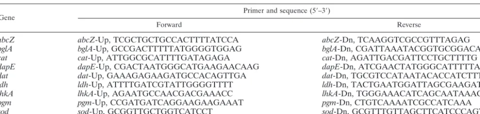
To identify the most polymorphic regions within each locus, several internal fragments were analyzed in 10L. monocytogenesstrains not related by PFGE (27). Those fragments showing the highest number of alleles were chosen for the analysis. The most variable ones were selected to be analyzed for the remaining isolates. DNAStar (Madison, Wis.) was used to design the primers for the amplification and sequencing of the selected fragments (Table 1).
Bacterial cell lysates were obtained by sonication (10 min) followed by cen-trifugation for 5 min. The PCR conditions were initial denaturation at 94°C for 4 min followed by 25 cycles of denaturation at 94°C for 30 s, annealing at 52°C for 30 s (except forpgmandbglA, which had an annealing temperature of 45°C), and extension at 72°C for 2 min followed by a final extension step of 72°C for 10 min. The DNA fragments were purified by using a PCR purification kit (Qiagen) and were sequenced in each direction with Big Dye fluorescent terminators (PE Applied Biosystems) on an Applied Biosystems Prism 377 automated sequencer.
Data analysis.The analysis of sequences obtained for each gene fragment was performed with the DNAStar program. For each fragment, the sequences ob-tained from the 62 strains were compared and allele numbers were assigned to each unique sequence. Each isolate was defined by the combination of numbers corresponding to the alleles at the loci analyzed, which is an allele profile or sequence type (ST). Sequences different even at a single nucleotide site were considered distinct alleles.
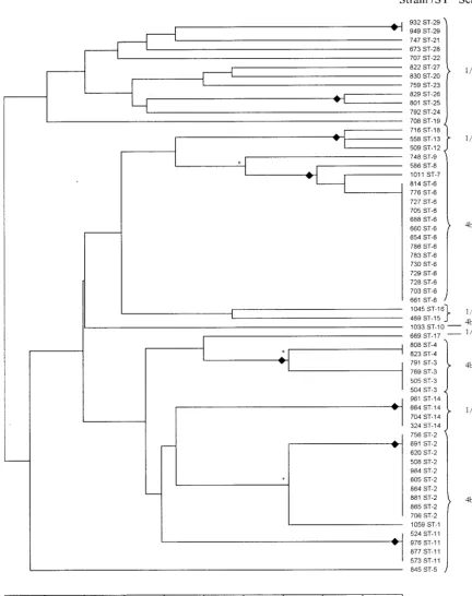
A dendrogram displaying the genetic linkage distance between each ST was constructed from the matrix of pairwise differences among the allelic profiles by the unweighted pair group cluster method with arithmetic mean (Fig. 1). This data analysis was performed by using the START software (http://www.mlst.net). The index of association (IA) (18) was used to test for linkage disequilibrium
among alleles at the housekeeping loci. The significance of theIAwas calculated
by using the program at the MLST website (http://www.mlst.net).
Nucleotide sequence accession numbers.The nucleotide sequences of each allele at the nine loci analyzed in the present work have been assigned the following GenBank accession numbers: AY 158265 to AY 158276 (abcZ), AY 158286 to AY158295 (bglA), AY158249 to AY 158264 (cat), AY 158303 to AY 158316 (dapE), AY158277 to AY 158284 (dat), AY 160115 to AY 160122 (ldh), AY 158296 to AY 158302 (lhkA), AY 158322 to AY 158326 (pgm), and AY 158317 to AY158321 (sod).
RESULTS
Diversity of the housekeeping genes analyzed.The length of
the analyzed fragments ranged between 354 bp (ldh) and 552
bp (abcZ) (Table 2).
The sequence diversity found in these fragments was quite
heterogeneous. The loci sodand pgm showed the lowest
se-quence diversity. So after the analysis of 40 strains, only one allele was found among the strains of serotypes 4b and 1/2b. Those genes were finally discarded due to a low contribution to the discrimination degree of this method. The locus with the
highest diversity wascatwith 16 alleles.
The proportion of variable sites present in the alleles ranged
from 2.9% (sod) to 12.4% (dat). Ratios of nonsynonymous
(dN) to synonymous (dS) changes derived from pairwise
se-quence comparison were much less than 1 for all studied loci (Table 2).
Relationship ofL.monocytogenesisolates by sequence
anal-ysis of housekeeping loci.A total of 29 allelic profiles or STs
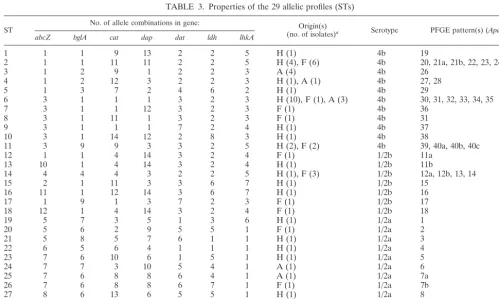
were identified, and 22 of these (75.8%) were represented by only one strain. The most common STs were ST6 and ST2, which grouped together 14 and 10 isolates, respectively (Table 3).
Those isolates that shared five or more loci in the allelic profile were considered members of the same clonal complex (9). The clonal complex that contained the largest number of isolates (17 isolates of serotype 4b) was the ST6 complex, which included ST6 (as the prevalent ST) and three other allelic profiles (ST7, ST8, and ST9) represented by a single isolate that differed from it at only one or two loci (Fig. 1). This lineage included 11 of the 21 serotype 4b human isolates stud-ied.
Significant linkage disequilibrium was detected when a
com-plete data set or STs were used in the analysis (IA, 2.195 [P⬍
0.01] andIA, 1.183 [P⬍0.01], respectively). However, evidence
for linkage equilibrium was found when theIAwas calculated
at the level of STs separately for the two genetic divisions (serotype 1/2a strains and serotype 1/2b and 4b strains). Values
ofIAobtained for these two divisions were 0.28 (P⬎0.001) for
division I and 0.34 (P⬎0.001) for division II.
Relationship between allelic profiles and serotypes. The
analysis of the housekeeping genes revealed that the sequences of serotype 1/2b and 4b isolates were more closely related to each other, or even identical, and showed significant diver-gence from the sequences of serotype 1/2a isolates. For all analyzed loci, alleles common to serotype 4b and 1/2b STs were found, whereas alleles present in serotype 1/2a STs were only found in this serotype (Table 3).
Congruence between STs and PFGE fingerprints.The
sim-ilarity of PFGE profiles among isolates belonging to same ST or to closely related STs was analyzed. Nevertheless, because both markers are based on very different principles, this com-parison should be carefully interpreted.
We found a high number of different cases with STs com-posed of strains showing a unique PFGE profile (ST3 or ST29) and also STs consisting of isolates with different PFGE
pat-TABLE 1. Primers used for PCR amplification and sequencing ofL. monocytogeneshousekeeping genes
Gene Primer and sequence (5⬘–3⬘)
Forward Reverse
abcZ abcZ-Up, TCGCTGCTGCCACTTTTATCCA abcZ-Dn, TCAAGGTCGCCGTTTAGAG
bglA bglA-Up, GCCGACTTTTTATGGGGTGGAG bglA-Dn, CGATTAAATACGGTGCGGACATA
cat cat-Up, ATTGGCGCATTTTGATAGAGA cat-Dn, AGATTGACGATTCCTGCTTTTG
dapE dapE-Up, CGACTAATGGGCATGAAGAACAAG dapE-Dn, ATCGAACTATGGGCATTTTTACC
dat dat-Up, GAAAGAGAAGATGCCACAGTTGA dat-Dn, TGCGTCCATAATACACCATCTTT
ldh ldh-Up, ATTTTGATCGTATTGGGGTTTT ldh-Dn, TACTGAATGGATTAGCGAAGATGA
lhkA lhkA-Up, AGAATGCCAACGACGAAACC lhkA-Dn, TGGGAAACATCAGCAATAAAC
pgm pgm-Up, CCGATGATCAGGAAGAAGAAAT pgm-Dn, CTGTCAAAATCGCCATCAAA
sod sod-Up, GCGGTTGCTGGTCATCCT sod-Dn, GCGTTTGTTAGCTTCATCCCAGTT
on May 15, 2020 by guest
http://jcm.asm.org/
[image:2.603.47.542.80.198.2]terns (ST2, ST4, or ST6) (Table 3). However, the consistency of the data between both methods was good in general, and the clustering of the strains was similar. Those STs defined as closely related (only one allele of difference) grouped together
[image:3.603.70.503.79.626.2]isolates with related or closely related PFGE pattern profiles (less than five band differences) (ST25 and ST26, or ST12, ST13, and ST18) (Fig. 1). Only one isolate of ST2 showed a higher number of fragment differences.
FIG. 1. Dendrogram showing cluster analysis (unweighted pair group cluster method with arithmetic mean) of the 62L. monocytogenesisolates. The serotype associated with each ST is shown. *, clonal complexes that were identified;}, branches containing isolates that showed related PFGE
patterns. Only one exception was found in ST2 (strain 706).
on May 15, 2020 by guest
http://jcm.asm.org/
DISCUSSION
The present study was conceived as a first step in the develop
of MLST for L. monocytogenes, with the first objective being
the selection of a set of housekeeping genes that were suited according to the MLST requirements. To evaluate the se-quence diversity of these loci, we considered it appropriate to use a genetically diverse group of strains. With this purpose, sequence analysis of the housekeeping genes was carried out in a group of isolates previously characterized by PFGE. These strains were isolated from different sources and geographic
locations of Spain during recent years. Isolates representing different PFGE patterns were included in this MLST project. Generally, a good congruence was found among groupings obtained by sequence analysis of housekeeping genes and those obtained by using PFGE. Thus, isolates that were iden-tical by MLST showed the same PFGE profile or patterns that differed at 1 to 5 fragments (27). We only found one exception: one isolate belonging to ST2 which showed greater differences with regard to other PFGE profiles associated with this ST. Similar patterns were also observed in those isolates that shared six of the seven alleles. The similarity was lower in those isolates that shared five of the seven loci.
Two isolates with the same allelic profile descending from a common ancestor might show PFGE profiles that are relatively different because PFGE is a highly discriminative method which is sensible to microvariation (17). It is important to take into account that a single nucleotide change that supposes a gain or loss of a restriction site can cause even three fragment differences (27), whereas MLST detects variation that accumu-lates slowly in housekeeping genes (26).
The validity of this molecular method is also supported by the congruence of the genetic data with previous MLEE
stud-ies that suggest that the genetic structure ofL. monocytogenes
is basically clonal (21). Thus, the value of the IAindicated a
significant linkage disequilibrium among the alleles at each of
the seven housekeeping loci (P ⬍ 0.01) (18). However, no
[image:4.603.42.284.88.217.2]evidence for linkage was detected when the analysis was per-formed separately in division I (serotype 1/2a isolates) and
TABLE 2. Genetic diversity of theL. monocytogeneshousekeeping genes analyzed
Gene fragmentSize of (bp)
No. of alleles
No. of variable
sites
% Variable nucleotide
sites
dN/dS
ratio
abcZ 552 12 34 6.1 0.058
bglA 417 10 21 5.0 0.095
cat 501 16 39 7.8 0.153
dapE 480 14 40 8.3 0.225
dat 484 8 60 12.4 0.216
ldh 354 8 19 4.3 0.000
lhkA 488 7 17 3.5 0.117
pgma 364 5 16 4.4 0.000
soda 420 5 12 2.9 0.166
aLoci that were only analyzed in 40 strains. These loci were not considered for
the final MLST scheme because of low sequence diversity.
TABLE 3. Properties of the 29 allelic profiles (STs)
ST No. of allele combinations in gene: (no. of isolates)Origin(s) a Serotype PFGE pattern(s) (ApaI)
abcZ bglA cat dap dat ldh lhkA
1 1 1 9 13 2 2 5 H (1) 4b 19
2 1 1 11 11 2 2 5 H (4), F (6) 4b 20, 21a, 21b, 22, 23, 24, 25
3 1 2 9 1 2 2 3 A (4) 4b 26
4 1 2 12 3 2 2 3 H (1), A (1) 4b 27, 28
5 1 3 7 2 4 6 2 H (1) 4b 29
6 3 1 1 1 3 2 3 H (10), F (1), A (3) 4b 30, 31, 32, 33, 34, 35
7 3 1 1 12 3 2 3 F (1) 4b 36
8 3 1 11 1 3 2 3 F (1) 4b 31
9 3 1 1 1 7 2 4 H (1) 4b 37
10 3 1 14 12 2 8 3 H (1) 4b 38
11 3 9 9 3 3 2 5 H (2), F (2) 4b 39, 40a, 40b, 40c
12 1 1 4 14 3 2 4 F (1) 1/2b 11a
13 10 1 4 14 3 2 4 H (1) 1/2b 11b
14 4 4 4 3 2 2 5 H (1), F (3) 1/2b 12a, 12b, 13, 14
15 2 1 11 3 3 6 7 H (1) 1/2b 15
16 11 1 12 14 3 6 7 H (1) 1/2b 16
17 1 9 1 3 7 2 3 F (1) 1/2b 17
18 12 1 4 14 3 2 4 F (1) 1/2b 18
19 5 7 3 5 1 3 6 H (1) 1/2a 1
20 5 6 2 9 5 5 1 F (1) 1/2a 2
21 5 8 5 7 6 1 1 H (1) 1/2a 3
22 6 5 6 4 1 1 1 H (1) 1/2a 4
23 7 6 10 6 1 5 1 H (1) 1/2a 5
24 7 7 3 10 5 4 1 A (1) 1/2a 6
25 7 6 8 8 6 4 1 A (1) 1/2a 7a
26 7 6 8 8 6 7 1 F (1) 1/2a 7b
27 8 6 13 6 5 5 1 H (1) 1/2a 8
28 9 10 15 6 8 1 1 A (1) 1/2a 9
29 7 10 16 7 5 1 1 H (1), A (1) 1/2a 10
aOrigin codes: H, human infections; A, animal infections; F, food products.
on May 15, 2020 by guest
http://jcm.asm.org/
[image:4.603.41.541.416.715.2]division II (serotype 1/2b and 4b strains). These data suggest that the recombination should be rare between strains belong-ing to different genetic lineages, but the evidence for clonal structure disappears when the division I and II alleles are analyzed separately. Bacteriophage might probably play an
important role in the genomic plasticity of Listeria(28), and
this factor might be involved in some of the differences found in this study. In addition, similarities in the genome sequence between serotype 4b and 1/2b isolates might explain a higher prevalence of homologous recombination between these strains. The extension of the analysis to a higher number of strains will offer information about the relative contribution of recombination and mutation to the allele variation found.
In addition, the genetic relationships among different sero-types showed by this gene allelic analysis were also consistent
with those previously established by MLEE, which dividesL.
monocytogenes into the two genetic divisions already men-tioned. However, an additional division has been proposed after analysis of the sequence of genes associated with viru-lence (22). Strains grouped in this third division consist of serotypes 4a and 4c, which were not included in our study. We think that those markers which are focused on housekeeping genes (MLEE) or random genetic characteristics (PFGE) are not able to distinguish more than two divisions, with the third division being evident when specific genes associated with vir-ulence are analyzed. However, the future analysis by MLST of serotype 4a and 4c strains might confirm this hypothesis.
The utility of MLST and MLEE (25) for the analysis of the genetic structure of bacterial populations is mainly based on the characteristic of housekeeping genes to have a selectively neutral variability (9). Analysis of synonymous and nonsynony-mous changes in the allele sequences of a locus can be used to
determine if it is subject to positive selection, so adN/dSratio
of greater than 1 implies selection for amino acid changes (4).
In our genetic analysis, the seven loci haddN/dSratios
signif-icantly lower than 1 (Table 2). Another important character-istic in relation to this fact is that the location of loci on the chromosome was distant enough to make the joint horizontal
transfer of two loci unlikely. Recently, the sequencing of theL.
monocytogenesgenome has been finished and published (11), which has allowed us to know the chromosomal locations of the loci selected for this study. The analysis has revealed that all of these loci are unlinked on the chromosome, with the minimum distance being 41.3 kb.
Previous studies with the use of MLEE and other molecular methods (1, 3, 21) have revealed that despite the high diversity
in natural populations of L. monocytogenes, only two clones
have been responsible for most major outbreaks detected dur-ing the last decades in Europe and North America (10, 13, 14, 16, 23). These results suggested the hypothesis that most of the clinical cases are caused by only a few clones that either are particularly common in the environment or have an unusually high level of pathogenicity (20, 21). In this epidemiological context, MLST could be a useful tool for the surveillance systems for listeriosis that might allow the identification and
analysis of the distribution of theseL. monocytogenesclones in
the environment. In fact, the isolates from ST2 and ST6 might be those strains belonging to the major clones, and the future
running of a central database for the Listeria STs with the
associated epidemiological data will allow us to confirm this and other questions.
The extension of the present analysis to a higher number of isolates could contribute to a better knowledge of the structure
of theL. monocytogenespopulation. Besides, the future genetic
analysis of other housekeeping loci with enough sequence di-versity might improve the discrimination level of this new typ-ing method.
ACKNOWLEDGMENTS
B.A. and C.S. were supported by a postdoctoral grant from Instituto de Salud Carlos III and a predoctoral grant from CYANAMID, re-spectively.
We are grateful to Brian G. Spratt for helpful comments on the manuscript.
REFERENCES
1. Bibb, W. F., B. G. Gellin, R. Weaver, B. Schwartz, B. D. Plikaytis, M. W. Reeves, R. W. Pinner, and C. V. Broome.1990. Analysis of clinical and food-borne isolates ofListeria monocytogenesin the United States by mul-tilocus enzyme electrophoresis and application of the method to epidemio-logic investigations. Appl. Environ. Microbiol.56:2133–2141.
2. Brosch, R., J. Chen, and J. B. Luchansky.1994. Pulsed-field fingerprinting of listeriae: identification of genomic divisions forListeria monocytogenesand their correlation with serovar. Appl. Environ. Microbiol.60:2584–2592. 3. Buchrieser, C., R. Brosch, B. Catimel, and J. Rocourt.1993. Pulsed-field gel
electrophoresis applied for comparing Listeria monocytogenesstrains in-volved in outbreaks. Can. J. Microbiol.39:395–401.
4. Dingle, K. E., F. M. Colles, D. R. A. Wareing, R. Ure, A. J. Fox, F. E. Bolton, H. J. Bootsma, R. J. L. Willems, R. Urwin, and M. C. J. Maiden.2001. Multilocus sequence typing system forCampylobacter jejuni.J. Clin. Micro-biol.39:14–23.
5. Enright, M. C., and B. G. Spratt.1998. A multilocus sequence typing scheme forStreptococcus pneumoniae: identification of clones associated with serious invasive disease. Microbiology144:3049–3060.
6. Enright, M. C., and B. G. Spratt.1999. Multilocus sequence typing. Trends Microbiol.7:482–487.
7. Enright, M. C., N. P. J. Day, and B. G. Spratt.2000. Multilocus sequence typing for characterization of methicillin-resistant and methicillin-suscepti-ble clones ofStaphylococcus aureus. J. Clin. Microbiol.38:1008–1015. 8. Enright, M. C., B. G. Spratt, and D. E. Bessen.2001. Multilocus sequence
typing ofStreptococcus pyogenesand the relationships betweenemmtype and clone. Infect. Immun.69:2416–2427.
9. Feil, E. J., M. C. J. Maiden, M. Achtman, and B. G. Spratt.1999. The relative contributions of recombination and mutation to the divergence of clones ofNeisseria meningitidis.Mol. Biol. Evol.16:1496–1502.
10. Fleming, D. W., S. L. Cochi, K. L. McDonald, J. Brondum, P. S. Hayes, B. D. Plikaytis, M. B. Holmes, A. Audurier, C. V. Broome, and A. L. Reingold.
1985. Pasteurized milk as a vehicle of infection in an outbreak of listeriosis. N. Engl. J. Med.312:404–407.
11. Glaser, P., L. Frangeul, C. Buchrieser, C. Rusniok, A. Amend, F. Baquero, P. Berche, H. Bloecker, P. Brandt, T. Chakraborty, A. Charbit, F. Chetouani, E. Couve, A. de Daruvar, P. Dehoux, E. Domann, G. Dominguez-Bernal, E. Duchaud, L. Durant, O. Dussurget, K. D. Entian, H. Fsihi, F. G. Portillo, P. Garrido, L. Gautier, W. Goebel, N. Gomez-Lopez, T. Hain, J. Hauf, D. Jackson, L. M. Jones, U. Kaerst, J. Kreft, M. Kuhn, F. Kunst, G. Kurapkat, E. Maduen˜o, A. Maitournam, J. M. Vicente, E. Ng, H. Nedjari, G. Nordsiek, S. Novella, B. de Pablos, J. C. Perez-Diaz, R. Purcell, B. Remmel, M. Rose, T. Schlueter, N. Simoes, A. Tierrez, J. A. Vazquez-Boland, H. Voss, J. Wehland, and P. Cossart.2001. Comparative genomics of Listeria species. Science294:849–852.
12. Graves, L. M., and B. Swaminathan.2001. PulseNet standarized protocol for subtypingListeria monocytogenesby macrorestriction and pulsed-field gel electrophoresis. Int. J. Food Microbiol.65:55–62.
13. Ho, J. L., K. N. Shands, G. Friedland, P. Eckind, and D. W. Fraser.1986. An outbreak of type 4bListeria monocytogenesinfection involving patients from eight Boston hospitals. Arch. Intern. Med.146:520–524.
14. Jacquet, C., B. Catimel, R. Brosch, C. Buchrieser, P. Dehaumont, V. Goulet, V. Lepoutre, P. Veit, and J. Rocourt.1995. Investigations related to the epidemic strain involved in the French listeriosis outbreak in 1992. Appl. Environ. Microbiol.61:2242–2246.
15. Lan, Z., F. Fiedler, and S. Kathariou.2000. A sheep in wolf’s clothing:
Listeria innocuastrains with teichoic acid-associated surface antigens and genes characteristic ofListeria monocytogenesserogroup 4. J. Bacteriol.182:
6161–6168.
16. Linnan, M. J., L. Mascola, X. D. Lou, V. Goulet, S. May, C. Salminen, D. W. Hird, M. L. Yonekura, P. Hayes, R. Weaver, A. Audurier, B. D. Plikaytis,
on May 15, 2020 by guest
http://jcm.asm.org/
S. L. Fannin, A. Kleks, and C. V. Broome.1988. Epidemic listeriosis asso-ciated with Mexican-style cheese. N. Engl. J. Med.319:823–828. 17. Maiden, M. C. J., J. A. Bygraves, and B. G. Spratt.1998. Multilocus
se-quence typing: a portable approach to the identification of clones within populations of pathogenic microorganisms. Proc. Natl. Acad. Sci. USA95:
3140–3145.
18. Maynard Smith, J., N. H. Smith, M. O’Rourke, and B. G. Spratt.1993. How clonal are bacteria? Proc. Natl. Acad. Sci. USA90:4384–4388.
19. Mazurier, S. I., and K. Wernars.1992. Typing ofListeriastrains by random amplification of polymorphic DNA. Res. Microbiol.143:499–505. 20. McLauchlin, J.1990. Distribution of serovars of Listeria monocytogenes
isolated from different categories of patients with listeriosis. Eur. J. Clin. Microbiol. Infect. Dis.9:210–213.
21. Piffaretti, J. C., H. Kressebuch, M. Aeschbacher, J. Bille, E. Bannerman, J. M. Musser, R. K. Selander, and J. Rocourt.1989. Genetic characterization of clones of the bacteriumListeria monocytogenescausing epidemic disease. Proc. Natl. Acad. Sci. USA86:3818–3822.
22. Rasmussen, O. F., P. Skouboe, L. Dons, L. Rossen, and J. E. Olsen.1995.
Listeria monocytogenesexists in at least three evolutionary lines: evidence from flagellin, invasive associated protein and listeriolysin O genes. Micro-biology 141:2053–2061.
23. Schlech, W. F., III, P. M. Lavigne, R. A. Bortolussi, A. C. Allen, E. V. Haldane, A. J. Wort, A. W. Hightower, S. E. Johnson, S. H. King, E. S. Nicholls, and C. V. Broome.1983. Epidemic listeriosis-evidence for trans-mission by food. N. Engl. J. Med.308:203–206.
24. Seeliger, H. P. R., and K. Ho¨hne.1979. Serotyping ofListeria monocytogenes
and related species. Methods Microbiol.13:31–49.
25. Selander, R. K., D. A. Caugant, H. Ochman, J. M. Musser, M. N. Gilmour, and T. S. Whittam.1986. Methods of multilocus enzyme electrophoresis for bacterial populations genetics and systematics. Appl. Environ. Microbiol.
51:873–884.
26. Spratt, B. G.1999. Multilocus sequence typing: molecular typing of bacterial pathogens in an era of rapid DNA sequencing and the internet. Curr. Opin. Microbiol.2:312–316.
27. Tenover, F. C., R. D. Arbeit, R. V. Goering, P. A. Mickelsen, B. E. Murray, D. H. Persing, and B. Swaminathan.1995. Interpreting chromosomal DNA restriction patterns produced by pulsed-field electrophoresis: criteria for bacterial typing. J. Clin. Microbiol.33:2233–2239.
28. Va´zquez-Boland, J. A., G. Domínguez-Bernal, B. Gonza´lez-Zorn, J. Kreft, and W. Goebel.2001. Pathogenicity islands and virulence evolution in Lis-teria.Microbes Infect.3:571–584.
29. Vela, A. I., J. F. Ferna´ndez-Garayzabal, J. A. Vazquez, M. V. Latre, M. M. Blanco, M. A. Moreno, L. De La Fuente, J. Marco, C. Franco, A. Cepeda, A. A. Rodríguez Moure, G. Sua´rez, and L. Domínguez.2001. Molecular typing by pulsed-field gel electrophoresis of Spanish animal and human
Listeria monocytogenesisolates. Appl. Environ. Microbiol.67:5840–5843. 30. Wiedmann, M., J. L. Bruce, R. Knorr, M. Bodis, E. M. Cole, C. I. McDowell,
P. L. McDonough, and C. A. Batt.1996. Ribotype diversity ofListeria mono-cytogenesstrains associated with outbreaks of listeriosis in ruminants. J. Clin. Microbiol.34:1086–1090.