Structural characteristics and functional consequences of
lateral ankle sprains
Rawan Hesham Abdeen
School of Health Sciences University of Salford, Manchester, UK
Submitted in Partial Fulfilment of the Requirement
of the Degree of Doctor of Philosophy (PhD)
Supervisors
1.
Professor Christopher Nester
Research programme leaderSchool of health Sciences
Room PO.32, Brian Blatchford Building, University of Salford, Salford, M6 6PU
2.
Dr. Paul Comfort
Senior lecturer
Programme Leader MSc Strength and Conditioning School of health Sciences
Room C701, Allerton Building, University of Salford, Salford, M5 4WT
3.
Dr. Chelsea Starbuck
Post Doc Research fellow School of health SciencesI
Table of contents
Table of contents ... I
List of table ... IIX
List of figures ... X
Publication, conferences paper and poster ... XV
Trainings undertaken during the course of the PhD ... XVI
Acknowledgement ... XX
List of abbreviation ... XXI
Abstract ... XXV
Chapter one: Introduction ... 1
1.1 Overview of the problem of lateral ankle sprains ... 1
1.2 The research problem ... 2
1.3 Overview and structure of the Thesis ... 3
2
Chapter two: Background/Literature review ... 5
2.1 Search strategy ... 5
2.2 Prevalence of ankle injury and lateral ankle sprain ... 6
2.3 Ankle sprain in health care ... 7
2.4 Structural and functional anatomy of selected ankle structures related to the ankle joint 9 2.4.1 Bones and joints ... 9
2.4.2 Muscles ... 17
2.5 Ligament injury ... 19
2.5.1 Aetiology of ankle sprain ... 20
2.5.2 Mechanism of ankle ligamentous sprain ... 21
2.5.3 Three grades of ankle sprain ... 24
II
2.5.5 Classification of ankle injury ... 28
2.6 Self-reported functional ankle instability measures ... 36
2.7 Risk factors of lateral ankle sprain ... 41
2.8 Diagnosis and evaluation of ankle injury ... 46
2.9 Ultrasound as diagnostic image modality ... 47
2.9.1 Ultrasound history and physics ... 47
2.9.2 Role of ultrasound in evaluation of ankle injury ... 52
2.9.3 Ultrasound imaging of healthy ankle ... 55
2.9.4 Ultrasound imaging of injured ankle ... 57
2.10 Subjective and objective evaluation of ankle injury using ultrasound ... 58
2.11 Ankle injury and postural control ... 63
2.11.1 Strategies of postural control ... 65
2.11.2 Measuring postural stability ... 66
2.11.2.1Star excursion balance test (SEBT) ... 68
2.11.2.2Ankle kinematics ... 76
2.12 Rationale for the study ... 81
2.13 Aim of the PhD ... 86
3
Chapter three: Ultrasound characteristic of selected ankle structures in
healthy, coper and chronic ankle instability ... 88
3.1 Chapter overview ... 88
3.2 Aims, objectives, and hypothesis of the study ... 89
3.3 Pilot study ... 91
3.4 Reliability study ... 92
3.4.1 Aim of the reliability study ... 92
III
3.4.3 Reliability study participants ... 96
3.4.4 Reliability study data collection ... 97
3.4.5 Reliability statistical analyses ... 97
3.4.6 Reliability study results ... 97
3.4.7 Reliability study conclusion ... 100
3.5 Method ... 100
3.5.1 Study design ... 100
3.5.2 Ethical considerations ... 100
3.5.3 Sample size for the main study ... 101
3.5.4 Recruitment strategy ... 101
3.5.5 Inclusion and exclusion criteria ... 102
3.5.6 Participants information ... 103
3.5.6.1 Demographic data for comparison between healthy, coper and CAI participants 105 3.5.6.2 Demographic data for comparison between right and left limbs among healthy participants ... 105
3.5.6.3 Demographic data for comparison between male and female healthy participants 106 3.5.6.4 Demographic data for comparison between normal weight and overweight healthy participants ... 106
3.5.7 Medical ultrasound machine ... 106
3.5.8 Assessment procedure ... 108
3.5.8.1 Ultrasound techniques and measurements ... 109
IV
3.5.8.1.2 Calcaneofibular Ligament (CFL) ... 114
3.5.8.1.3 Peroneal Tendons ... 115
3.5.8.1.4 Tibialis Posterior Tendon (TPT) ... 117
3.5.8.1.5 Achilles Tendon (AT) ... 119
3.5.8.1.6 Peroneal Muscles... 120
3.6 Image analysis ... 121
3.7 Statistical analyses ... 122
3.8 Results ... 124
3.8.1 Participants ... 124
3.8.2 Comparison of length, thickness and CSA of selected ankle structures between healthy, coper and CAI ... 125
3.8.3 Comparison between neutral and tension position among healthy participants 127 3.8.4 Comparison between right and left limbs among healthy participants ... 129
3.8.5 Comparison between male and female healthy participants ... 130
3.8.6 Comparison between normal weight and overweight participants ... 132
3.9 Discussion ... 134
3.9.1 Comparison of the length and thickness of the ATFL between healthy, coper and CAI groups ... 134
3.9.2 Comparison of the thickness of CFL between healthy, coper and CAI groups 136 3.9.3 Comparison of the thickness and CSA of selected ankle structures between healthy, coper and CAI groups ... 136
V
3.9.5 Comparison between right and left limbs of healthy participants ... 141
3.9.6 Comparison between female and male healthy participants ... 142
3.9.7 Comparison between normal weight and overweight healthy participants .... 143
3.10 Limitation ... 144
3.11 Conclusion ... 144
4
Chapter four: Quantitative evaluation of ultrasound images to
compare healthy and injured anterior talofibular ligaments ... 146
4.1 Chapter overview ... 146
4.2 Aims, objectives, and hypothesis of the study ... 146
4.3 Methods ... 147
4.3.1 Image dataset ... 147
4.3.2 Quantification echogenicity of ATFL ... 147
4.4 Statistical Analysis ... 149
4.5 Results ... 149
4.6 Discussion ... 150
4.7 Limitation ... 154
4.8 Conclusion ... 154
5
Chapter five: SEBT and 3D kinematics as measure balance
performance in injured ankles compare to healthy controls ... 155
5.1 Chapter overview ... 155
5.2 Aim, objectives, and hypothesis of the study ... 156
5.3 Method ... 158
5.3.1 Participants ... 158
5.3.2 The motion analysis system ... 158
5.3.3 System calibration ... 159
VI
5.3.5 SEBT ... 163
5.3.6 Protocol of the study ... 164
5.3.7 Reliability of the SEBT ... 167
5.4 Data processing ... 170
5.5 Statistical analysis ... 172
5.5.1 Participants demographic and questionnaires ... 173
5.5.2 Reach distances and 3D kinematics ... 173
5.5.3 Correlation between the thickness of lateral ligaments and anterolateral direction of the SEBT ... 173
5.6 Results ... 174
5.6.1 Participants demographic and questionnaires ... 174
5.6.2 SEBT reach distances ... 174
5.6.3 3D kinematics ... 177
5.6.4 Correlation between the thickness of lateral ligaments and the most affected direction of the SEBT ... 184
5.7 Discussion ... 185
5.7.1 Comparison of the reach distance of the SEBT between CAI compared to healthy participants ... 185
5.7.2 Comparison of the reach distance of the SEBT between coper and healthy participants ... 189
5.7.3 Comparison of the reach distance of the SEBT between coper and CAI participants ... 190
VII
5.7.6 The inconsistency of the results between this study and previous studies ... 193
5.7.7 Correlation between the thickness of lateral ligaments and the most affected direction of the SEBT ... 194
5.8 Limitation ... 195
5.9 Conclusion ... 195
6
Chapter
six:
Overall
summary,
conclusion,
limitation
and
recommendations for future work ... 196
6.1 Chapter overview ... 196
6.2 Overall summary of the thesis... 196
6.3 Thesis novelty ... 198
6.4 Clinical relevance ... 199
6.5 Limitations ... 200
6.6 Recommendations for future work... 201
Appendix 1- Measuring peroneal tendons at three different locations ... 203
Appendix 2- Ethical approval letter for reliability study ... 204
Appendix 3- Consent form for reliability study... 205
Appendix 4- Data collection sheet ... 206
Appendix 5- The CAIT Questionnaire ... 207
Appendix 6- Ethical approval letter for main study ... 208
Appendix 7- Participants information sheet for main study ... 209
Appendix 8- Consent form of main study ... 213
Appendix 9- Risk Assessment Summary of Student Projects ... 214
Appendix 10- Flyer/poster of the main study ... 216
Appendix 11- General Practice Physical Activity Questionnaire ... 218
Appendix 12- Ultrasound measurements of selected ankle structures between
healthy, coper and CAI ... 219
VIII
Appendix 14- Ultrasound measurements of selected ankle structures between
right and left limbs ... 221
Appendix 15- Ultrasound measurements of selected ankle structures between
male and female ... 222
Appendix 16- Ultrasound measurements of selected ankle structures between
normal weight and overweight participants ... 223
Appendix 17- Score sheet for SEBT & limb length ... 224
Appendix 18- Reliability results of SEBT ... 225
Appendix 19- Bland and Altman plots for several directions of healthy and
injured participant with representation of limit of agreements ... 226
Appendix 20- Differences of kinematics data between the 3 groups in sagittal
plane ... 227
Appendix 21- Differences of kinematics data between the 3 groups in transverse
plane ... 228
IX
List of table
X
List of figures
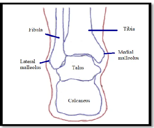
Figure 2-1: The bones of ankle joints ... 10
Figure 2-2: The movement of foot and ankle joint. A: Frontal plane components of inversion/eversion. B: Sagittal plane components of dorsiflexion/plantarflexion. C: Transverse plane components of lateral rotation/medial rotation. The red dot and tube demonstrates the axis for that motion (Muscolino, 2016) ... 10
Figure 2-3: Example of articular cartilage at the ankle joint Ligaments and tendons ... 11
Figure 2-4: Schematic diagram presenting hierarchical structure of ligament in cross section ... 11
Figure 2-5: Lateral view demonstrates the lateral ligaments ... 12
Figure 2-6: CFL throughout the movements of ankle. a: neutral position. b: Dorsal flexion c: Plantar flexion ... 13
Figure 2-7: Medial side of the ankle demonstrating deltoid ligament ... 13
Figure 2-8: The peroneus longus and brevis tendons ... 14
Figure 2-9: Medial tendons of the ankle; TPT (tibialis posterior tendon), FDL (flexor digitorum longus), and FHL (flexor halluces longus) ... 15
Figure 2-10: Anterior ankle tendons ... 16
Figure 2-11: Achilles tendon ... 16
Figure 2-12: Peroneal tendons and muscles... 17
Figure 2-13: Tibialis posterior muscle ... 18
Figure 2-14: Lateral and medial head of the gastrocnemius and soleus muscles ... 18
Figure 2-15: Mechanism of inversion ankle sprain (Al-Mohrej & Al-Kenani, 2016). ... 21
Figure 2-16: Classical mechanism for lateral ligament injury in football (Andersen et al., 2004) ... 22
Figure 2-17: Grading of lateral ligaments sprain ... 25
Figure 2-18: Lateral ankle ligaments ... 26
Figure 2-19: Diagram of mechanical and functional ankle instability that contributes to chronic ankle instability ... 30
Figure 2-20: Three different modes of ultrasound. (A) 2D of tibialis anterior muscles (Pillen, 2010). (B) M-mode presenting the mitral valve leaflets of the heart (Gill, 2012). (C) Doppler mode of carotid artery: (1) colour Doppler and (2) pulsed Doppler (Merritt, 2017). ... 49
XI
Figure 2-22: Creation of ultrasound images. (1) Electricity is applied to the probe. (2) Piezoelectric crystals vibrate quickly, creating sound waves. (3) Ultrasound beam penetrates tissues. (4) Sound waves reflected (echo) and returned to the probe. (5) Echoes are turn into
electrical signals which are processed into grey-scale image ... 51
Figure 2-23: Normal appearance of peroneal muscles. (A) Cross sectional plane. (B) Longitudinal plane. ... 56
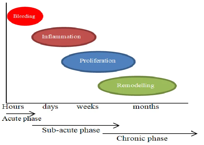
Figure 2-24: Three phases of healing process (inflammation, proliferation, and remodelling) during acute, sub-acute, and chronic phases ... 59
Figure 2-25: Glossary of US echogenicity terms (Das, 2016) ... 60
Figure 2-26: Image analysis region of interest selections and the corresponding greyscale histogram values. Yellow rectangular represented the region of interest of the longitudinal image of rectus femoris. The corresponding greyscale histogram showed the mean echo (Harris-Love, Seamon, Teixeira, & Ismail, 2016) ... 62
Figure 2-27: Human postural control ... 66
Figure 3-1: Flowchart demonstrating the structural of this study ... 90
Figure 3-2: designed wedge to dorsiflexed the foot to 15° ... 92
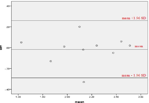
Figure 3-3: Bland and Altman plot for CSA of AT in normal position with representation of limit of agreements. The middle line demonstrates the mean of the differences between the day 1 and day 2 and the side lines demonstrate mean differences ± 1.96 times the SD of the difference between the two. ... 95
Figure 3-4: Bland and Altman plot for length of ATFL in tension position with representation of limit of agreement. The middle line demonstrates the mean of the differences between the day 1 and day 2 and the side lines demonstrate mean differences ± 1.96 times the SD of the difference between the two. ... 95
Figure 3-5: portable ultrasound machine ... 107
Figure 3-6: Linear array transducer ... 107
Figure 3-7: An ultrasound image demonstrating the depth scale to the right of the screen which define by red arrows; Green arrow defines the focus of the image; Blue circle defines the gain ... 110
Figure 3-8: Right feet hold on AFO for neutral position ... 110
Figure 3-9: Transducer held and positioning (Jacobson, 2012). ... 112
Figure 3-10: Transducer position to scan ATFL... 112
Figure 3-11: US image for ATFL in neutral position ... 113
Figure 3-12: Tension position for ATFL ... 113
XII
XIII
XIV
XV
Publication, conferences paper and poster
- Presented a poster at SPARC (Salford Postgraduate Annual Research Conference) at University of Salford on 15-14/06/2016.
- Demonstrated an ultrasound workshop at congress of a EUROPEAN network of Podiatry Schools on 22/03/2018
- Presented a poster at Annual conference of American Institute of Ultrasound in Medicine in the United States of America (USA) on 24/03/2018.
- Presented a presentation at International Foot and Ankle Biomechanics (i-FAB2018) Meeting in the USA on 09/04/2018.
- Published an article in Journal of Ultrasound in Medicine on 12/09/2018.
OBJECTIVE: Ankle sprains constitute approximately 85% of all ankle injuries, and up to 70% of people experience residual symptoms. While the injury to ligaments is well understood, the potential role of other foot and ankle structures has not been explored. The objective was to characterize and compare selected ankle structures in participants with and without a history of lateral ankle sprain.
METHODS: A total of 71 participants were divided into 31 healthy, 20 coper, and 20 chronic ankle instability groups. Ultrasound images of the anterior talofibular and calcaneofibular ligaments, fibularis tendons and muscles, tibialis posterior, and Achilles tendon were obtained. Thickness, length, and cross-sectional areas were measured and compared among groups.
RESULTS: When under tension, the anterior talofibular ligament (ATFL) was longer in copers and chronic ankle instability groups compared to healthy participants (P < .001 and P = .001, respectively). The chronic ankle instability group had the thickest ATFL and calcaneofibularligamentamongthe3groups(p < 0.001).Nosignificantdifferences(P > .05) in tendons and muscles were observed among the 3 groups.
XVI
Trainings undertaken during the course of the PhD
Date Title of training course Key learning aim
11-11-2015 Excel: Formulas and function How to use the formula and the function and applied that in your work.
17-11-2015 Musculoskeletal ultrasound lectures1 Lecture was given about Musculoskeletal ultrasound
18-11-2015 Musculoskeletal ultrasound lectures2 Lecture was given about Musculoskeletal ultrasound
24-11-2015 Tackling literature review -structuring a review.
-Critically assessing literature. 30-11-2015 Critical and Analytical Skills -how to be a critical student. 30-11-2015 Organising and synthesising your
work
-research practices.
-how knowledge is used to construct an academic argument.
01-12-2015 Doing a literature review -help the student get started with literature review.
-explain what it is, what it is not. 10-12-2015 Becoming a researcher: realizing
your potential and raising your profile
This session help you to describe characteristics of excellent researchers and map your own current performance.
16-12-2015 PGR seminar series that spans the research themes of: Rehab, Gait, Knee, Foot & Ankle biomech, and Activity Monitoring
PGR seminar session.
05-02-2016 The Seven Secrets of Highly Successful Research Students
This workshop describes the key habits that our research and experience with thousands of student’s shows will make a difference to how quickly and easily you complete your PhD. Just as importantly, these habits can greatly reduce the stress and increase the pleasure involved in completing a PhD.
08-02-2016 Advanced Search: Health & Social Care Databases
Locating databases for Health and Social Care
XVII
Research and how it affects academic use.
-Be able to assess the risks involved in breaching copyright.
16-02-2016 Googlescholar for research It is a Hands-on session on making effective academic use of Google scholar, aimed at Postgraduate Researchers.
18-02-2016 ResearchEthicsforPGR’s This session will discuss the issues and procedures around statutory requirements and professional codes for maintaining the highest possible ethical standards.
26-02-2016 PHD students meeting In this meeting there will be a discussion on critical reading/writing and the use of a data synthesis matrix
01-03-2016 LEAP higher writing session This session is designed to help PGRS in academic writing
02-03-2016 Understanding how skin contributing to balance control
PGR seminar session.
08-03-2016 LEAP higher writing session This course designed to give support with academic writing, including grammar, vocabulary and how to organize this information for an assessment.
10-03-2016 Creating academic poster This workshop provides an introduction to presenting at academic conferences
18-03-2016 ResearchEthicsforPGR’s This session will discuss the issues and procedures around statutory requirements and professional codes for maintaining the highest possible ethical standards.
23-03-2016 Intro to Endnote X7 How to use the EndNote X7 (bibliographic software) to organise and manage my reference.
19-04-2016 Locating and using historical
archives for researchers
To learn how to find unique historical materials for your research
20-04-2016 Introduction to SPSS -This introductory session to SPSS will provide participants with an overview of the capabilities of this common statistical package.
XVIII
25-04-2016 Presenting at Academic Conferences This workshop provides an introduction to presenting your research at academic conferences
26-04-2016 Ultrasound scanning session Practice the new ultrasound protocol 29-04-2016 PGR monthly meeting Discusses about the IA and IE. 03-05-2016 Ultrasound scanning session Practice the new ultrasound protocol 05-05-2016 LEAP (academic writing) This session focuses on PhD students to
help them writing in academic way.
10-05-2016 LEAP (critical writing) To help PhD student to be critique in their writing.
18-05-2016 Critical Thinking and Critical Writing at Doctoral Level
This session focuses on research practices and how knowledge is used to construct an academic argument
20-05-2016 Rehearsal and coaching session for poster
Practise before shooting the film next week, and the facilitator will be able to go through some tips with us and show us how the autocue works in the studio. 27-05-2016 PGR monthly meeting
15-06-2016 SPARC Present my poster in the poster session of SPARC.
07-10-2016 Word: formatting your thesis To learn more about the specific feature of word in design the writing.
02-11-2016 A Survival Guide to Doing a PhD An essential guide to surviving your PhD 09-11-2016 The Interview: its place in social
scientific research strategies
The session will explore both the theoretical and practical issues associated with interviewing as a data gathering technique
10-11-2016 Being critical Bases for critique throughout the thesis 17-11-2016 Excel: Analysing Data -To learn how to sort and filter the data.
-To construct a pivot table and pivot chart from a table of data.
17-11-2016 Building the argument -Structure of an argument. -keeping the thread. -Gaps in the literature.
18-01-2017 Doctoral Training seminar -Getting your first paper published 25-01-2017 Electronic Resources for Researchers
27-01-2017 Pathway to Professional: Time management
XIX
15-02-2017 Doctoral Training seminar -Statistics and data analysis of kinematic/kinetic data
15-03-2017 Doctoral Training seminar - Critical/peer review
19-04-2017 Doctoral Training seminar - Translating research into industry 26-04-2017 Ankle sprain webinar
17-05-2017 Doctoral Training seminar -Research governance 02-11-2017 Building resilience and
bouncebackability
-Improve your resilience to ‘knockbacks’ and criticism.
02-11-2017 Goal setting and staying on tack -To set effective and realistic goals to progress your research.
02-11-2017 How to submit a conference paper -Strategies to approaching writing abstracts for conferences.
03-11-2017 Your thesis: structure -Introduction to different types of thesis structure
- Planning your own thesis structure
03-11-2017 Your thesis: the thesis of your thesis A practical session where the researchers will produce their own clear thesis statement to help us focus our PhD work around a central proposition.
03-11-2017 Formatting and submitting your thesis- getting it done
What a thesis should look like and how to format it
- Supervisory role and getting other sources of feedback
- Submission process
- Staying motivated in the late stages 30-01-2018 Publishing during your PhD The quick guide to publishing whilst doing
your PhD – navigating the process, publishers, peer review and
procrastination.
15-02-2018 Intensive data SPSS This is a practical workshop using SPSS --This session aims to explore the
experimental design of your research. 14-05-2018 Radiation dose and image quality in
XX
Acknowledgement
The opportunity to live in Manchester and to study at the University of Salford have been one of the most exciting experiences of my life, which I will treasure its memories forever. I would like to express my sincere gratitude to my supervisor Prof. Chris Nester for giving me the chance to undertake this research and for his unwavering belief in my ability. It was my pleasure to have supervisor like you with your kindness attitude and your great experience in biomechanics and research. I always appreciate your guidance, your support, your critical comments and your time. I would like also to extend a huge thanks to my co-supervisors, Dr. Paul Comfort and Dr. Chelsea Starbuck for their guidance, support, assistance, and helpful suggestions. Thank you my supervisory team to whom I owe the completion of this thesis.
Great thanks to my friends and postgraduates colleagues especially Basmah Allarakia and Kholoud Alzyoud who have supported me through my PhD journey. Thanks to everyone who took part in the studies of this thesis, your time was much appreciated. This thesis could not completed without you.
Special thanks go to my wonderful family, in particular my loving parents, thank you for supporting me to pursue what I loved, and thank you for your endless motivation and your unconditional love. You are always the source of my power and strength and I am forever grateful to you. I wish you always feel proud of me.
XXI
List of abbreviation
ACL Anterior cruciate ligament AD Anterior drawer
AFO Ankle and foot orthosis AL Anterolateral
ALARA As low as reasonably achievable ALS Amyotrophic lateral sclerosis
AII Ankle Instability Instrument AM Anteromedial
AMTI Advanced Mechanical Technology Incorporation ANOVA Analysis of variance
ANT Anterior
ATFL anterior talofiblar ligament AT Achilles tendon
B-mode Brightness mode BMI Body mass index C Celsius
CAI Chronic ankle instability
CAIT Cumberland Ankle Instability Tool CAST Calibration anatomical system technique CFL Calcenofibular Ligament
CINAL Cumulative Index to Nursing and Allied Health Literature CLAHE Contrast Limited Adoptive Histogram Equalization cm centimetre
CNS Central Nervous System CoM Centre of Mass
XXII DAQ Data acquisition
DICOM Digital imaging and communication in medicine DLT Direct-linear transformation
EDL Extensor digitorum longus EHL Extensor halluces longus EMG Electromyography
FAAM Foot and Ankle Ability Measure FADI Foot and Ankle Disability Index FAI Functional ankle instability
FAOS Foot and Ankle Outcome Score FI Functional instability
FCM Fuzzy c-mean FFCM Fast fuzzy c-mean FDL Flexor digitorum longus FHL Flexor halluces longus GE General electric
GPPAQ The general practice physical activity questionnaire GRFs Ground Reaction Forces
ICC Intraclass correlation coefficient
IdFAI Identification of Functional Ankle Instability IL Illinois
JPEG Joint Photographic Experts Group Kg Kilogram
kHz Kilohertz
K-S Kolmogorov-Smirnov L Lateral
XXIII LT Left
M Medial
m meter mm millimeter MAI Mechanical ankle instability MATLAB Matrix laboratory
MBIM Model-based image-matching (MBIM)
MEDLINE Medical Literature Analysis and Retrieval System Online MD Maryland
MHz Megahertz
MI Mechanical instability MM Medial malleolus
MRI Magnetic resonance image MSKUS Musculoskeletal ultrasound N Neutral
NHS National Health Service NWB Non-weight bearing PAI Physical Activity Index PBT peroneal brevis tendon PBM Peroneal brevis muscle PF Plantar fascia
PhD Doctor of philosophy PL Posterolateral
PLT peroneal longus tendon PLM Peroneal longus muscle PM Posteromedial
POST Posterior
PubMed Public/Publisher Medical Literature Analysis and Retrieval System Online QI Quetelet Index
XXIV RBF-NN Radial Basic Function Neural Network
RT Right
RNA Ribonucleic acid ROI Region of interest ROM Range of Motion SD Standard deviation
SEBT Star Excursion Balance Test
SPARC Salford Postgraduate Annual Research Conference SPSS Statistical Package for the Social Sciences
SRAD Speckle Reducing Anisotropic Diffusion T Tension
TAT Tibialis anterior tendon TAB Tibialis anterior muscle TPT Tibialis posterior tendon TPM Tibialis posterior muscle TT Talar tilt
TTB time-to-boundary
TTBMM time-to-boundary mean minima UK United kingdom
US Ultrasound
USA United states of America WA Washington
WB Weight bearing
WBLT Weight bearing lung test Y Year
XXV
Abstract
Ankles injuries account for 8% of health care consultations and ankle sprains constitute about 85% of all ankle injuries. Lateral ankle sprain is a type of injury that affects both the general and the sporting population. Lateral ankle sprain has a high rate of recurrence which can often lead to individual developing chronic ankle instability. This chronicity contributes to continue deficits of sensorimotor and constrained functioning) which could have a decrease effect on the health-related quality of life, the level of physical activity, and absence from training or competition for athletes, thus create a substantial global healthcare burden. With such negative consequences and related financial burden associated with LAS and CAI enhanced effort to understand structure and function differences between those that develop CAI and those that do not following an initial acute LAS is needed.
Understanding the relationships between the integrity of the ankle structures pre and post sprain, and functional ability of the ankle is important to inform our understanding of the long-term effects of sprains and consider better targeting of interventions. This thesis aims to study the structural characterisation and functional consequences of lateral ankle sprain. Three studies were undertaken.
The first study used ultrasound to characterise and compare selected ankle structures between healthy (n=48), coper (n=22) and chronic ankle instability (n=32) groups. Participants with prior injury had significantly longer anterior talofibular ligament when the ligament was under tension (by 6% when compared CAI to healthy participants), and thicker anterior talofibular ligament and calcaneofibular ligament compared to healthy participants (by 54.21% and 8.3% respectively when compared CAI to healthy participants). These gross structural differences are evidence of residual structural damage. However, they do not indicate whether the quality and nature of the ligament tissue is similarly affected.
XXVI
damage. However, it does not provide any insight into any functional consequences of these changes.
In a third study, the dynamic balance was evaluated in healthy (n=28), coper (n=18) and chronic ankle instability (n=22) ankles during the star excursion balance tests, using force plate and ankle kinematic analysis. This sought to investigate the functional consequences of the structural changes identified in studies 1 and 2. Participants with chronic ankle instability demonstrated poorer dynamic balance and altered ankle kinematics compared to healthy and coper participants, and copers also had altered kinematics. There was a significant negative relationship between the thickness of the ligament and the distance achieved when reaching in the anterolateral direction of the balance test (r = -0.53, p<0.001and r = -0.40, p<0.001 respectively). Characterisation of normal and injured ligaments appears to differentiate post sprain functionally. Balance tests reveal functional balance deficits and altered kinematic strategies that relate to the lateral ankle structures previously injured.
1
Chapter one: Introduction
This PhD thesis is focussed on changes in foot and ankle structures that occur due to lateral ankle sprains and the functional consequences of these. This first chapter will introduce the background to the PhD thesis and set out the various chapters, including the individual contributions made by each study to the overarching purpose of the PhD.
1.1
Overview of the problem of lateral ankle sprains
In the United States of America (USA) there are more than three million emergency room visits each year for foot and ankle injuries and the highest portion of self-reported musculoskeletal injuries concern the ankle (Gribble et al., 2014). More than 628,000 injuries to the ankle are treated annually in emergency rooms, accounting for approximately 20% of the total treated injuries in the USA’s emergency departments (Gribble et al., 2014). Furthermore, ankle sprains account for approximately 3% to 5% of the United Kingdom (UK emergency room visits, consuming a considerable amount of healthcare resources (Gribble et al., 2014). More generally, ankle sprains represent one of the most common sources of musculoskeletal joint pain and disability faced in primary care (Doherty, Bleakley, Delahunt, & Holden, 2017).
2
Brown, 2014). Thus, it seems important to fully understand and seek to prevent CAI in order to decrease the burden of disease and the cost of healthcare.
1.2
The research problem
There are three aspects to the research problem that this thesis contributes to.
1- we do not have a complete understanding of structural differences between healthy ankle and those injured by a lateral ankle sprain;
2- we do not have quantitative measure of tissue quality for the damaged ligament structures;
3- we do not know whether the structural changes at the ankle relate to functional changes.
Lateral ankle sprains (LAS) are the most common form of musculoskeletal injuries encountered both in clinical practice and in the sporting community (Fong et al., 2007). Radiography is part of the initial diagnostic test in many cases of apparent ankle sprain, but ligaments do not show up clearly on radiographs. This can lead to ligament tears being missed and false diagnoses of ankle sprains (Hauser et al., 2013). A number of authors have shown that Magnetic Resonance Imaging (MRI) offers a high level of sensitivity in diagnosing a ligament sprains or rupture (Hauser et al., 2013; Polzer et al., 2012; Slimmon & Brukner, 2010). However, in a systematic review evaluating MRI versus arthroscopy, the authors demonstrated that MRI does not have the ability to reveal ligament damage when it is stretched or lax (Crawford, Walley, Bridgman, & Maffulli, 2007). In other words, there is no difference in appearance between an injured ligament that has been stretched many times and an uninjured ligament in MRI (Hauser et al., 2013).
Musculoskeletal ultrasound (MSKUS) on the other hand can provide more detailed images of the structure of ankle ligaments (Hauser et al., 2013), and has been shown to be reliable in scanning foot-related structures (Crofts, Angin, Mickle, Hill, & Nester, 2014). However, little is known about ultrasound’s characterisation of the structures relevant to ankle sprains.
3
the returning ultrasound sound waves (Das, 2016). Most of the prior literature has demonstrated that subjective measurement of echogenicity is sensitive to the sonographer’s experience and may not be appropriate for a study of ankle structures involving different sonographers. Therefore, there is a need to evaluate the structural integrity of the ligaments quantitatively and provide more objective evaluation of echogenicity such that changes in structure can be better understood.
In addition to the structural changes associated with ankle sprains, functional deficits in postural stability are believe to occur (Mettler, Chinn, Saliba, McKeon, & Hertel, 2015). Meehan, Martinez-Salazar, and Tprriani (2017) reported that injury to lateral ankle ligaments was linked to lateral ankle instability and thus functional ankle instability can result from unbalanced loading of the ankle joint. These deficits may decrease health-related quality of life and impact on an individual’s long-term mobility and thereafter health. Continuing to improve our understanding of ankle sprains is important in the evaluation of treatment strategies to assist people with sprained ankle to overcome the related health difficulties (Hoch, Gaven, & Weinhandl, 2016).
1.3
Overview and structure of the Thesis
The focus of this PhD thesis is to investigate the characterisation of ankle sprain that could explain the differences between people in their structural and biomechanical response to a dynamic balance test. Therefore, the thesis is comprised of six chapters. Chapter one, the introduction chapter, is to introduce an overview of the problem of ankle sprains and its effect on patients and the healthcare support provided. The research problem is also discussed which introduces the research being conducted and places the ankle sprain issue in the radiology and biomechanics context, and why it is important to study the structural and functional of people with lateral ankle sprain.
4
Chapter three builds on prior use of ultrasound to evaluate selected ankle structures and presents the method, results, and discussion of the first study. The study begins with a pilot study followed by an investigation of the reliability of the ultrasound measurement. Statistics are used to compares the length, thickness, and cross sectional area of selected ankle structures between healthy vs injured participants (coper and chronic ankle instability), chronic ankle instability vs coper participants, neutral vs tension positions of the ankle. Part of this chapter was accepted as journal paper in Journal of Ultrasound in Medicine.
In chapter four quantitative analysis is conducted on ultrasound images of anterior talofibular ligament in healthy, coper, and chronic ankle instability participants. A computer-aided grayscale analysis was used to provide a numerical value of the echo intensity of the ATFL, and compare this between healthy, coper, and chronic ankle instability participants.
Work in Chapter five investigates the functional consequences of lateral ankle sprain using force plate and ankle kinematics to explain changes in strategies to maintain balance during a dynamic balance test. The star excursion balance test is used to challenge the maintenance of balance using ankle strategies and ankle kinematics measured whilst balance is maintained. The performance of the balance tests is compared between healthy, coper, and chronic ankle instability participants, and any correlation between structural changes in the ATFL and CFL and balance.
5
2
Chapter two: Background/Literature review
In this chapter, the results of a literature search on the prevalence of lateral ankle sprain, mechanism and classification of ankle injury, the role of ultrasound in evaluation the ankle injury as well as measuring the postural stability of people with injured ankles will be presented. The literature will be reviewed and critiqued for the risk factors for lateral ankle sprain, methods of radiographic assessment, the subjective and objective evaluation of ultrasound images of ankle injury, and use of the star excursion balance test (SEBT) and kinematic assessment to investigate the effects of sprain on measuring the postural stability. The chapter will conclude with a discussion on the potential effectiveness of using ultrasound as a screening tool for evaluating the structural part of ankle sprain, and using SEBT as functional test to evaluate the function consequences of ankle sprain. The gap in the radiographic and biomechanics literature on ankle sprain, which leads to setting the rationale for the subsequent studies.
2.1
Search strategy
6
the search results. The search used Boolean operators (OR, AND & NOT) to further narrow the results. To ensure that the knowledge and information obtained in the literature review chapter is accurate, only submissions from peer-reviewed journals were included. Moreover, only texts related to ankle ultrasound and SEBT were included. The search was limited to English language articles and undertaken iteratively between October 2015 and September 2018.
2.2
Prevalence of ankle injury and lateral ankle sprain
Lower limb disorders are common in developed countries and foot and ankles injuries account for 8% of all health care consultations (Lobo et al., 2016). Whilst few detailed epidemiological studies are available, the evidence suggests that a great number of people experience ankle sprains and ankle instability.
Ankle injuries rank among the most frequent musculoskeletal injuries that affect both athletes and the general population (Fong et al., 2007; Meehan et al. 2017; Sun et al., 2018; Wade, Mok, & Fong, 2018). A systematic review on ankle injury and ankle sprain in sports found that among 70 different types of sport, ankle classified as the first injured body site in most studies (Fong et al., 2007), with ankle sprains accounting for approximately 85% of all ankle injuries even as much as 100% of ankle injury obtained in some sports such as soccer, volleyball, rugby, and handball (Fong et al., 2007). Verhagen and cholleagues (2004a) found that ankle sprain was the most common injury accounting for 41% of all volleyball related injuries. Critically, 80% to 90% of all ankle sprains are injuries to the lateral ligament complex and the result of an inversion injury (Funk, 2011; Jain, 2014; Meehan et al., 2017; Rein, Hagert, Schneiders, Fieguth, & Zwipp, 2015).
7
Ankle sprains demonstrated a high incidence also in general population attending to the emergency departments. Three studies in the United States conducted in the emergency department. Waterman, Owens, Davey, Zacchilli, & Belmont (2010) found that during four years study period, the incidence rate of 2.15 per 1000 person-years occur in general population attending to the emergency departments. Lambers, Ootes, & Ring (2012) found that ankle sprain was the greatest incidence of lower extremity injuries (36%) with the incidence rate of 2.06 per 1000 person-year during one year. Moreover, Shah and colleagues (2016) reported that ankle sprains demonstrated a common injury in the emergency departments with incidence rate of 3.02 lateral ankle sprains per 1000 person-year. In the United Kingdom there are approximately 5600 ankle sprains per day, which account for almost 3% - 5% of all admissions to hospital emergency departments with a significant draw upon healthcare resources (Cooke, Lamb, Marsh, & Dale, 2003). A study has been done to investigate the incidence of ankle sprains in four health districts in the West Midlands of England found that attendance rate of patients with ankle sprains to the Accident and Emergency units was 52.7 per 10,000 person yearly (Bridgman, Clement, Wallety, Phair, & Maffulli, 2003).
These numbers are getting higher each year; with population growth, the active life style and with the increasing number of people taking part in sporting activities (Jung, 2016). Up to 55% of individuals who experience an ankle sprain do not ask for assessment from healthcare professional or hospital treatment for ankle sprain, the accurate prevalence is probably higher (Gribble et al., 2014; Hiller et al., 2012). Ankle sprains have a high rate of reoccurrence, resulting in a high incidence of persistent chronic ankle problems (Pourkazemi et al., 2016).
2.3
Ankle sprain in health care
8
as eversion injuries and are far less common, accounting for approximately 5% -15% of ankle sprain injuries; these involve damage to the deltoid ligament (McCriskin et al., 2015). A third type of ankle sprain is syndesmosis sprains which occurs in the ligaments above the ankle joint between fibula and tibia which involves the anterior tibiofibular, posterior tibiofibular, and transverse tibiofibular ligaments (high ankle sprain). Syndesmosis sprains account for around 10% of all ankle sprains (Rein et al., 2015).
Following the first lateral ankle sprain, residual symptoms occur in up to 73% of people (Pionnier, Découfour, Barbier, Popineau, & Simoneau-Buessinger, 2016). In addition, it has been reported that the risk of re-injury increases by twofold in the following year of a LAS (Gribble et al., 2016b). Several previous studies have evaluated the consequences of ankle sprains in various population settings. Braun (1999) reported that almost 73% of patients in general practice had residual symptoms six to 18 months after the ankle sprain. In a prospective study, Konradsen and colleagues (2002) observed that 32% of individuals who had been seen in the emergency rooms and diagnosed with sprained ankle suffer from recurrent and chronic ankle pain seven years post ankle injury.
9
Ankle sprains could have an effect on an athlete’s performance, and cause their absence from training or competition, and thereafter impact their quality of life (Doherty et al., 2014). LAS accounts for almost one-sixth of all the time people are unable to be involved in their sport activities (Jain, 2014). Indeed, Audenaert et al. (2010) reported that 29 days was found to be the average period of being away from normal work duties as a consequence of an ankle sprain.
It follows that ankle sprains have a major negative financial effect on communities. Gribble et al. (2016b) reported in his evidence review for 2016 International Ankle Consortium, that societal cost of ankle sprains in British population to be around £940, and in the Netherlands the costs of ankle sprains attending at an emergency departments are €823 with an estimated costof€208millionper year due to impact on sport alone. Furthermore, it has been reported that the cost of treatment and rehabilitation of LAS is about $2 billion annually in the USA (McCriskin et al., 2015). A study in 2016 reported that the average cost of lateral ankle sprain per emergency room visit was $1,008 per incident in the United States (Shah, Thomas, Noone, Blamchette, & Wikstrom, 2016).
2.4
Structural and functional anatomy of selected ankle structures
related to the ankle joint
The ankle joint is where the leg and foot segments meet (Fong et al., 2009a). It has been reported that the ankle joint is considered to be a very stable joint because of its design (Pal, 2014) which tolerates more weight per unit area than any other joint in the body (Gu, Ren, Ruan, Zeng, & Li, 2011) and is able to resist 1.5 times an individual’s body weight during walking, and up to 8 times body weight during running at high speed or jumping (Pal, 2014). The ankle joint can be divided structurally into: bones and joints, ligaments and tendons, muscles, nerves, and blood vessels (Pal, 2014).
2.4.1 Bones and joints
10
Figure 2-1: The bones of ankle joints
The talus acts similar to hinge inside the socket to permit the foot to move up toward the chest (dorsiflexion) and down (plantar flexion) (Figure 2.2) (Parkin, Logan, & McCarthy, 2007).
Figure 2-2: The movement of foot and ankle joint. A: Frontal plane components of inversion/eversion. B: Sagittal plane components of dorsiflexion/plantarflexion. C: Transverse plane components of
lateral rotation/medial rotation. The red dot and tube demonstrates the axis for that motion (Muscolino, 2016)
11
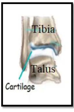
Figure 2-3: Example of articular cartilage at the ankle joint Ligaments and tendons
[image:39.595.249.378.71.265.2]The main function of the ankle ligaments is force transmission from bone to bone and to provide joint stability, and tendons enable load transmission from muscle to bone. Ligaments and tendons consist of small collagen fibres which are bundled together to create a stiff but flexible structure (Figure 2.4) (Pal, 2014).
Figure 2-4: Schematic diagram presenting hierarchical structure of ligament in cross section
12
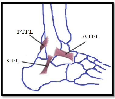
Figure 2-5: Lateral view demonstrates the lateral ligaments
The ATFL controls the anterior displacement and the plantar flexion of the talus (Al-Mohrej & Al-Kenani, 2016). ATFL is quadrilateral and flat in structure (Yildizgoren et al., 2017) and originates from the fibula at the anterior edge of the lateral malleolus (LM) (Meehan et al., 2017). It runs anteromedially and slightly downwards from its origin to insert into the articular cartilage of talar dome (Figure 2.5) (Saxena 2012; Yildizgoren et al. 2017). The ATFL has 16-20 mm in length and 2 mm in thickness (Al-Mohrej & Al-Kenani, 2016; de Asla, Kozánek, Wan, Rubash, & Li 2009). In a neutral position (i.e. 90 degree between foot and leg) the ATFL is almost horizontal to the ankle; however, the ATFL inclines downward and upward in plantar flexion and dorsiflexion position respectively (Golanó et al., 2010). It remodels based on the applied stress and motion (Yildizgoren et al., 2017). The ATFL comes under strain in plantar flexion position and susceptible to injury, especially when the foot is inverted (Golanó et al., 2010).
13
Figure 2-6: CFL throughout the movements of ankle. a: neutral position. b: Dorsal flexion c: Plantar flexion
The PTFL is the strongest of the lateral ankle ligaments (Martin et al., 2013) and originates from the back of the lateral malleolus. It runs horizontally from the posteromedial part of the fibula to the posterior aspect of the talus (Figure 2.5) (Bonnel, Toullec, Mabit, & Tournne, 2010). The PTFL comes under strain in extreme dorsiflexion and external rotation of the foot (Martin et al., 2013). The PTFL acts primary to produce rotatory stability in the transverse plane (Martin et al., 2013).
The medial part of the ankle contains the deltoid ligament, which consists of four ligaments (Figure 2.7) (Al-Mohrej & Al-Kenani, 2016). The ligament has a triangular or a fan shape (Silvestri, Muda, & Sconfienza, 2012) and originates from the distal end of the medial malleolus before dividing into four bundles. Two ligaments run anteriorly; the tibionavicular ligament, which is the more superficial of the two and inserts into the dorsal surface of the scaphoid, and the anterior tibiotalar ligament, which is deeper and inserts into the medial aspect of the talus (Silvestri et al., 2012). Medially, tibiocalcaneal ligament inserts into the substentaculum tali of the calcaneus. The posterior tibiotalar ligament runs posteriorly and inserts into the medial aspect of the talus (Silvestri et al., 2012).
14
[image:42.595.222.403.399.564.2]In addition to the ligaments, tendons around the ankle play a significant part in supporting the ankle joint. Two peroneal tendons are sited on the lateral aspect of the ankle; these are the peroneus longus tendon (PLT) and peroneus brevis tendon (PBT), which lie directly superior to the CFL (Dimmick et al., 2009). The PLT originates from the peroneus longus muscle (Silvestri et al., 2012). It runs diagonally beneath the foot (inferior to the cuboid bone) to insert into the plantar aspect of the first cuneiform and the base of the first metatarsal (Figure 2.8) (Dubin, Comeau, McClelland, Dubin, & Ferrel, 2011; Hodgson, O’Connor, & Grainger, 2012). The PBT, meanwhile, originates from the peroneus brevis muscle (Silvestri et al., 2012). It runs anteriorly to insert into the dorsal aspect of the lateral aspect of the fifth metatarsal base (Figure 2.8) (Hodgson et al., 2012). The main function of the peroneus longus and peroneus brevis tendons is to provide dynamic stabilisation of the lateral ankle and to play a role in eversion of the foot. Furthermore, both tendons allow plantarflexion of the ankle joint as well as abduction and pronation of the foot (Neustadter, Raikin & Nazarian, 2004).
Figure 2-8: The peroneus longus and brevis tendons
15
second, third, and fourth metatarsals (Precerutti et al., 2014). The main function of the TPT is to support the arch and to permit the plantarflexion and supination of the ankle (Pal, 2014).
The FDL tendon is more slender and is located posterior to the TPT (Precerutti et al., 2014). It passes underneath the medial malleolus, over the internal part of the sustentaculum tali, and continues into plantar surface, reaching the plantar fascia (Figure 2.9). The distal part of the FDL terminates with insertions into the plantar parts of the distal phalanges of the second, third, fourth, and fifth toes (Precerutti et al., 2014).
The FHL tendon is the deepest and most posterior among the three medial tendons (Precerutti et al., 2014). It is passes between the medial and lateral tuberosities along the posterior part of the talus. It continues into the plantar surface, intersecting the FDL tendon underneath the sustentaculum tali, and inserting into the distal phalanx of the big toe (Figure 2.9) (Precerutti et al., 2014).
Figure 2-9: Medial tendons of the ankle; TPT (tibialis posterior tendon), FDL (flexor digitorum longus), and FHL (flexor halluces longus)
The anterior ankle includes three tendons arranged in the following order from medial to lateral: tibialis anterior tendon (TAT), extensor halluces longus (EHL), and extensor digitorum longus (EDL) tendons (Demetracopulos & Deland, 2013). TAT runs obliquely and inferomedially to insert on the medial surface of the first cuneiform (Figure 2.10) (Precerutti et al., 2014). The main function of TAT is to dorsiflex the ankle and it also inverts the foot (Demetracopulos & Deland, 2013).
16
interphalangeal joints of the great toe. Furthermore, EHL helps in ankle dorsiflexion (Demetracopulos & Deland, 2013).
EDL is the most lateral tendon of the anterior component. The proximal part of the ligament is thin and broad (Precerutti et al., 2014). Furthermore, EDL splits into four distinct tendons just inferior to the neck of the talus, which insert on the dorsum of the distal phalanges of the second, third, fourth, and fifth toes (Figure 2.10) (Precerutti et al., 2014). The EDL tendon works to assess the ankle dorsiflexion (Demetracopulos & Deland, 2013).
Figure 2-10: Anterior ankle tendons
On the other hand, the posterior part of the ankle has the largest and strongest tendon in the body, which is known as the Achilles tendon (AT) (Doral et al., 2010). It originates in the middle part of the calf, from the medial and lateral gastrocnemius muscles, and merges with the soleus muscle in the distal part of the calf (Doral et al., 2010). The AT inserts into the posterosuperior aspect of the calcaneus (Figure 2.11) (Jane & Marriott, 2009). It allows individuals to stand up on their toes (Pal, 2014).
17 2.4.2 Muscles
Movement of the ankle during walking, running, and jumping is due to muscle contraction in response to forces from the ground (Pal, 2014). The peroneal (longus and brevis) muscles are located on the lateral aspect of the leg and have a significant role in the lateral stability of the ankle and foot (Mansfield & Neumann, 2014). The peroneal longus muscle (PLM) is the more superficial of the two muscles, originating from the upper two-thirds of the lateral aspect of the fibula and the lateral condyle of the tibia (Figure 2.12) (Taljanovic et al., 2015).The PLM acts in eversion and plantarflexion of the foot (Bisschops & Lavallee, 2016). The peroneal brevis muscle (PBM) is located just anterior to the longus (Bisschops & Lavallee, 2016). It originates from the distal two-thirds of the lateral aspect of the fibula (Figure 2.12) (Jung, 2016). The primary function is to evert the foot, with a secondary role in stabilisation of the lateral ankle (Taljanovic et al., 2015).
Figure 2-12: Peroneal tendons and muscles
18
Figure 2-13: Tibialis posterior muscle
The calf (gastrocnemius and soleus) muscles are situated in the posterior aspect of the leg (Doral et al., 2010). The gastrocnemius muscle has medial and lateral heads which provide the bulging shape of the calf (Figure 2.14) (Doral et al., 2010). The medial head arises from the popliteal aspect of the femur, whereas the lateral head originates from the posterolateral surface of the lateral femoral condyle and shorter than the medial head (Figure 2.14) (Doral et al., 2010). Both heads extend to the posterior calcaneus through the Achilles tendon (Doral et al., 2010). The gastrocnemius muscle generates the plantarflexion of the ankle (Mansfield & Neumann, 2014). The soleus muscle is located under the gastrocnemius muscle, and arises from the posterior part of the head of the fibula and the middle third of the medial boundary of the tibia (Figure 2.14) (Doral et al., 2010). It unites with the gastrocnemius muscle to insert into the calcaneus via the Achilles tendon. Along with the gastrocnemius muscle, it makes the three-headed triceps surae which works to provide the plantarflexion movement of the ankle joint through its conjoint tendon (Achilles tendon) (Doral et al., 2010).
19
2.5
Ligament injury
Ligament injuries are one of the predominant causes of musculoskeletal joint pain in acute care (Hauser et al., 2013). Injury to the ligaments often creates an imbalance between mobility and stability of the joint and abnormal loading on the joint may disrupt other skeletal or soft tissues in and around a joint, resulting in pain (Jung, Fisher, & Woo, 2009). In 2012, Bartlett and Bussey identified three different types of ligament failures that are based on the loading rate of the ligament, which represents the speed at which load is applied to the ligament (Puddle & Maulder, 2013):
Mid-substance tears. Bony avulsion.
Cleavage at the ligament-bone interface.
Mid-substance tears of the ligamentous tissues which occur in the mid length of the ligaments are the most common mechanism of ankle ligament injury (Bartlett & Bussey, 2012). This injury is characteristic of fast loading rate failures where the bundle of ligament fibres fails through shear and tension mechanisms, and is typical in sports such as football. This leads to a partial or complete rupture of the ankle ligaments, which represents Grade II and Grade III ankle sprains respectively (Myrick, 2014).
By contrast, bony avulsion (rupture of the bony attachment of the ligament) failure is characteristic of slow loading rate failures. The failure takes place underneath the insertion site of the spongy bone. This type is more prominent in young athletes, whose ligaments are stronger than their bones (Bartlett & Bussey, 2012).
20 2.5.1 Aetiology of ankle sprain
It has been proposed that most ankle sprain injuries were occurred because of increased supination moment at subtalar joint, which was often as consequence of the extent and the position of the vertically projected ground reaction force (GRF) at early foot contact (Fong et al., 2009a). An ankle sprain is possibly caused by the deviation of such GRF vector from the centre of the ankle joint, commonly at the lateral plantar edge acting to the medial aspect during a sideway cutting movement, creating a large moment arm and therefore an explosive ankle supination torque (Fong et al., 2011). This torque would quickly trigger joint twisting motion and stretch the lateral ligaments in robust way (Fong et al., 2011).
It has been suggested that incorrect foot positioning at landing is one of the common aetiology of ankle sprain. Wright and colleagues (2000) reported that increased touchdown plantar flexion caused increased in the occurrence of ankle sprain. In other words, if the foot was plantarflexed at touchdown, the foot contact the ground with the forefoot, therefor the ground reaction force moment arm about the subtalar joint axis might increase (Wright, Neptune, van den Bogert, & Nigg, 2000) and increase the resultant joint torque to create abrupt explosive twisting motion and thus ankle sprain injury (Fong et al., 2009a).
21 2.5.2 Mechanism of ankle ligamentous sprain
Lateral ankle ligamentous sprain is the most common sports injuries, however the mechanism of the sprain is not clear (Fong, Ha, Mok, Chan, & Chan, 2012b). Acute ankle sprain has been defined by Delahunt et al. (2010) and endorsed by the International Ankle Consortium (Gribble et al., 2016) as “an acute traumatic injury to the lateral ligament complex of the ankle joint as a result of excessive inversion of the rear foot or a combined plantarflexion and adduction of the foot. This usually results in some initial deficits in function and disability”. Understanding the mechanism of ankle injury with quantitative analysis of ankle biomechanics is important for the design of protective tool and the improvement of injury prevention protocols (Fong et al., 2012).
Ankle sprain does not happen at only one single plane; however, it is accompanied by the other two planes. It is commonly known that the most prominent mechanism of lateral ankle sprain is supination (Seah & Mani-Babu, 2011) accounting for 84% of all sport-related ankle injuries (Fong et al., 2007). The supination is defined as a combination of ankle inversion and forefoot adduction in plantar flexion, resulting in a lateral inversion sprain (Seah & Mani-Babu, 2011). Excessive inversion of the ankle shifts the centre of gravity to the lateral edge of the weight-bearing leg, resulting in a twisting of the ankle at high velocity (Figure 2.15) (Myrick, 2014). Damage to the ligaments is dependent on the foot and ankle positions at the time of injury and the velocity of the mechanism of injury determine the ligament’s injury (Martin et al., 2013). The supination mechanism of an ankle sprain causes a partial tear or complete rupture of the lateral ankle ligaments (Martin et al., 2013).
22
Several methods were published in the literature to quantitatively evaluate the mechanism of ankle sprain such as motion analysis of non-injury simulations, injuries during biomechanical experiments, cadaver studies, video analysis, and athlete interviews (Ha, Fong, & Chan, 2015). Kinematics of sprained ankles have been investigated through simulated sub-injury or close-to-injury where an inversion perturbation device abruptly inverts the ankle to simulate the mechanism of LAS (Chan, Fong, Yung, Fung, & Chan, 2008; Myers, Riemann, Hwang, Fu, & Lephart, 2003). Fong et al. (2009b) mentioned that since these experiments did not create real injury, they might only represent the kinematics to a limited degree.
The most appropriate method to evaluate the mechanism of the injury is to investigate actual injury incidents; however, it is unethical and impracticable to make tests that generate a high risk of injury for the subjects (Ha et al., 2015). Real life mechanisms of ankle sprain have been described based on video recordings of injuries from Norwegian and Icelandic elite football (Andersen et al., 2004). They concluded that about half of all ankle injuries was due to player to player contact, with collision with an opponent on the medial side of the leg, just prior to or at the moment of foot strike, creates a laterally directed force and the player lands with the ankle in a vulnerable inverted position (Figure 2.16) (Andersen et al., 2004). In addition, authors observed some cases where the injured player hits theopponent’sfoot when trying to shoot or clear the ball, positioning the ankle in forced plantar flexion. It seems that these mechanisms are specific to football injury. Moreover, the study was done on elite male football players. The mechanism of injury could be different for different players, such as female or different types of sport.
23
Very occasionally, sprains occur during biomechanical testing (Fong et al., 2009b; Gehring et al., 2013; Kristianslund et al., 2011). Fong et al., (2009b) published the first kinematic analysis of inversion ankle sprain, which occurred accidentally on one male athlete who was performing a sequence of cutting motion trials in the laboratory. The motion of the injury was videotaped by 3 synchronized and calibrated high-speed cameras. Fong et al. (2009b) analysed the video sequences of the injury utilising model-based image-matching (MBIM) technique described by Krosshaug and Bahr (2005). This motion analysis technique was introduced to reconstruct 3D (three dimensional) human motion from uncalibrated video sequences and applied to define the mechanism of anterior cruciate ligament (ACL) ruptures that occur during real world events (Krosshaug, Slauterbeck, Engebretsen, & Bahr, 2007). However, this technique was only validated for knee and hip joints. Therefore, Mok et al. (2011a) conducted a study evaluate the validity and reproducibility of the MBIM method to estimate the kinematic of ankle joint. The authors concluded the validity, inter-rater, and intra-rater reliability was excellent and MBIM motion analysis method could give excellent estimates of ankle joint kinematics (Mok et al., 2011a).
The results ofFong’sstudy(2009b) indicated that at the time of injury, the ankle was inverted to 48°, internally rotated to 10º and about 18° dorsiflexed (Fong et al., 2009b). Moreover, Kristianslund et al. (2011) described the kinematic of accidental lateral ankle sprain that happened in a female elite handball player during sidestep cutting in a motion analysis laboratory. The sprain occurred at 23° ankle inversion, 46° internal rotation, and 22° dorsiflexion. There was a substantial degree of ankle dorsiflexion in both studies which indicated that plantar flexion is not required for sprain to occur (Kristianslund et al., 2011) and there could be several possible mechanisms that create an inversion sprain injury (Fong et al., 2012b).