INTRODUCTION
During development, Hedgehog (Hh) proteins can act as morphogens that form concentration gradients to specify distinct fates in a concentration-dependent manner (Hooper and Scott, 2005; Jiang and Hui, 2008; Tabata and Takei, 2004; Wang et al., 2007). Hh proteins are dually lipid modified and are expected to be associated with the cell membrane (Guerrero and Chiang, 2007; Mann and Beachy, 2004). How Hh proteins are secreted from their producing cells and form concentration gradients is one of the key issues in the Hh research field. Various studies have shown that Hh signaling strength and gradient ranges are regulated by a complex feedback network that acts at the level of ligand binding (Kang et al., 2007; Wilson and Chuang, 2006). For example, in both Drosophila and vertebrates, the Hh receptor Patched (Ptc) is upregulated by Hh signaling and subsequently sequesters Hh, thus limiting the range of the Hh gradient (Chen and Struhl, 1996). Here, we address the roles of two cell-surface proteins, Dally-like (Dlp) and Interference hedgehog (Ihog), in Hh signaling and distribution.
Dlp and its family member Dally are glypican-type heparan sulfate proteoglycans (HSPGs), comprising a core protein and attached heparan sulfate (HS) glycosaminoglycan (GAG) chains (Lin, 2004). Glypicans are linked to the plasma membrane by a
glycosylphosphatidylinositol (GPI) anchor. Previous studies have shown that Hh signaling and distribution are defective in animals mutant for GAG biosynthesis enzymes, demonstrating the crucial roles of GAG chains in Hh signaling (Lin, 2004). A subsequent RNAi screen identified Dlp as an essential HSPG involved in Hh signaling (Lum et al., 2003). Genetic studies demonstrated further that Hh signaling is diminished in dlp-RNAior dlpmutant embryos (Desbordes and Sanson, 2003; Franch-Marro et al., 2005; Han et al., 2004b). Recent studies suggest that the GPI anchor of Dlp is required for its transcytosis and for its activity in Hh signaling (Gallet et al., 2008).
Ihog is a member of the conserved Ig/fibronectin superfamily, which includes Boi (Brother of ihog) in Drosophila and the mammalian homologs Boc and Cdo (Tenzen et al., 2006; Yao et al., 2006; Zhang et al., 2006). ihogwas initially identified in an RNAi screen as a gene essential for Hh signaling (Lum et al., 2003). Further studies demonstrated that Ihog family members are Hh-binding proteins essential for the reception of Hh signal (Yao et al., 2006). Recent experiments showed that Ihog can bind to heparin, which induces Ihog dimerization and is required to mediate high-affinity interactions between Ihog and Hh (McLellan et al., 2006). Importantly, ihog mutants exhibit strong genetic interaction with dlp mutants in embryos, suggesting that Ihog might work together with Dlp in regulating Hh signaling (Yao et al., 2006).
Here, we have examined the mechanisms by which Dlp and Ihog regulate Hh signaling. Our results suggest that the Dlp core protein is essential for its function in Hh signaling, whereas the GAG chains are important for its non-cell-autonomous activity. We provide evidence that Ihog has a biphasic effect in Hh signaling depending on its level, and that Ihog is required to maintain Hh on the cell surface. In the wing disc, Dlp promotes Hh signaling strength, whereas Ihog extends Hh signaling range. Our results Development 137, 2033-2044 (2010) doi:10.1242/dev.045740
© 2010. Published by The Company of Biologists Ltd
1Division of Developmental Biology, Cincinnati Children’s Hospital Medical Center, and The Graduate Program in Molecular and Developmental Biology, University of Cincinnati College of Medicine, Cincinnati, OH 45229, USA. 2State Key Laboratory of Biomembrane and Membrane Biotechnology, and Key Laboratory of Stem Cell, Institute of Zoology, Chinese Academy of Sciences, Beijing, 100101, China.
*Author for correspondence ([email protected])
Accepted 21 April 2010 SUMMARY
Hedgehog (Hh) acts as a morphogen in various developmental contexts to specify distinct cell fates in a concentration-dependent manner. Hh signaling is regulated by two conserved cell-surface proteins: Ig/fibronectin superfamily member Interference hedgehog (Ihog) and Dally-like (Dlp), a glypican that comprises a core protein and heparan sulfate glycosaminoglycan (GAG) chains. Here, we show in Drosophilathat the Dlp core protein can interact with Hh and is essential for its function in Hh signaling. In wing discs, overexpression of Dlp increases short-range Hh signaling while reducing long-range signaling. By contrast, Ihog has biphasic activity in Hh signaling in cultured cells: low levels of Ihog increase Hh signaling, whereas high levels decrease it. In wing discs, overexpression of Ihog represses high-threshold targets, while extending the range of low-threshold targets, thus showing opposite effects to Dlp. We further show that Ihog and its family member Boi are required to maintain Hh on the cell surface. Finally, Ihog and Dlp have complementary expression patterns in discs. These data led us to propose that Dlp acts as a signaling co-receptor. However, Ihog might not act as a classic co-receptor; rather, it may act as an exchange factor by retaining Hh on the cell surface, but also compete with the receptor for Hh binding.
KEY WORDS: HSPG core protein, Hh signaling, Interference hedgehog, Biphasic activity, Glypican, Morphogen, Drosophila
The cell-surface proteins Dally-like and Ihog differentially
regulate Hedgehog signaling strength and range during
development
Dong Yan1, Yihui Wu1,2, Yongfei Yang1,2, Tatyana Y. Belenkaya1, Xiaofang Tang1and Xinhua Lin1,2,*
D
E
V
E
LO
P
M
E
N
suggest that Dlp acts as an Hh co-receptor, whereas Ihog is unlikely to act as a classic co-receptor; instead, we propose an exchange factor model for its activity in Hh signaling.
MATERIALS AND METHODS Drosophilastrains
Mutant lines were dlpA187, dally80(Han et al., 2004b), sgll(3)08310(Hacker et al., 1997), sfl9B4(Lin and Perrimon, 1999), which are all null alleles. dlp, dally, sgl and sfl germline clones were generated by the FLP-DFS technique (Chou and Perrimon, 1996).ptc-lacZand dpp-lacZP10638lines were used to visualize ptcand dppexpression (Han et al., 2004b). The ihog
mutant was generated by imprecise excision of a P-element, P(SUPor-P) CG10158KG05348. It has a 3305 bp deletion extending from 214 bp downstream of the ihogATG start codon to the 3⬘end of the gene and is therefore likely to be a null allele.
Transgenic lines were prd-Gal4, en-Gal4, ap-Gal4, ptc-Gal4, dpp-Gal4,
tub-Gal80ts(FlyBase), Ci-Gal4(Croker et al., 2006), ywhs-flp, UAS-GFP; FRT40, tub-GAL80; tub-GAL4/TM6B(Janody and Treisman, 2006), UAS-Rab5S43N (dominant-negative Rab5) (Entchev et al., 2000), UAS-dlp
(Baeg et al., 2001), UAS-dlp-GFP(Baeg et al., 2004) and UAS-GFP-dlp-CD2(Gallet et al., 2008). The following lines were generated in this study:
UAS-dlp(–HS),UAS-dlp(–HS)-CD2,UAS-dlp-⌬N,UAS-dlp-⌬C, UAS-dlp-⌬GAG,UAS-dlp(–HS)-GFP,UAS-dlp(–HS)-GFP-CD2,UAS-ihog, UAS-ihog-RNAiandUAS-boi-RNAi.
Plasmid construction
Dlp(–HS) was generated using the Gene-Tailor Mutagenesis Kit (Invitrogen). All five potential GAG attachment sites – Ser625, Ser629, Ser631, Ser643 and Ser686 – were converted to Ala. Dlp(–HS)-CD2 contains Dlp(–HS) amino acids 1-713 fused with the rat CD2 gene (excluding the signal peptide and starting from amino acid 24, Asp) (Strigini and Cohen, 1997). Dlp-⌬N deletes amino acids 42-553 of Dlp, Dlp-⌬GAG deletes amino acids 611-694, and Dlp-⌬C deletes all of the region C-terminal of amino acid 610. dlp(–HS)-GFPwas generated by inserting the GFP fragment from pEGFP-N1 (Clontech) at a unique NdeI site in dlp(–HS). This insertion site is the same as in dlp-GFP(Baeg et al., 2004). dlp(–HS)-CD2-GFPalso has the GFP inserted in the same NdeI site of dlp(–HS)-CD2. GFP-GPI contains EGFP sequence followed by the GPI signal from Dlp (amino acids 695-765). All of the dlpDNA fragments were inserted into pUASTvectors. For UAS-ihog, the complete ihogcoding sequence was amplified from ihogcDNA clone GH03927. UAS-ihog-V5
contains a V5 tag at its C-terminus. UAS-connectin-V5has a V5 tag inserted between amino acids 93 and 94, shortly after the signal sequence (Nose et al., 1992). UAS-GFP-Dlp-CD2is from Gallet et al. (Gallet et al., 2008) and UAS-Ptc-GFPfrom Lu et al. (Lu et al., 2006). For UAS-ihog-RNAiand UAS-boi-RNAiconstructs, the corresponding gene fragments were cloned as inverted repeats into the pWIZvector. Primers for ihog
RNAi were 5⬘-CCAAAACCAGCACCACAG-3⬘and 3⬘GATTACACG -CCAACGCTG-5⬘. Primers for boiRNAi were 5⬘CGAGCATGGTGC -CTCCG-3⬘and 3⬘-TTCTGGTCCTCGCTTGCCG-5⬘.
Immunostaining
Wing disc and embryo staining procedures were preformed as described (Han et al., 2004b). The following antibodies were used: mouse anti-Wg, rat anti-E-cadherin, mouse anti-Ptc, mouse anti-Dlp (all DSHB), rabbit anti-Hh (Taylor et al., 1993), mouse anti-V5 (Invitrogen), rabbit anti-GFP (Molecular Probes), mouse anti-⌬HS 3G10 (Seikagaku), rabbit anti-Bap (Lee and Frasch, 2000), rabbit anti-Dlp (Baeg et al., 2001), rat anti-Ci (Motzny and Holmgren, 1995), rabbit anti-Col (Vervoort et al., 1999), rabbit anti--Gal (Cappel), rat anti-Ihog (Yao et al., 2006) and guinea pig anti-Rab5 (made in our laboratory). For extracellular Hh staining, discs were incubated for 45 minutes on ice with triple the usual antibody concentration before fixation. To detect HS, discs were fixed and treated with 500 mU/ml heparinase III (Sigma) at 37°C for 6 hours and stained with 3G10 antibody. For quantification of confocal images, the raw data were exported in tiff format. The plot values were measured from selected regions using ImageJ (NIH). The plot values were then used to generate plot profiles in Microsoft Excel.
Cell binding assay and co-immunoprecipitation
Drosophila S2 cells were transfected with pAc5.1-Hh-N-V5 and the conditioned medium was collected and concentrated. The medium was then applied to S2 cells grown on coverslips and transfected with the indicated plasmids. The cells were incubated in Hh-N-V5 medium for 3 hours on ice, then fixed and stained with V5 and GFP antibodies.
For co-immunoprecipitation experiments, S2 cells were transfected using Effectene (Qiagen) and pArmadillo-Gal4 was co-transfected to induce pUAST plasmids. Cells were lysed in 150 mM NaCl, 20 mM Tris-HCl (pH 7.5), 2% Triton X-100, 1 mM EDTA and proteinase inhibitors (Roche) on ice for 1 hour. After pre-clearance with protein G-Sepharose beads (Amersham), the lysate was incubated with antibodies for 4 hours at 4°C and then for an additional 2 hours in the presence of beads. Beads were washed three times and proteins eluted in Laemmli sample buffer. Western blotting was conducted as described (Han et al., 2004a). Primary antibodies were rabbit anti-Hh, guinea pig anti-GFP, guinea pig anti-Dlp (made in our laboratory) and goat anti--Tubulin (Santa Cruz).
Luciferase reporter assay
Drosophilacl-8 cells were transfected with double-stranded (ds) RNA or plasmid DNA together with ptcluciferase reporter and the normalization vector (copia-Renilla) (50:1). After 48 hours, transfected cells were split into control or Hh-N-containing medium and incubated for an additional 24 hours. Cells were then lysed and luciferase activities measured using the Dual-Luciferase Assay Kit (Promega). dsRNAs were synthesized using the MEGAscript In Vitro Transcription Kit (Ambion). The primers for ptc
and dlp5⬘UTRwere as described (Lum et al., 2003); the primers for ihog
are indicated above and the dlp/dlp(–HS)constructs used for rescue do not contain the 5⬘UTR region.
RESULTS
The Dlp core protein can directly activate Hh signaling, whereas the attached GAG chains provide non-cell-autonomous activity of Dlp We attempted to define the mechanisms by which Dlp regulates Hh signaling in Drosophilaembryos. Removing both the maternal and zygotic contribution of Dlp produces embryos with a segment polarity phenotype, with a lawn of denticle belts and an absence of naked cuticle (Fig. 1B) (Desbordes and Sanson, 2003; Franch-Marro et al., 2005; Han et al., 2004b). This phenotype could be derived from a failure in Hh or Wingless (Wg) signal transduction, as Hh signaling is required to maintain Wg expression, which in turn is required for naked cuticle specification via repression of shaven baby(ovo– FlyBase) (Hatini and DiNardo, 2001). Previous studies found that dlp RNAi is able to block Hh, but not Wg, signaling in the embryonic epidermis, suggesting that the segment polarity phenotype associated with dlpembryos is due to a failure of Hh signal transduction (Desbordes and Sanson, 2003).
We confirmed this RNAi result using a null allele of dlp. Ectopic expression of Wg by prd-Gal4was able to restore the naked cuticle in dlpembryos (Fig. 1C), whereas ectopic expression of Hh was not (Fig. 1D). Consistent with the cuticle defects, overexpression of Hh failed to restore the expression of Wg, an Hh signaling target, in dlp embryos (see Fig. S1E-H in the supplementary material). This result is surprising because ectopic expression of Hh was able to restore naked cuticle in embryos mutant for sugarless (sgl) (Hacker et al., 1997) or sulfateless (sfl) (Lin and Perrimon, 1999) (see Fig. S1A-D in the supplementary material). As sgland sflare essential for the biosynthesis of HS GAG chains attached to Dlp and other HSPGs, our data imply that specific functions associated with the Dlp core protein are involved in Hh signaling.
To test this hypothesis, we constructed a Dlp core protein that lacks all of the GAG attachment sites [Dlp(–HS); see Materials and methods]. Ectopic expression of Dlp(–HS) does not enhance
D
E
staining by the 3G10 antibody, which recognizes an HS epitope produced by enzymatic digestion with heparitinase (see Fig. S2B-C⬙in the supplementary material), suggesting that Dlp(–HS) is indeed devoid of HS chains (David et al., 1992). Expression of Dlp(–HS) by prd-Gal4 effectively rescued naked cuticle patterning cell-autonomously in dlp embryos (Fig. 1E, yellow arrows indicate the prd-Gal4expression domain). The activity of Dlp(–HS) is not due to Dally, as virtually identical rescue was observed when Dlp(–HS) was expressed in dally dlpdouble-mutant embryos (Fig. 1F). Interestingly, expression of wild-type Dlp by prd-Gal4 restored cuticle patterning in entire segments in a non-cell-autonomous manner (Fig. 1G, red arrows indicate the prd-Gal4 non-expression domain). Consistent with this, Wg expression was restored by Dlp(–HS) in its expression domain (Fig. 1H,H⬘), whereas Wg expression was restored non-autonomously by wild-type Dlp (Fig. 1I,I⬘).
We further determined whether Dlp(–HS) could replace the function of Dlp in other Hh signaling-mediated events. Expression of the Hh target gene bagpipe (bap) in the mesoderm is defective in
dlpembryos (Han et al., 2004b) (Fig. 1J,K). Ectopic expression of Dlp by prd-Gal4fully rescuedbapexpression in all of the segments (Fig. 1L), whereas expression of Dlp(–HS) could only restore bap in every other segment, as expected from the expression domain of prd-Gal4(Fig. 1M). We also assayed Dlp activity in Hh signaling using Drosophilacl-8 cells (Lum et al., 2003). Depletion of Dlp by RNAi in cl-8 cells abolished Hh signaling, whereas Dlp and Dlp(–HS) expression restored the response of dlp-depleted cl-8 cells to Hh signal (Fig. 1N). Together with the cuticle data, these results strongly argue that the core protein is essential for Dlp activity in Hh signaling, whereas the attached GAG chains confer additional non-cell-autonomous activity to Dlp.
The Dlp core protein interacts with Hh and its signaling activity requires the N-terminal domain anchored to the cell membrane
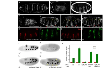
[image:3.612.53.489.57.349.2]We further examined the roles of the Dlp core protein in Hh signaling. To test whether the Dlp core protein participates in Hh signaling by interacting with Hh, we incubated Hh-N-containing Fig. 1. Dlp core protein can restore Hh signaling autonomously in dlpembryos and Drosophila cell lines.Embryos are oriented anterior to the left. Mutated genes are indicated in the left-hand corner and ectopically expressed transgenes are indicated in the right-hand corner.
(A-G)Ventral cuticles of embryos of different genotype. (A)Wild-type (WT); (B) dlp(all dlpmutant embryos in this study are derived from germline clones of allele dlpA187); (C-E,G)dlpembryos rescued by prd-Gal4, UAS-wg(C), prd-Gal4, UAS-hh(D), prd-Gal4, UAS-dlp(–HS)(E) andprd-Gal4, UAS-dlp(G); (F) dally, dlpembryo rescued by prd-Gal4, UAS-dlp(–HS). prd-Gal4drives gene expression in every other segment. Yellow arrows indicate prd-Gal4expression segments, and red arrows indicate prd-Gal4non-expression segments. In prd-Gal4, UAS-dlp-rescued embryos, the naked cuticles in prd-Gal4non-expression segments are also rescued (G, red arrows). We analyzed 58 such embryos and found that 92% of prd-Gal4non-expression segments are rescued. By contrast, prd-Gal4, UAS-dlp(–HS)mostly only rescues naked cuticles in the prd-Gal4expression domain (E). We examined 56 such embryos and found that only 3% of non-expression segments rescued. (H-I⬘) In prd-Gal4, UAS-dlp(–HS)-rescued
dlpembryos, Wg expression is restored in the Dlp(–HS) expression domain. In prd-Gal4, UAS-dlp-rescued embryos, Wg expression is restored also in the Dlp non-expression domain. Dlp(–HS) and Dlp expression was detected by Dlp antibody. (J-M)Bap staining in a stage 10 wild-type embryo (J),
dlpembryo (K), dlpembryo rescued by prd-gal4, UAS-dlp(L) or prd-gal4, UAS-dlp(–HS) (M). Yellow arrows indicate recovered Bap staining in every other segment. (N)An Hh signaling reporter assay was performed in cl-8 cells. lacZdsRNA serves as a control. The addition of ptcdsRNA leads to increased reporter activity, whereas dsRNA against dlp5⬘UTRreduces the pathway activation. This reduction is fully rescued by addition of dlpor
dlp(–HS)expression vectors. The error bars represent s.d.
D
E
V
E
LO
P
M
E
N
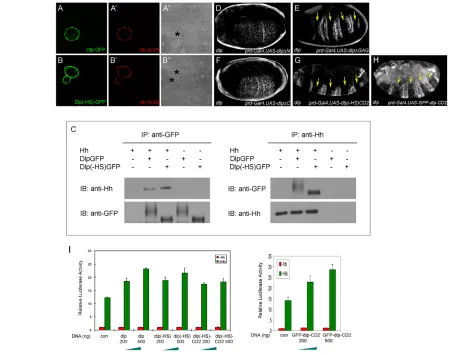
conditioned media with DrosophilaS2 cells transfected with Dlp-GFP, Dlp(–HS)-GFP or GFP-GPI. Similar levels of Hh-N (the N-terminal domain of Hh) accumulated on the surface of cells transfected with Dlp(–HS)-GFP or Dlp-GFP, but not on GFP-GPI cells (Fig. 2A-B⬙; see Fig. S3A,A⬘in the supplementary material). Similarly, Hh co-immunoprecipitated with Dlp-GFP or Dlp(–HS)-GFP, but not GFP-GPI, when co-expressed in S2 cells (Fig. 2C; see Fig. S3B in the supplementary material). Collectively, these data argue that the Dlp core protein can interact with Hh.
Next, we determined the Dlp domain required for its activity in Hh signaling. Glypican is composed of three domains: a GPI anchor, a GAG attachment domain and an N-terminal globular cysteine-rich domain (see Fig. S2A in the supplementary material for Dlp mutant constructs). In rescuing experiments, Dlp⌬GAG, which lacks the GAG attachment domain, was functional (Fig. 2E), whereas Dlp⌬N, which lacks the N-terminal domain, was defective (Fig. 2D), suggesting that the N-terminal domain of Dlp is essential for its function. The N-terminal domain of Dlp alone (Dlp⌬C) was not active (Fig. 2F); however, when it was fused with the transmembrane protein CD2, as Dlp(–HS)CD2, its
cell-autonomous activity in Hh signaling was restored (Fig. 2G). Finally, we tested another CD2-linked form of Dlp that contains intact GAG chains, GFP-Dlp-CD2 (Gallet et al., 2008). Interestingly, GFP-Dlp-CD2 was only able to rescue naked cuticle cell-autonomously in dlp embryos (Fig. 2H). It is worthwhile mentioning that the ectopic expression of three other HSPGs [Dally, Syndecan and Perlecan (Trol – FlyBase)] failed to rescue Hh signaling defects in dlpembryos (data not shown).
Together, our data suggest that the N-terminal domain and its retention on the cell surface are essential for Dlp activity in Hh signaling. Furthermore, the function of the GPI anchor can be replaced by CD2, indicating that the specific activity of GPI is not essential for Dlp function in Hh signaling.
Expression of Dlp enhances Hh signaling
magnitude but reduces signaling range in wing discs
[image:4.612.55.525.296.651.2]We further examined whether elevated levels of Dlp can promote Hh signaling in vitro and in vivo. cl-8 cells were transfected with increasing amounts of Dlp constructs and an Hh-responsive
Fig. 2. Dlp core protein interacts with Hh.(A-B⬙) Transfection of Dlp-GFP or Dlp(–HS)-GFP intoDrosophilaS2 cells causes accumulation of exogenous Hh-N-V5 at the cell surface. Asterisks mark transfected cells. (C)Dlp or Dlp(–HS) and Hh can precipitate each other. Dlp-GFP or Dlp(–HS)-GFP and Hh expression vectors were transfected individually or together into S2 cells. Cell lysates were immunoprecipitated and analyzed by western blotting with the antibodies indicated. Dlp-GFP forms a smear typical of HSPG, whereas Dlp(–HS)-GFP displays a sharp band indicating that it is non-glycanated. (D-H)Cuticle preparations from dlpembryos rescued by various deletion isoforms of the dlptransgene. See Fig. S2A in the supplementary material for a detailed map of these constructs. Yellow arrows indicate restored naked cuticle. (I)Drosophilacl-8 cells transfected with the indicated amounts of Dlp, Dlp(–HS), Dlp(–HS)-CD2 or GFP-Dlp-CD2 expression vectors show a concentration-dependent increase in the Hh signaling response. The error bars represent s.d. The GFP-Dlp-CD2 assay was performed separately and so the results are shown as a separate chart.
D
E
V
E
LO
P
M
E
N
luciferase reporter. Hh signaling was enhanced in a dose-dependent manner when Dlp, Dlp(–HS), Dlp(–HS)-CD2 or GFP-Dlp-CD2 were transfected (Fig. 2I). This result is consistent with the view that Dlp binds Hh on the cell surface and facilitates its interaction with Ptc.
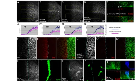
In the wing disc, Hh produced in the posterior (P) compartment forms a concentration gradient into the anterior (A) compartment abutting the anterior-posterior (A/P) boundary. Hh signaling activates expression of the high-threshold targets Ptc and Collier (Col; Knot – FlyBase) and of the low-threshold target decapentaplegic (dpp), and stabilizes another low-threshold target, Cubitus interruptus (Ci) (Aza-Blanc et al., 1997; Strigini and Cohen, 1997; Vervoort et al., 1999). We further examined the roles of Dlp in Hh signaling and gradient activity in discs. In wild-type discs, the levels of Ptc, Ci and dpp-lacZ were virtually identical in the dorsal (D) and ventral (V) compartments (Fig. 3A-C, the fluorescence intensity in selected areas of each image is quantified in Fig. 3A⬘-C⬘). Interestingly,
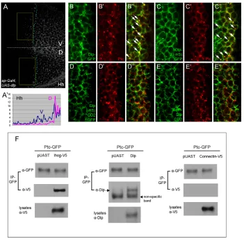
when Dlp was ectopically expressed at the D compartment by ap-Gal4, levels of Ptc, Ci and dpp-lacZwere elevated in cells closer to the P compartment; however, their ranges towards more anterior cells were reduced (Fig. 3D-F⬘). Ectopic expression of Dlp(–HS), Dlp(–HS)-CD2 or GFP-Dlp-CD2 produced similar results (Fig. 3G-O⬘). These results suggest that Dlp might act as a co-receptor to present Hh for Ptc, thereby enhancing Hh signaling strength. Increased interaction of Hh with Ptc might further reduce available Hh, thereby reducing the signaling range. Indeed, we found that Hh spread over shorter distances in Dlp-overexpression tissues (Fig. 4A,A⬘).
[image:5.612.56.506.279.666.2]If Dlp is a co-receptor of Ptc, then one possibility is that the two proteins interact. We tested this hypothesis in co-immunoprecipitation experiments in S2 cells (Fig. 4F). As a positive control, Ihog co-precipitated with Ptc-GFP, consistent with a recent study (Zheng et al., 2010). By contrast, Connectin, a GPI-linked protein that has not been implicated in Hh signaling, does not co-precipitate with Ptc-GFP (Nose et al., 1992). Importantly,
Fig. 3. Dlp expression enhances Hh signaling strength while reducing signaling range. (A-O⬘) Ptc, Ci and dpp-lacZstaining in Drosophila
third instar larvae wing discs, oriented ventral (V) up and anterior left. (A-C⬘) Wild-type discs. (D-O⬘) Ectopic expression of Dlp (D-F⬘), Dlp(–HS) (G-I⬘), Dlp(–HS)CD2 (J-L⬘) or GFP-Dlp-CD2 (M-O⬘) in the dorsal (D) half of the disc by ap-gal4leads to enhanced Hh signaling strength but reduced signaling range. The fluorescence intensities from selected areas (boxed) in these images are quantified in A⬘-O⬘. The images used for
quantifications were from single confocal sections. The y-axis indicates relative fluorescence intensity and the x-axis indicates the distance from the
anterior (left) to posterior (right) compartment of the wing discs.
D
E
V
E
LO
P
M
E
N
we found that Dlp can also co-precipitate with Ptc-GFP, suggesting that Dlp might facilitate interaction between Ptc and Hh by interacting with both of them.
Our experiments show that CD2 forms of Dlp can rescue the dlp embryo and promote Hh signaling in the disc, suggesting that the GPI anchor is not essential for the activity of Dlp. We further analyzed the subcellular localization of the GFP-tagged forms of Dlp, Dlp(–HS), Dlp(–HS)-CD2 and GFP-Dlp-CD2 in discs. Interestingly, whereas both Dlp-GFP and Dlp(–HS)-GFP formed many intracellular vesicles that colocalized well with Ptc vesicles (Fig. 4B-C⬙, arrows indicate colocalized vesicles), Dlp(–HS)-CD2-GFP and Dlp(–HS)-CD2-GFP-Dlp-CD2 formed very few intracellular vesicles and these were not colocalized with Ptc vesicles (Fig. 4D-E⬙). The lack of colocalization of GFP-Dlp-CD2 with Ptc vesicles has also been observed by Gallet et al. (Gallet et al., 2008). These results argue that the GPI anchor of Dlp can affect its endocytic trafficking but is not essential for Dlp signaling activity, which only requires that Dlp be on the cell surface.
Overexpression of Ihog fails to rescue Hh
signaling defects in dlpmutants
Although Ihog has been shown to be involved in Hh signaling, the mechanism of its action remains unclear. One important issue is whether Ihog and Dlp play similar roles. We examined whether overexpression of Ihog could rescue Hh signaling defects in dlp embryos and discs. As shown in Fig. S4A-B⬘in the supplementary material, ectopic expression of Ihog failed to restore naked cuticle or Wg expression in dlp embryos. In addition, Ptc expression was
reduced in dlphomozygous mutant discs and this defect was not rescued by expression of Ihog (see Fig. S4C-D⬘ in the supplementary material). These results differ from the previous observation that Ihog overexpression can rescue Hh signaling defects in dlp-RNAicl-8 cells (Yao et al., 2006). One explanation for this discrepancy is that residual Dlp is still present in dlp-RNAi cl-8 cells, whereas our experiment is performed in dlpnull mutants.
Ihog has a biphasic activity in Hh signaling that depends on its level
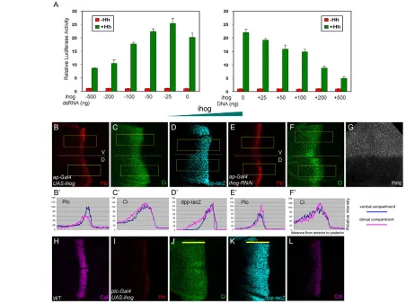
[image:6.612.52.403.59.406.2]The above experiment implies that the function of Ihog might be distinct from that of Dlp. We analyzed the function of Ihog by luciferase reporter assay in cultured cells. As shown previously, increasing levels of Dlp enhance Hh signaling in a dose-dependent manner. However, we found that increasing levels of Ihog actually reduce Hh signaling (data not shown). This result is unexpected because Ihog has only been shown to positively regulate Hh signaling (Yao et al., 2006). We examined how Hh signaling changes with different Ihog levels by transfecting cells with varying amounts of ihog RNAi or DNA. ihog dsRNA or UAS-ihogDNA effectively reduced or increased, respectively, Ihog levels in cultured cells, as demonstrated by western blot (see Fig. S5 in the supplementary material). Interestingly, Hh signaling showed a biphasic response depending on the level of Ihog: increasing the amount of Ihog reduced Hh signaling, whereas decreasing Ihog by a small amount actually enhanced signaling, but further reductions in the Ihog level decreased Hh signaling again (Fig. 5A).
Fig. 4. Dlp may act as a co-receptor for Ptc on the cell surface. (A,A⬘) Hh staining in ap-Gal4, UAS-dlpdiscs shows that Hh spreads over shorter distances in Dlp-overexpression dorsal tissue. (B-E⬙) The GPI anchor of Dlp affects its cellular trafficking. Dlp-GFP, Dlp(–HS)-GFP, Dlp(–HS)-CD2-GFP or GFP-Dlp-CD2 was expressed in Hh-receiving cells by
ptc-Gal4and analyzed for their distribution. The GPI-anchored forms, Dlp-GFP and Dlp(–HS)-GFP, colocalize extensively with Ptc punctate staining (arrows), whereas the CD2 forms, Dlp(–HS)-CD2-GFP and GFP-Dlp-CD2, form far fewer vesicles and show almost no colocalization with Ptc vesicles. (F)Dlp co-immunoprecipitates with Ptc-GFP. (Top and middle) DrosophilaS2 cells were transfected with the indicated expression vectors, and cell lysates were
immunoprecipitated and analyzed by western blotting with the antibodies indicated. (Bottom) The amount of protein in cell lysates as assessed by western blot.
D
E
V
E
LO
P
M
E
N
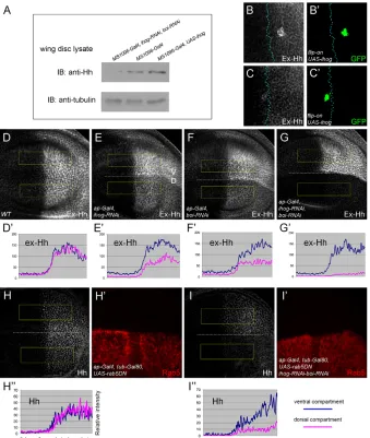
We further examined whether Ihog has similar biphasic activity in wing discs. Overexpression of Ihog in the D compartment by ap-Gal4caused reduced levels of Ptc expression (Fig. 5B,B⬘), but increased ranges of Ptc, Ci and dpp-lacZexpression (Fig. 5B-D⬘). Similarly, expression of Ihog in Hh-receiving cells by ptc-Gal4 strongly repressed Ptc and Col expression levels (high-threshold targets) (Fig. 5I,L), but extended the range of Ci and dpp-lacZ (low-threshold targets) (Fig. 5J,K). By contrast, the reduction of Ihog levels by RNAi generated the opposite effect (Fig. 5G): induction of ihog RNAi in the D compartment enhanced Ptc and Ci expression levels, but reduced their ranges (Fig. 5E-F⬘). Furthermore, the Ptc level was slightly reduced in ihog homozygous mutant clones (Fig. 6J-J⬙) (see below for ihogallele), consistent with a previous report (Yao et al., 2006). Thus, a high level of Ihog leads to reduced Hh signaling in the disc. Reducing Ihog levels by RNAi enhances Hh signaling, but complete absence of Ihog in the mutant clones reduces signaling again in the disc. These data suggest that Ihog has biphasic activity in Hh signaling both in vivo and in vitro.
[image:7.612.53.505.62.400.2]Ihog and Boi function redundantly in Hh signaling Hh signaling is only slightly reduced in ihogmutant clones in wing discs and in embryos (Yao et al., 2006), which is likely to be due to functional redundancy with another Ig/fibronectin superfamily member, Boi. We examined whether these two proteins indeed play redundant roles. Depletion of Ihog or Boi alone by RNAi in Hh-receiving cells did not reduce Ptc, Col or Ci expression; however, induction of both ihogand boiRNAi led to a very strong reduction in the expression of these Hh target genes (Fig. 6A-I). We also generated a mutant allele of ihogby imprecise excision of a P-element insertion (KG05348). This mutation deletes all of the ihog sequence starting 214 bp downstream of the ATG start codon to the 3⬘ end of the gene and is therefore likely to be a null allele. Because the original P-element is also inserted into the 5⬘UTR of the nearby gene CG10158, our mutant allele is very likely to affect the function of CG10158. However, as discussed by Yao et al. (Yao et al., 2006), CG10158 is unlikely to be required for the Hh signal response. We found that Ptc expression is only mildly reduced in ihogmutant clones (Fig. 6J-J⬙), but is significantly reduced in ihog Fig. 5. Ihog exhibits biphasic activity in Hh signaling depending on its levels.(A)Drosophilacl-8 cells were transfected with the indicated amounts of ihog-RNAior ihogexpression vector DNA. The Hh signaling luciferase reporter shows a biphasic response that depends on Ihog levels: too little or too much Ihog reduces Hh signaling. The error bars represent s.d. (B-G)Ectopic expression of Ihog in the D pouch by ap-gal4causes a reduction in Ptc expression strength (B), but an expansion of Ptc (B), Ci (C) and dpp-lacZ (D) ranges to more anterior cells. Induction of ihog-RNAiin the D compartment enhances Ptc (E) and Ci (F) expression levels, but reduces their expression ranges. The fluorescence intensities from selected areas (boxed) in these images are quantified in B⬘-F⬘. The ihog-RNAiline can effectively knockdown Ihog levels as assessed by Ihog antibody staining (G). (H-L)Ectopic expression of Ihog in Hh-receiving cells by ptc-Gal4strongly diminishes Ptc (I) and Col (L) expression, while expanding the Ci (J) and dpp-lacZ(K) expression range (yellow bars).
D
E
V
E
LO
P
M
E
N
clones that co-expressboiRNAi (Fig. 6K-K⬙). Together, our results argue that both Ihog and Boi are functionally redundant in Hh signaling.
Ihog is required to retain Hh on the cell surface How do we explain the biphasic activity of Ihog? As Ihog interacts with Hh with high affinity in vitro (McLellan et al., 2006; Yao et al., 2006), we examined its effect on Hh distribution in vivo. Knockdown of Ihog or Boi by RNAi in the D compartment clearly reduced Hh levels, whereas removal of both caused an even more dramatic reduction of Hh in its expression cells (Fig. 7A-D⬘). This reduction of Hh occurred in both apical and basolateral regions (Fig. 7E). Similarly, expression of ihog, boiRNAi by ptc-Gal4 strongly reduced Hh levels in signal-receiving cells (compare Fig. 7F with 7G). By contrast, when Ihog was overexpressed in clones, either in Hh expression or signal-receiving cells, Hh levels were
strikingly enhanced in those clones (Fig. 7H-I⬘). The elevated Hh was apparent in both apical and basolateral regions, indicating that Ihog does not simply relocalize Hh (Fig. 7J).
We further addressed whether overexpression of Ihog indeed increases Hh levels by western blot. As shown in Fig. 8A, ubiquitous expression of UAS-ihogby MS1096-Gal4in the wing disc increased the Hh level in the disc lysate, whereas expression of ihog, boi RNAi decreased the Hh level. Next, we examined whether Ihog causes Hh to accumulate on the cell surface by extracellular staining of Hh. Ihog expression clones accumulated Hh on the cell surface, both in Hh expression and signal-receiving tissue (Fig. 8B-C⬘). In addition, the extracellular Hh level was reduced in ihogor boiRNAi discs, and was almost completely absent in double-RNAi discs (Fig. 8D-G⬘). The reduction of extracellular Hh in ihog, boiRNAi discs (Fig. 8G,G⬘) was even stronger than that of total Hh staining (Fig. 7D-D⬘), suggesting that the reduction in total Hh levels is a consequence of changes in extracellular Hh. Together, these experiments suggest that the function of Ihog is to retain Hh on the cell surface.
One mechanism for Ihog to stabilize Hh on the cell surface is to prevent Hh internalization and degradation. We tested this possibility by blocking endocytosis in wing disc tissue that lacks Ihog and Boi. We usedap-Gal4, tub-Gal80tsto express dominant-negative Rab5 (Rab5DN) for a short space of time so as to block endocytosis. On lateral surfaces, expression of Rab5DN alone did not affect Hh distribution (Fig. 8H-H⬙); however, the Hh level still decreased in ihog, boiRNAi discs even when endocytosis was blocked (Fig. 8I-I⬙). On the basal surface, the Hh level was elevated in Rab5DN expression domains, with an especially large amount of Hh accumulating in Hh-receiving cells (see Fig. S6A,A⬘in the supplementary material, arrow) (Eugster et al., 2007). In ihog, boi RNAi discs, Hh on the basal surface of Hh expression cells was reduced compared with control tissue (see Fig. S6B,B⬘ in the supplementary material). In Hh-receiving cells, the Hh accumulation was also reduced (compare Fig. S6B with S6A in the supplementary material, arrows). These results suggest that Ihog does not simply stabilize Hh on the cell surface by preventing endocytosis. We favor the model that Ihog is required to trap Hh on the disc tissue, restricting its diffusion to two, instead of three, dimensions.
Our results suggest that the Hh signaling defect in ihog, boi double-mutant discs is likely to be due to the absence of Hh on the cell surface. However, a high level of Ihog causes a large amount of Hh to accumulate on the cell surface, but also reduces Hh signaling strength. A possible explanation is that although Ihog binds Hh on the cell surface, rather than acting as a co-receptor, it actually competes with Ptc for Hh binding.
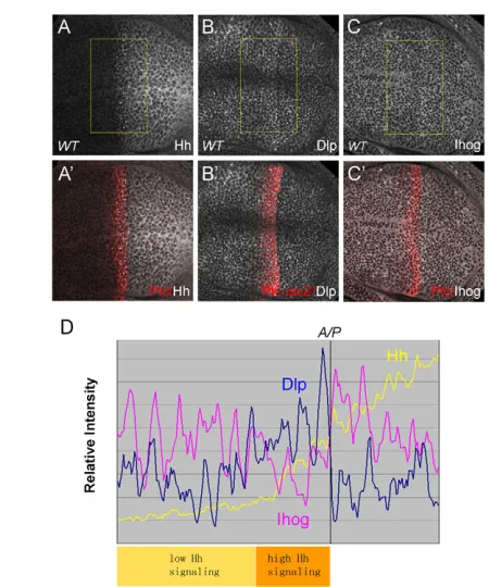
Dlp and Ihog show distinct expression patterns in wing discs
[image:8.612.52.276.58.435.2]We have shown that Dlp and Ihog play distinct functions in Hh signaling. Finally, we examined their expression patterns in the wing disc. Interestingly, we found that Dlp is upregulated in the Ptc expression domain (Gallet et al., 2008), whereas Ihog is downregulated in that domain (Fig. 9). The reduced level of Ihog is not due to changes in subcellular localization, as the Ihog protein level was low in high Hh signaling domains in all apical, basal and lateral sections (see Fig. S7 in the supplementary material). In addition, this reduction of Ihog is not due to enhanced internalization and degradation in response to Hh binding because Ihog, unlike Ptc, does not accumulate in endocytosis-defective shibiremutant clones (see Fig. S8 in the supplementary material). Fig. 6. Ihog and Boi function redundantly in Hh signaling.
(A-F)Expression of ihog-RNAior boi-RNAiby ptc-Gal4does not reduce Ptc or Col expression, but knockdown of both proteins results in a strong reduction of Ptc and Col expression. (G-I)Induction of ihog-RNAi
or boi-RNAiby ci-Gal4does not reduce Ci stabilization, but knockdown of both leads to a strong reduction of Ci stabilization. (J-K⬙) Ptc expression is slightly reduced in ihogmutant clones (J-J⬙, arrow), but is significantly reduced in ihogclones that also express boi-RNAi(K-K⬙, arrow). Clones are marked by the presence of GFP.
D
E
V
E
LO
P
M
E
N
The complementary expression patterns of Dlp and Ihog might highlight their opposing roles: the elevated level of Dlp may be needed to provide the strong Hh signaling required to activate high-threshold target genes such as ptc, whereas the negative regulation of Ihog may ensure that a strong Hh signal is reached in this area.
DISCUSSION
The core protein is essential for Dlp function in Hh signaling
Previous studies have shown that Dlp is specifically required for Hh signaling in cell-based assays and in embryos (Desbordes and Sanson, 2003; Han et al., 2004b; Lum et al., 2003). However, the molecular basis of this specificity was unknown. Here, we show that overexpression of Hh can restore naked cuticle in sgl and sfl embryos, but not in dlpembryos. We further demonstrated that the specificity of Dlp in Hh signaling results from its core protein. The Dlp core protein can restore Hh signaling autonomously in dlp embryos and in dlp-RNAicells. In addition, the Dlp core protein interacts with Hh and promotes Hh signaling in the disc. Overexpression of Dlp increases Hh signaling strength, but reduces signaling range as well as Hh gradient range. These data suggest that the Dlp core protein could act as a classic co-receptor in Hh signaling by facilitating Hh-Ptc interaction.
Recent studies have shown that the vertebrate glypican-3 core protein directly promotes Wnt signaling in cancer cells, but inhibits sonic hedghog signaling during development (Capurro et al., 2005; Capurro et al., 2008). That the same glypican has opposite effects on Wnt and Hh is interesting because Dlp can inhibit
high-threshold Wg signaling when overexpressed in discs (Yan et al., 2009). In addition to the essential role of the core protein, the attached GAG chains are important for the non-cell-autonomous functions of Dlp. Our results demonstrated that wild-type Dlp can rescue non-cell-autonomously in dlpembryos, whereas the core protein mainly acts in its expression domains. Interestingly, the CD2 forms of Dlp also lose the non-autonomous activity in dlp embryos. Several studies suggest that the GPI anchor of Dlp can be cleaved by the hydrolase Notum and that the GAG chains of Dlp can recruit lipoprotein particles (Eugster et al., 2007; Kreuger et al., 2004). Thus, it will be interesting to determine the mechanism of Dlp non-autonomous activity.
[image:9.612.55.506.61.331.2]Our study suggests that the GPI anchor of Dlp is not essential for its activity in Hh signaling. Most importantly, two CD2 forms of Dlp, Dlp(–HS)-CD2 and GFP-Dlp-CD2, can effectively rescue Hh signaling in dlpembryos. In addition, CD2 forms of Dlp can also signal in cultured cells and discs. It is important to note that although the GPI, but not the CD2, form of Dlp is colocalized with Ptc in intracellular vesicles, both forms have similar activities in promoting Hh signaling in the disc. These data argue that colocalization of Dlp with Ptc in endocytic vesicles is not essential for Dlp function in Hh signaling. Consistent with this view, several studies have shown that endocytosis is not essential for Hh signaling in Drosophilawing discs (Callejo et al., 2008; Han et al., 2004b; Torroja et al., 2004). Our conclusion differs from a recent publication arguing that the GPI anchor of Dlp is required for its function in Hh signaling (Gallet et al., 2008). In their study, it was shown that overexpression of GFP-Dlp-CD2 can reduce Hh Fig. 7. Role of Ihog and Boi in Hh distribution.(A-D⬘) Knockdown of Ihog or Boi in the D compartment by RNAi clearly reduces Hh levels in Hh expression cells. Knockdown of both proteins causes an even more dramatic Hh reduction. (E)z-section of discs expressing ihog, boi RNAi by ap-Gal4. The Hh level is uniformly reduced at all surfaces. E-cadherin marks the sub-apical junctions. The section is from the Hh expression domain. (F-G⬙) Hh staining in a wild-type disc shows punctate and diffusive staining in Hh-receiving cells. Knockdown of Ihog and Boi leads to much reduced Hh levels in receiving cells. The A/P boundary is marked by dotted lines. (H-I⬘) Ihog-expressing clones (marked by GFP) in either the Hh expression or receiving domain cause elevated Hh levels. (J)z-section of clones expressing Ihog in the Hh expression domain. Hh accumulates at all the surfaces.
D
E
V
E
LO
P
M
E
N
signaling in the wing discs. However, we did not observe any dominant-negative effect of GFP-Dlp-CD2, which can rescue dlp mutant embryos and enhance Hh signaling strength in cells and discs. A possible explanation for the discrepancy is that expression of Dlp enhances signaling strength but also reduces signaling range. We always use ap-Gal4, which allows us to use the ventral disc as an internal control. The observed dominant-negative effect might reflect the reduced signaling range rather than signaling strength.
Ihog exhibits biphasic activity in Hh morphogen signaling
Previous studies in both Drosophila and vertebrates have demonstrated positive roles of the Ihog family proteins in Hh signaling (Tenzen et al., 2006; Yao et al., 2006; Zhang et al., 2006). Ihog and Ptc synergize in mediating Hh binding to cells. These observations suggest that Ihog functions as a co-receptor for Hh (Yao et al., 2006). However, our new data argue that Ihog does not simply act as a classic co-receptor that only increases the binding of ligand to the signaling receptor. By altering Ihog levels in vivo and in vitro, we show that Ihog has biphasic activity in Hh signaling, with too much or too little Ihog leading to reduced signaling. Overexpression of Ihog leads to the accumulation of Hh,
[image:10.612.53.392.334.735.2]and knockdown of Ihog results in reduced Hh levels, suggesting that one major activity of Ihog is to retain Hh on the cell surface. Moreover, knockdown of both Ihog and Boi dramatically reduces Hh levels and signaling. This reduction of Hh signaling activity is likely to be due to an absence of Hh on the cell surface of the double-mutant tissue. However, a high level of Ihog causes a large amount of Hh to accumulate on the cell surface, but also reduces signaling strength. One explanation for this result is that Ihog can compete with Ptc for Hh binding. Thus, a low level of Ihog is required to maintain Hh on the cell surface, whereas a high level of Ihog can sequester Hh from its receptor. In other words, depending on the context, Ihog can either provide Hh for the receptor by retaining Hh on the cell surface, or compete with the receptor for Hh binding. This activity of Ihog is very similar to the recently proposed ‘exchange factor’ model, which allows the exchange of Hh between Ptc and Ihog (Serpe et al., 2008; Yan et al., 2009). A recent study has demonstrated that Ihog also interacts with Ptc and that Ihog, Ptc and Hh form a triple complex (Zheng et al., 2010). The close association between the Ihog and Ptc receptors may thus allow them to exchange Hh ligand. It will be important to determine whether the triple complex has a greatly reduced ability to signal, a prediction from the mathematical exchange factor model (Umulis et al., 2009).
Fig. 8. Ihog and Boi are required to retain Hh on the cell surface. (A)Drosophilawing disc lysates from
MS1096-Gal4, MS1096-Gal4/UAS-ihog
andMS1096-Gal4, ihog-RNAi, boi-RNAi
larvae were probed with Hh and
anti--Tubulin. The Hh level is reduced in ihog, boi RNAidiscs, but increased in Ihog-expressing discs. -Tubulin provides a loading control. (B-C⬘) Extracellular Hh staining shows that Ihog-expressing clones (marked by GFP) accumulate Hh on the cell surface in both the Hh expression and receiving domains. The A/P boundary is marked by dotted lines. (D-G⬘) Expression of ihog-RNAior boi-RNAiby ap-Gal4
causes a reduction in extracellular Hh in the D compartment. Double RNAi causes a striking reduction in the extracellular Hh level. (H-I⬙) Induction of Rab5DN in the dorsal part of the wing disc by ap-Gal4, tub-Gal80tsfor 10 hours does not affect Hh distribution on the lateral surface (H-H⬙). Induction of Rab5DNand ihog, boi
RNAi together still leads to a reduction in Hh levels (I-I⬙). Rab5DN is detected by Rab5 antibody.
D
E
V
E
LO
P
M
E
N
Although to our knowledge this is the first demonstration of a biphasic co-factor in Hh signaling, similar biphasic co-factors have been reported. DrosophilaCv-2 enhances BMP signaling at low concentrations, but inhibits signaling at high concentrations (Serpe et al., 2008). Syndecan-1 shows a similar concentration-dependent activation or inhibition of BMP signaling in Xenopus(Olivares et al., 2009). Interestingly, Dlp has biphasic activity in Wg signaling, depending on its protein levels (Yan et al., 2009). All these co-factors are likely to act by a similar mechanism, suggesting that the biphasic co-factor is a recurring motif in different morphogen systems.
We have shown that Dlp and Ihog play distinct roles in Hh signaling. Expression of Dlp enhances Hh signaling strength, but reduces signaling range. By contrast, expression of Ihog reduces Hh signaling strength, but extends signaling range. In addition, the Dlp level is elevated in the high Hh signaling area, whereas the Ihog level is reduced in that region. It is important to consider from a system point of view what these two co-factors provide for the Hh morphogen. For morphogens to work, they should be able to generate sharp boundaries between target genes with different thresholds. They also need to diffuse over a certain range without being lost in the extracellular space. The positive feedback of Dlp expression in the high Hh signaling areas helps to sharpen the boundaries between high- and low-threshold target genes (Lander, 2007). The negative-feedback regulation of Ihog might ensure that a strong Hh signal is attained in the areas close to the Hh source and also allow the Hh gradient to diffuse to those areas distant from the source.
Acknowledgements
We thank G. Baeg, P. Beachy, S. Cohen, S. Eaton, M. Frasch, E. Furlong, M. Gonzalez-Gaitan, I. Guerrero, R. Holmgren, P. Ingham, T. Kornberg, P. Therond, J. Treisman, A. Vincent and J. Vincent for reagents; P. Beachy for sharing results before publication; and L. Ray for comments on the manuscript. This work was supported by grants from NIH (2R01 GM063891), American Cancer Society (RSG-07-051), and Chinese Academy of Sciences (KSCX2-YW-R-263) to X.L. D.Y. was an Albert J. Ryan fellow and was supported by a predoctoral fellowship from American Heart Association. Y.W. is supported by a grant from Chinese Academy of Sciences (KSCX2-YW-R-240). Deposited in PMC for release after 12 months.
Competing interests statement
The authors declare no competing financial interests.
Supplementary material
Supplementary material for this article is available at
http://dev.biologists.org/lookup/suppl/doi:10.1242/dev.045740/-/DC1
References
Aza-Blanc, P., Ramirez-Weber, F. A., Laget, M. P., Schwartz, C. and Kornberg, T. B.(1997). Proteolysis that is inhibited by hedgehog targets Cubitus interruptus protein to the nucleus and converts it to a repressor. Cell89, 1043-1053.
Baeg, G. H., Lin, X., Khare, N., Baumgartner, S. and Perrimon, N.(2001). Heparan sulfate proteoglycans are critical for the organization of the extracellular distribution of Wingless. Development128, 87-94.
Baeg, G. H., Selva, E. M., Goodman, R. M., Dasgupta, R. and Perrimon, N.
(2004). The Wingless morphogen gradient is established by the cooperative action of Frizzled and Heparan Sulfate Proteoglycan receptors. Dev. Biol. 276, 89-100.
Callejo, A., Culi, J. and Guerrero, I.(2008). Patched, the receptor of Hedgehog, is a lipoprotein receptor. Proc. Natl. Acad. Sci. USA105, 912-917.
Capurro, M. I., Xiang, Y. Y., Lobe, C. and Filmus, J.(2005). Glypican-3 promotes the growth of hepatocellular carcinoma by stimulating canonical Wnt signaling. Cancer Res.65, 6245-6254.
Capurro, M. I., Xu, P., Shi, W., Li, F., Jia, A. and Filmus, J.(2008). Glypican-3 inhibits Hedgehog signaling during development by competing with patched for Hedgehog binding. Dev. Cell14, 700-711.
Chen, Y. and Struhl, G.(1996). Dual roles for patched in sequestering and transducing Hedgehog. Cell87, 553-563.
Chou, T. B. and Perrimon, N.(1996). The autosomal FLP-DFS technique for generating germline mosaics in Drosophila melanogaster. Genetics144, 1673-1679.
Croker, J. A., Ziegenhorn, S. L. and Holmgren, R. A.(2006). Regulation of the Drosophila transcription factor, Cubitus interruptus, by two conserved domains.
Dev. Biol. 291, 368-381.
David, G., Bai, X. M., Van der Schueren, B., Cassiman, J. J. and Van den Berghe, H.(1992). Developmental changes in heparan sulfate expression: in situ detection with mAbs.J. Cell Biol. 119, 961-975.
Desbordes, S. C. and Sanson, B.(2003). The glypican Dally-like is required for Hedgehog signalling in the embryonic epidermis of Drosophila. Development 130, 6245-6255.
Entchev, E. V., Schwabedissen, A. and Gonzalez-Gaitan, M.(2000). Gradient formation of the TGF-beta homolog Dpp. Cell103, 981-991.
Eugster, C., Panakova, D., Mahmoud, A. and Eaton, S.(2007). Lipoprotein-heparan sulfate interactions in the Hh pathway. Dev. Cell13, 57-71.
Franch-Marro, X., Marchand, O., Piddini, E., Ricardo, S., Alexandre, C. and Vincent, J. P.(2005). Glypicans shunt the Wingless signal between local signalling and further transport. Development132, 659-666.
Gallet, A., Staccini-Lavenant, L. and Therond, P. P.(2008). Cellular trafficking of the glypican Dally-like is required for full-strength Hedgehog signaling and wingless transcytosis. Dev. Cell14, 712-725.
Guerrero, I. and Chiang, C.(2007). A conserved mechanism of Hedgehog gradient formation by lipid modifications. Trends Cell Biol. 17, 1-5.
Hacker, U., Lin, X. and Perrimon, N.(1997). The Drosophila sugarless gene modulates Wingless signaling and encodes an enzyme involved in polysaccharide biosynthesis. Development124, 3565-3573.
Han, C., Belenkaya, T. Y., Khodoun, M., Tauchi, M., Lin, X. and Lin, X.
(2004a). Distinct and collaborative roles of Drosophila EXT family proteins in morphogen signalling and gradient formation. Development131, 1563-1575.
Han, C., Belenkaya, T. Y., Wang, B. and Lin, X.(2004b). Drosophila glypicans control the cell-to-cell movement of Hedgehog by a dynamin-independent process. Development131, 601-611.
Hatini, V. and DiNardo, S.(2001). Divide and conquer: pattern formation in Drosophila embryonic epidermis. Trends Genet.17, 574-579.
[image:11.612.52.277.56.326.2]Hooper, J. E. and Scott, M. P.(2005). Communicating with Hedgehogs.Nat. Rev. Mol. Cell Biol. 6, 306-317.
Fig. 9. The expression patterns of Dlp and Ihog in the Drosophila
wing disc.(A-C⬘) Antibody staining of Hh/Ptc (A,A⬘), Dlp/Ptc--Gal (B,B⬘) and Ihog/Ptc (C,C⬘) in wild-type wing discs. The Dlp protein level is elevated in the Ptc expression domain, whereas the Ihog level is reduced in this domain. (D)The intensity of Hh (A), Dlp (B) and Ihog (C) staining from selected areas (boxed) are plotted along the A/P axis. In the high Hh signaling area, the Dlp level is upregulated and the Ihog level is downregulated, suggesting distinct functions in Hh signaling.
D
E
V
E
LO
P
M
E
N
Janody, F. and Treisman, J. E.(2006). Actin capping protein alpha maintains vestigial-expressing cells within the Drosophila wing disc epithelium.
Development133, 3349-3357.
Jiang, J. and Hui, C. C.(2008). Hedgehog signaling in development and cancer.
Dev. Cell15, 801-812.
Kang, J. S., Zhang, W. and Krauss, R. S.(2007). Hedgehog signaling: cooking with Gas1. Sci. STKE2007, pe50.
Kreuger, J., Perez, L., Giraldez, A. J. and Cohen, S. M.(2004). Opposing activities of Dally-like glypican at high and low levels of Wingless morphogen activity. Dev. Cell7, 503-512.
Lander, A. D.(2007). Morpheus unbound: reimagining the morphogen gradient.
Cell128, 245-256.
Lee, H. H. and Frasch, M.(2000). Wingless effects mesoderm patterning and ectoderm segmentation events via induction of its downstream target sloppy paired. Development127, 5497-5508.
Lin, X.(2004). Functions of heparan sulfate proteoglycans in cell signaling during development. Development131, 6009-6021.
Lin, X. and Perrimon, N.(1999). Dally cooperates with Drosophila Frizzled 2 to transduce Wingless signalling. Nature400, 281-284.
Lu, X., Liu, S. and Kornberg, T. B.(2006). The C-terminal tail of the Hedgehog receptor Patched regulates both localization and turnover. Genes Dev.20, 2539-2551.
Lum, L., Yao, S., Mozer, B., Rovescalli, A., Von Kessler, D., Nirenberg, M. and Beachy, P. A.(2003). Identification of Hedgehog pathway components by RNAi in Drosophila cultured cells. Science299, 2039-2045.
Mann, R. K. and Beachy, P. A.(2004). Novel lipid modifications of secreted protein signals. Annu. Rev. Biochem.73, 891-923.
McLellan, J. S., Yao, S., Zheng, X., Geisbrecht, B. V., Ghirlando, R., Beachy, P. A. and Leahy, D. J.(2006). Structure of a heparin-dependent complex of Hedgehog and Ihog. Proc. Natl. Acad. Sci. USA103, 17208-17213.
Motzny, C. K. and Holmgren, R.(1995). The Drosophila cubitus interruptus protein and its role in the wingless and hedgehog signal transduction pathways.
Mech. Dev.52, 137-150.
Nose, A., Mahajan, V. B. and Goodman, C. S.(1992). Connectin: a homophilic cell adhesion molecule expressed on a subset of muscles and the motoneurons that innervate them in Drosophila. Cell70, 553-567.
Olivares, G. H., Carrasco, H., Aroca, F., Carvallo, L., Segovia, F. and Larrain, J.
(2009). Syndecan-1 regulates BMP signaling and dorso-ventral patterning of the ectoderm during early Xenopus development. Dev. Biol. 329, 338-349.
Serpe, M., Umulis, D., Ralston, A., Chen, J., Olson, D. J., Avanesov, A., Othmer, H., O’Connor, M. B. and Blair, S. S.(2008). The BMP-binding protein
Crossveinless 2 is a short-range, concentration-dependent, biphasic modulator of BMP signaling in Drosophila. Dev. Cell14, 940-953.
Strigini, M. and Cohen, S. M.(1997). A Hedgehog activity gradient contributes to AP axial patterning of the Drosophila wing. Development124, 4697-4705.
Tabata, T. and Takei, Y.(2004). Morphogens, their identification and regulation.
Development131, 703-712.
Taylor, A. M., Nakano, Y., Mohler, J. and Ingham, P. W.(1993). Contrasting distributions of patched and hedgehog proteins in the Drosophila embryo.
Mech. Dev.42, 89-96.
Tenzen, T., Allen, B. L., Cole, F., Kang, J. S., Krauss, R. S. and McMahon, A. P.
(2006). The cell surface membrane proteins Cdo and Boc are components and targets of the Hedgehog signaling pathway and feedback network in mice. Dev. Cell10, 647-656.
Torroja, C., Gorfinkiel, N. and Guerrero, I.(2004). Patched controls the Hedgehog gradient by endocytosis in a dynamin-dependent manner, but this internalization does not play a major role in signal transduction. Development 131, 2395-2408.
Umulis, D., O’Connor, M. B. and Blair, S. S.(2009). The extracellular regulation of bone morphogenetic protein signaling. Development136, 3715-3728.
Vervoort, M., Crozatier, M., Valle, D. and Vincent, A.(1999). The COE transcription factor Collier is a mediator of short-range Hedgehog-induced patterning of the Drosophila wing.Curr. Biol. 9, 632-639.
Wang, Y., McMahon, A. P. and Allen, B. L.(2007). Shifting paradigms in Hedgehog signaling.Curr. Opin. Cell Biol. 19, 159-165.
Wilson, C. W. and Chuang, P. T.(2006). New ‘hogs’ in Hedgehog transport and signal reception. Cell125, 435-438.
Yan, D., Wu, Y., Feng, Y., Lin, S. C. and Lin, X.(2009). The core protein of glypican Dally-like determines its biphasic activity in wingless morphogen signaling. Dev. Cell17, 470-481.
Yao, S., Lum, L. and Beachy, P.(2006). The ihog cell-surface proteins bind Hedgehog and mediate pathway activation. Cell125, 343-357.
Zhang, W., Kang, J. S., Cole, F., Yi, M. J. and Krauss, R. S.(2006). Cdo functions at multiple points in the Sonic Hedgehog pathway, and Cdo-deficient mice accurately model human holoprosencephaly. Dev. Cell10, 657-665.
Zheng, X., Mann, R. K., Sever, N. and Beachy, P. A.(2010). Genetic and biochemical definition of the Hedgehog receptor. Genes Dev.24, 57-71.