R E S E A R C H A R T I C L E
Open Access
Distribution of ossified spinal lesions in
patients with severe ossification of the
posterior longitudinal ligament and
prediction of ossification at each segment
based on the cervical OP index classification:
a multicenter study (JOSL CT study)
Takashi Hirai
1,23*, Toshitaka Yoshii
1,23, Narihito Nagoshi
2, Kazuhiro Takeuchi
3,23, Kanji Mori
4,23, Shuta Ushio
1,23,
Akio Iwanami
2, Tsuyoshi Yamada
1,23, Shoji Seki
5,23, Takashi Tsuji
6,23, Kanehiro Fujiyoshi
7,23, Mitsuru Furukawa
8,23,
Soraya Nishimura
2,9,23, Kanichiro Wada
10,23, Takeo Furuya
11,23, Yukihiro Matsuyama
12,23, Tomohiko Hasegawa
12,23,
Katsushi Takeshita
13,23, Atsushi Kimura
13,23, Masahiko Abematsu
14,23, Hirotaka Haro
15,23, Tetsuro Ohba
15,23,
Masahiko Watanabe
16,23, Hiroyuki Katoh
16,23, Kei Watanabe
17,23, Hiroshi Ozawa
18,23, Haruo Kanno
19,23,
Shiro Imagama
20,23, Kei Ando
20,23, Shunsuke Fujibayashi
21,23, Masao Koda
22,23, Masashi Yamazaki
22,23,
Morio Matsumoto
2, Masaya Nakamura
2, Atsushi Okawa
1,23and Yoshiharu Kawaguchi
5,23Abstract
Background:In patients with ossification of the posterior longitudinal ligament (OPLL) in the cervical spine, it is well known that the thoracic ossified lesions often coexist with the cervical lesions and can cause severe myelopathy. However, the prevalence of OPLL at each level of the thoracic and lumbar spinal segments is unknown. The aims of this study were to investigate how often OPLL occurs at each level in the thoracolumbar spine in patients with a radiological diagnosis of cervical OPLL and to identify the spinal levels most likely to develop ossification.
Methods:Data were collected from 20 institutions in Japan. Three hundred and twenty-two patients with a diagnosis of cervical OPLL were included. The OPLL index (OP index), defined as the sum of the vertebral body and intervertebral disc levels where OPLL is present, was used to determine disease severity. An OP index≥20 was defined as severe OPLL. The prevalence of OPLL at each level of the thoracic and lumbar spinal segments was calculated.
(Continued on next page)
* Correspondence:[email protected]
1
Department of Orthopedic Surgery, Tokyo Medical and Dental University, 1-5-45 Yushima, Bunkyo-ku, Tokyo 113-8519, Japan
23Japanese Organization of the Study for Ossification of Spinal Ligament (JOSL), 1-5-45 Yushima, Bunkyo-ku, Tokyo 113-8519, Japan
Full list of author information is available at the end of the article
(Continued from previous page)
Results:Women were more likely to have ossified lesions in the thoracolumbar spine than men. Severe OPLL was significantly more common in women than in men (20% vs. 4.5%). For thoracic vertebral OPLL, the most frequently affected was the T1 segment in both men and women, followed by the T1/2 and T3/4 intervertebral levels in men and women, respectively. Ossified lesions were frequently seen at the intervertebral and vertebral levels around the cervicothoracic and thoracolumbar junctions in men with severe OPLL, whereas OPLL was more diffusely distributed in the thoracic spine in women with severe OPLL.
Conclusion:Thoracolumbar OPLL occurred most often at T1 in men and at T3/4 in women. In severe OPLL cases, although ossified lesions were frequently seen at the intervertebral and vertebral levels around the cervicothoracic and thoracolumbar junctions in men, OPLL could be observed more diffusely in the thoracic spine in women.
Keywords:OPLL, Computed tomography, Whole spine, Ossification predisposition, Prevalence,
Background
Ossification of the spinal ligaments is a form of het-erotopic ossification that occurs throughout the spine. Ossification of the posterior longitudinal ligament (OPLL) involves the spinal canal and often leads to myelopathy [1, 2]. Cervical OPLL, which can be de-tected easily on plain radiographs, has been investi-gated in detail as far back as the nineteenth century [3]. Ossified lesions in the thoracic spine sometimes coexist with cervical OPLL and cause severe symp-toms. However, it is difficult to diagnose pathologic conditions of the thoracic spine on radiographs alone, so the prevalence of thoracic OPLL remains unclear [4]. Recently, multidetector computed tomography (CT) has been found to be the most suitable modality for identifying ossification in the thoracic spine, re-gardless of any superimposed thoracic complexities. Nevertheless, there is no detailed multicenter research on the prevalence of thoracolumbar OPLL. The pur-pose of this study was to investigate the prevalence of
ossified lesions in the thoracolumbar spine in patients with cervical OPLL at multiple institutions. We also investigated how frequently OPLL occurred at each spinal level and attempted to clarify which levels of the spine are more predisposed to OPLL lesions.
Methods
Patients and methods
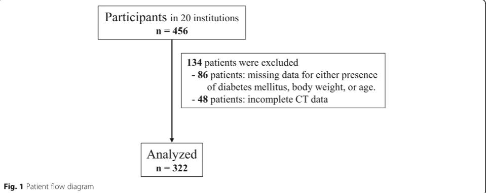
The study included patients with a radiographic diagno-sis of OPLL involving the cervical spine as well as symp-toms, such as neck pain, numbness in the upper and/or lower extremities, and clumsiness or gait disturbance. Each patient underwent whole-spine CT imaging at any one of the 20 institutions where members of the re-search team worked. Patients who had undergone anter-ior decompression surgery for OPLL and patients younger than 15 years of age were excluded. Four hun-dred and fifty-six patients with OPLL were identified, of whom only 322 had full demographic and anthropomet-ric data available for analysis (Fig.1). The study involved
[image:2.595.59.540.538.729.2]242 men and 80 women with a mean age of 64.6 (range 30–93) years. The study was approved by the institu-tional review board at each institution. Informed consent was obtained from all patients before enrollment in the study.
Evaluations
[image:3.595.56.546.99.261.2]Basic clinical data were collected for all patients, in-cluding for age, sex, presence of diabetes, and body mass index (BMI). CT images of the cervical, thoracic, and lumbosacral spine, from the occipital bone to the sacrum, were obtained in each patient. The prevalence of OPLL in the cervical spine from the clivus to C7 and in other spinal regions from T1 to S1 was evaluated on mid-sagittal CT images. The analysis was independently performed by five senior spine surgeons (TH, KT, KM, AI, TY). Prior to reviewing the images for this study, all of the readers interpreted the same images for 20 pa-tients to check interobserver agreement. The average kappa coefficient was 0.76 (0.71–0.81), which indicated good agreement and was consistent with the findings of a previous study [5]. Ossified lesions were recorded at each vertebral body and at the intervertebral disc level. An OPLL index (OP index) [5], defined as the sum of the vertebral body and intervertebral disc levels where OPLL is present, was calculated according to the method described in a previous report [5]. Patients with an OP index ≥20 were deemed to have severe OPLL. We also defined the sum of the levels at which OPLL was present in the cervical spine as the cervical OP index. Using a previously reported method [6], we divided patients into three groups according to their cervical OP index values, namely, grade 1 (≤5), grade 2 (6–9), and grade 3 (≥10). The physical and radiologic data were compared between the male and female pa-tient populations. Furthermore, we investigated whether there was an association between the cervical Table 1Demographics of male and female patients
Male + Female (n= 322)
Male (n= 242)
Female (n= 80)
P (M vs F)
Age (years old) 64.7 ± 11.2 64.7 ± 11.6 64.6 ± 10.0 0.90
Diabetes Mellitus (%) 31.7% 31.8% 31.3% 0.92
BMI 25.7 ± 4.8 25.8 ± 4.8 25.5 ± 4.7 0.62
JOSL CT classification
Grade 1 (1 < cervical OP-index≤5) 169 (52.5%) 125 (51.7%) 44 (55%) –
Grade 2 (6 < cervical OP-index≤9) 107 (33.2%) 83 (34.3%) 24 (30%) –
Grade 3 (10 < cervical OP-index) 46 (14.3%) 34 (14.0%) 12 (15%) –
Cervical OP-index 5.83 ± 2.9 5.86 ± 2.9 5.75 ± 3.0 0.78
OP-index 9.21 ± 6.8 8.24 ± 5.5 12.1 ± 9.0 < 0.01
No. of patients with OP-index of > = 20 (%) 27 (8.3%) 11 (4.5%) 16 (20%) < 0.01
Data are expressed as the mean ± standard deviation;BMIbody mass index,OP-indexossification index of OPLL
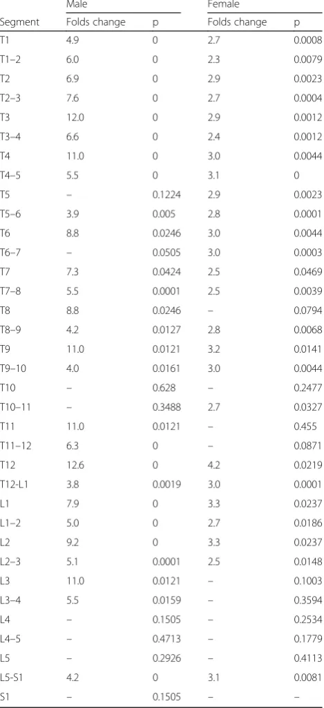
Table 2Incidence of ossified lesion in each level
low more significant different (0.01 <p< 0.05) moderate more significant different (0.001 <p< 0.01)
[image:3.595.303.537.300.706.2]OP index grade and the prevalence of thoracolumbar OPLL at each level.
Statistical analysis
Student’s unpairedttest was used to identify statistically significant differences in age, BMI, and the OP index for the cervical spine between men and women. Chi-squared test was used to test for a sex-related difference in the presence of diabetes. Tukey’s post hoc test was ap-plied to compare the three groups classified according to cervical OP grade. A forward stepwise logistic regression model was used to investigate whether the cervical OP grade could predict the presence of OPLL in the thora-columbar spine and the prevalence of patients with an OP index of≥20 [7]. All statistical analyses were performed using SPSS for Windows version 22.0 (IBM Corp., Armonk, NY). All data are expressed as the mean ± standard devi-ation. A p-value < 0.05 was considered to be statistically significant.
Results
Demographic data
Patient demographic characteristics are summarized in Table 1. Mean patient age was 64.7 years. Mean BMI was 25.7 and 31.7% (102/233 patients) of the patients
had diabetes mellitus. Using the CT classification, the cervical OP index was grade 1 in 169 patients (52.5%), grade 2 in 107 patients (33.2%), and grade 3 in 46 pa-tients (14.3%). The mean cervical and whole-spine OP index values were 5.83 and 9.21, respectively. Twenty-seven patients (8.3%) had an OP index ≥20. Severe OPLL was significantly more common in women than in men (16 patients, 20% vs. 11 patients, 4.5%, p< 0.01). There was no significant sex-related difference in age, prevalence of diabetes mellitus, or BMI. Interestingly, the OP index for the whole spine was significantly higher in women than in men (9.2 vs. 8.2, p< 0.01). However, there was no statistically significant sex-related difference in the cervical OP index.
Pattern of distribution of ossified lesions throughout the spine in patients with severe OPLL
The prevalence of OPLL at each level of the spine is shown in Table 2. Thoracic OPLL occurred most often at T1 (14.9%) and at T1/2 (13.6%) in men and at T1 (27.5%) and T3/4 (33.8%) in women. Lumbar OPLL was most common at L1 (5.8%) and T12/L1 (14.5%) in men and at L1 and L2 (both 11.3%) and T12/L1 (27.5%) in women.
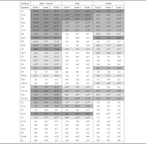
[image:4.595.58.537.398.703.2]We then compared the prevalence of OPLL at each level in all patients and that in patients with an OP index≥20. Overall, the patients with severe OPLL were more likely to have OPLL at the level of the upper cer-vical spine and at levels from the lower cercer-vical spine to the upper lumbar spine. In men with severe OPLL, ossi-fied lesions were frequently seen at the intervertebral and vertebral levels from C6/7 to T4/5 and from T11/12 to L3/4. However, severe OPLL appeared to be distrib-uted more diffusely in the thoracic spine in women. There were no statistically significant differences in the prevalence of thoracic OPLL at T5, T6/7, T10, and T10/ 11 in men or at T8, T10, T11, and T11/12 in women. Interestingly, the only significant difference in preva-lence of OPLL in the area from the lower lumbar spine to the sacrum was at L5/S1 in both sexes. We also inves-tigated the fold difference in prevalence of ossified le-sions at each spinal segment (Fig. 2, Table 3). The prevalence of severe OPLL in the thoracic spine was 3. 9–12.6 fold higher in men and 2.3–4.2 fold higher in women. Of note, in men with OPLL affecting the upper thoracic spine, there was a 12.0 fold increase in likeli-hood at T3 and a 7.6 fold increase at T2/3, and in those with lower thoracic OPLL, there was a 12.6 fold in-creased likelihood at T12 and a 6.3fold increase at T11/ 12. Interestingly, although thoracic OPLL had a bimodal distribution in men, the distribution was uniform in women. The distribution pattern in the lumbar spine was very similar between the sexes. However, the preva-lence of ossified lesions increased to a greater extent in men (by 4.25–11.0 fold) than in women (by 2.5–3.3 fold). In the lumbosacral region, there was a significantly increased prevalence of ossified lesions only at L5/S1 in both men (4.2 fold) and women (3.1 fold).
Prevalence of OPLL at each level according to cervical OP index grade
The cervical OP index classification was originally de-signed to categorize cervical OPLL into three grades of severity according to the sum of all ossifications in the cervical spine. The prevalence of OPLL was investigated at each vertebral and intervertebral segment according to grade to evaluate the usefulness of this classification for prediction of the prevalence of OPLL at each seg-ment in the thoracic and lumbar spines (Table4). Over-all, the prevalence of ossified lesions in the upper thoracic spine and at the thoracolumbar junction in-creased significantly with increasing grade of severity. The distribution of prevalence of OPLL was similar in men and women. A weak or no association between the prevalence of OPLL and the cervical OP index classifica-tion was found in the middle thoracic spine in men. However, statistically significant differences were ob-served at T4/5, T5/6, T6, T6/7, T8/9, and T9/10 in
women. A significant correlation was found at the thora-columbar junction, except at L2 in men and at T11, L1, L1/2, and L2 in women.
JOSL-CT grading system for prediction of prevalence of OPLL at each thoracolumbar spinal level
[image:5.595.305.540.114.627.2]We further investigated the hazard ratios for the preva-lence of OPLL at each spinal level where there was a Table 3Hazard ratio of the incidence of ossified lesion at each level in patients with OP-index≧20 compared to total patients
Male Female
Segment Folds change p Folds change p
T1 4.9 0 2.7 0.0008
T1–2 6.0 0 2.3 0.0079
T2 6.9 0 2.9 0.0023
T2–3 7.6 0 2.7 0.0004
T3 12.0 0 2.9 0.0012
T3–4 6.6 0 2.4 0.0012
T4 11.0 0 3.0 0.0044
T4–5 5.5 0 3.1 0
T5 – 0.1224 2.9 0.0023
T5–6 3.9 0.005 2.8 0.0001
T6 8.8 0.0246 3.0 0.0044
T6–7 – 0.0505 3.0 0.0003
T7 7.3 0.0424 2.5 0.0469
T7–8 5.5 0.0001 2.5 0.0039
T8 8.8 0.0246 – 0.0794
T8–9 4.2 0.0127 2.8 0.0068
T9 11.0 0.0121 3.2 0.0141
T9–10 4.0 0.0161 3.0 0.0044
T10 – 0.628 – 0.2477
T10–11 – 0.3488 2.7 0.0327
T11 11.0 0.0121 – 0.455
T11–12 6.3 0 – 0.0871
T12 12.6 0 4.2 0.0219
T12-L1 3.8 0.0019 3.0 0.0001
L1 7.9 0 3.3 0.0237
L1–2 5.0 0 2.7 0.0186
L2 9.2 0 3.3 0.0237
L2–3 5.1 0.0001 2.5 0.0148
L3 11.0 0.0121 – 0.1003
L3–4 5.5 0.0159 – 0.3594
L4 – 0.1505 – 0.2534
L4–5 – 0.4713 – 0.1779
L5 – 0.2926 – 0.4113
L5-S1 4.2 0 3.1 0.0081
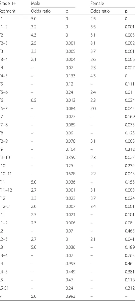
significant correlation between the prevalence of OPLL and the cervical OP index grade (Fig. 3, Table 5). The hazard ratio was 2.1–5.0 in the upper thoracic spine, 6.5 at T6 in the middle thoracic spine in men, and 2.0–4.3 from T4/5 to T6/7 in women. At the thoracolumbar junction in men, the hazard ratio was 2.0–5.0 at both the intervertebral and vertebral segments, but not at the L2 vertebral level. However, a significant correl-ation (2.1–3.7-fold) was found at T10/11, T11/12, T12, T12/L1, and L2/3 in women.
Discussion
[image:6.595.62.542.99.567.2]Various studies [5, 8–10] have reported high concomi-tance rate in spinal ligament ossification. In this study, we identified the prevalence of OPLL in the thoracic and lumbar spines in patients with cervical OPLL. Kawagu-chi et al. [5] demonstrated that more than 50% of pa-tients with cervical OPLL also had OPLL at the thoracic and/or lumbar spine. The prevalence of thoracic OPLL in the general population is reported to be 1.6–1.9% in Japan [4, 8]. This finding suggests that patients with Table 4Incidence of ossified lesion in each level according to cervical OP-index grading system
low more significant different (0.01 <p< 0.05)
moderate more significant different (0.001 <p< 0.01)
cervical OPLL have a predisposition to hyperostosis in the posterior longitudinal ligament throughout the spine. Therefore, when an ossified lesion is detected in the cer-vical spine, a whole-spine CT study should be performed to detect lesions in other spinal segments.
Several studies [11–13] have demonstrated that the most frequently involved thoracic site on plain radiog-raphy is T6. However, Fujimori et al. [8] reviewed whole-spine CT data for 1500 patients who underwent positron emission tomography and CT (PET-CT) and concluded that the most frequently affected level was T1/2 in men and T5/6 in women. Mori et al. [4] reviewed ossified lesions in patients undergoing chest CT and found that thoracic OPLL was identified most often at T3/4. Our study also demonstrated that thoracic OPLL was most common at T1/2 in men and at T3/4 in women. The likely explanation for the discrepancy be-tween the results of conventional radiographs and those of CT images is that information about the upper thor-acic spine is often masked by the superposed bony struc-tures such as the shoulders and ribs. Furthermore, it is known that the posterior longitudinal ligament is
thickest and widest in the transitional portion of the cer-vicothoracic junction [14], so ossification may occur in the upper thoracic spine.
We found that approximately 70–100% of ossified lesions in men with severe OPLL occurred at the cer-vicothoracic junction, whereas approximately 70–100% of ossified lesions in women with severe OPLL oc-curred in the middle thoracic spine and approxi-mately 55–75% occurred at the cervicothoracic and thoracolumbar junctions. Furthermore, in women with severe OPLL, ossified lesions tended to occur consist-ently from the upper thoracic spine to the lumbar spine at a rate approximately 2–3 times that in women with OPLL of any grade. However, the preva-lence of ossified lesions in men with severe OPLL was relatively higher at the T3 and T8 vertebral levels and at the thoracolumbar junction (Fig. 2, Table 3). These findings indicate that the presence of ossified lesions at the T3, T8 and thoracolumbar junction might be rare in men (Table 2).
In this study, we categorized patients with cervical OPLL into three groups according to the number of
[image:7.595.59.539.88.427.2]segments at which cervical OPLL could be confirmed. In a previous study [6], we demonstrated that this classification could predict not only the presence but also the degree of hyperostosis in the whole spine. Therefore, we investigated whether there was a cor-relation between this classification system and the prevalence of ossified lesions at each segment in the thoracolumbar spine. The prevalence of OPLL in the
thoracic and lumbar spines increases with increasing severity of cervical hyperostosis. Although OPLL in the upper thoracic spine is generally able to be de-tected on CT sagittal images, which are often recon-structed, ossification in the thoracolumbar junction cannot be found when patients with cervical OPLL are examined. Park et al. [9] reported coexistence of cervical and thoracic OPLL (tandem calcification) in 33.8% of patients with cervical OPLL, 8.9% of whom subsequently underwent surgery for deterioration of thoracic OPLL. Thoracic stenosis attributable to OPLL is often not recognized or misdiagnosed as lumbar canal stenosis, especially in patients with mye-lopathic symptoms mainly involving the lower ex-tremities [15]. These reports suggest that patients with a high cervical OP index value are at increased risk of ossified lesions that can cause a spinal dis-order. Therefore, whole-spine CT screening is recom-mended for a patient with cervical OPLL who presents with severe gait disturbance.
Cervical OPLL has occasionally been reported to coex-ist with not only thoracic OPLL but also ossification of the ligamentum flavum (OLF) [16, 17] and diffuse idio-pathic skeletal hyperostosis (DISH) [18, 19]. Fujimori et al. investigated the concomitance of spinal ligament ossi-fication in patients undergoing PET-CT and concluded that the frequencies in patients with cervical OPLL were 13, 34, and 36% for thoracic OPLL, OLF, and DISH, re-spectively. Mori et al. also reported prevalence of 36 and 8.7% for OLF and DISH in more than 3000 patients undergoing chest CT for investigation of pulmonary dis-ease. There have also been some reports on the preva-lence of ossification of other spinal ligaments in patients with cervical OPLL [5,6]. Kawaguchi et al. reviewed the CT data for 178 patients with cervical OPLL and re-ported that 64.6% had OLF, while Nishimura et al. evalu-ated whole-spine CT images for 234 patients with cervical OPLL and found that the prevalence rate of DISH was 48.7%. Thus, ossification of each ligament may influence hyperostosis of each ligament.
This study does have some limitations in that it is based on CT examination of patients with cervical OPLL rather than being a population-based study. Further, we could not evaluate quantitative measurements of clinical symptoms. In addition, we did not determine the distri-bution of ossification in spinal ligaments other than the OPLL. However, in spite of these limitations, we believe that this study provides important epidemiological infor-mation for not only patients with cervical OPLL but also radiologists and spine surgeons.
Conclusion
[image:8.595.59.293.111.627.2]This multi-institutional study represents the largest re-view of whole-spine CT images in patients with cervical Table 5Increased risk of OPLL in each level according to
cervical OP-index grading system
Grade 1+ Male Female
Segment Odds ratio p Odds ratio p
T1 5.0 0 4.5 0
T1–2 3.2 0 3.5 0.001
T2 4.3 0 3.1 0.003
T2–3 2.5 0.001 3.1 0.002
T3 3.3 0.005 3.7 0.001
T3–4 2.1 0.004 2.6 0.006
T4 – 0.07 2.3 0.027
T4–5 – 0.133 4.3 0
T5 – 0.12 – 0.111
T5–6 – 0.24 2.4 0.01
T6 6.5 0.013 2.3 0.034
T6–7 – 0.084 2.0 0.045
T7 – 0.077 – 0.169
T7–8 – 0.089 – 0.075
T8 – 0.09 – 0.123
T8–9 – 0.078 3.1 0.003
T9 – 0.104 – 0.312
T9–10 – 0.359 2.3 0.027
T10 – 0.25 – 0.234
T10–11 – 0.628 2.2 0.043
T11 5.0 0.036 – 0.153
T11–12 2.7 0.001 3.1 0.003
T12 3.3 0.023 3.7 0.024
T12-L1 2.0 0.007 3.4 0.001
L1 2.3 0.021 – 0.101
L1–2 2.3 0.006 – 0.08
L2 – 0.07 – 0.465
L2–3 2.7 0 2.1 0.041
L3 5.0 0.036 – 0.189
L3–4 – 0.07 – 0.763
L4 – 0.993 – 0.46
L4–5 – 0.449 – 0.381
L5 – 0.47 – 0.118
L5-S1 – 0.24 – 0.312
OPLL. The most frequent segment was T1 in both men and women for thoracic vertebral OPLL, and T1/2 and T3/4 intervertebral levels in men and women, respect-ively. Ossified lesions were frequently seen at the inter-vertebral and inter-vertebral levels around the cervicothoracic and thoracolumbar junctions in men with severe OPLL, but could be observed more diffusely in the thoracic spine in their female counterparts.
Abbreviations
BMI:Body mass index; CT: Computed tomography; DISH: Diffuse idiopathic skeletal hyperostosis; JOSL: Japanese Organization of the study for ossification of spinal ligament; OLF: Ossification of ligamentum flavum; OPLL: Ossification of posterior longitudinal ligament; PET-CT: Positron emission tomography-computed tomography
Acknowledgements
This study was supported by the grant of the Ministry of Health, Labour and Welfare. We greatly thank to Nobuko Nakajima and Yukiko Oya for data collection.
Funding
This work was supported by Health and Labour Science Research Grants. No other financial associations that may be relevant or seen as relevant to the submitted manuscript.
Availability of data and materials
The data and materials in current paper may be made available upon request through sending e-mail to first author.
Authors’contributions
TH, TY, KM, MM, MN, AO and YK designed the study; TH, TY, NN, KM, SU, AI, KT, TY, SS, TT, KF, MF, SN, KW, MK, TF, YM, TH, KT, AK, MA, HH, TO, MW, HK, KW, HO, HK, SI, KA, SF, MK, MY, MM, MN, and YK collected the data; KM, TY, TH, AI and KT analyzed and interpreted the data; KM and YK wrote the initial draft; TH, NN, KM and SU performed statistical analyses. KM, TY, TH, AI, KT, TY, SS, TT, KF, MF, SN, KW, MK, TF, YM, TH, KT, AK, MA, HH, TO, MW, HK, KW, HO, HK, SI, KA, SF, MK, MY, MM, MN, AO and YK participated in revising the manuscript. MM, MN, AO and YK supervise the study; MM and AO participated in acquisition of funding. All authors read and approved the final manuscript.
Ethics approval and consent to participate
Written informed consent was obtained from each participant before registration at each institution. The local ethics committee of each institute approved this study, namely, the ethics committees of Shiga University of Medical Science, Tokyo Medical and Dental University, Keio University, National Hospital Organization Okayama Medical Center, University of Toyama, Kitasato Institute Hospital, National Hospital Organization Murayama Medical Center, Hirosaki University Graduate School of Medicine, Chiba University Graduate School of Medicine, Hamamatsu University School of Medicine, Jichi Medical University, Kagoshima University, University of Yamanashi, Tokai University School of Medicine, Niigata University Medicine and Dental General Hospital, Tohoku Medical and Pharmaceutical University Tohoku University School of Medicine, Nagoya University Graduate School of Medicine, Kyoto University, and University of Tsukuba.
Consent for publication Not applicable.
Competing interests
The authors declare that they have no competing interests.
Publisher’s Note
Springer Nature remains neutral with regard to jurisdictional claims in published maps and institutional affiliations.
Author details
1Department of Orthopedic Surgery, Tokyo Medical and Dental University, 1-5-45 Yushima, Bunkyo-ku, Tokyo 113-8519, Japan.2Department of Orthopedic Surgery, School of Medicine, Keio University, 35 Shinanomachi, Shinjuku-ku, Tokyo 160-8582, Japan.3Department of Orthopedic Surgery, National Hospital Organization Okayama Medical Center, 1711-1 Tamasu, Okayama, Okayama 701-1154, Japan.4Department of Orthopedic Surgery, Shiga University of Medical Science, Tsukinowa-cho, Seta, Otsu, Shiga 520-2192, Japan.5Department of Orthopedic Surgery, Faculty of Medicine, University of Toyama, 2630 Sugitani, Toyama, Toyama 930-0194, Japan. 6Department of Orthopedic Surgery, Fujita Health University, 1-98 Dengakugakubo, Kutsukake, Toyoake, Aichi 470-1192, Japan.7Department of Orthopedic Surgery, National Hospital Organization Murayama Medical Center, 2-37-1 Gakuen, Musashimurayama, Tokyo 208-0011, Japan. 8Department of Orthopedic Surgery, Shizuoka City Shimizu Hospital, 1231 Miyakami, Shimizu-ku, Shizuoka 424-8636, Japan.9Department of Orthopedic Surgery, Keiyu Hospital, 3-7-3 Minatomirai, Nishi-ku, Yokohama, Kanagawa 220-0012, Japan.10Department of Orthopedic Surgery, Hirosaki University Graduate School of Medicine, 53 Honcho, Hirosaki, Aomori 036-8203, Japan. 11
Department of Orthopedic Surgery, Chiba University Graduate School of Medicine, 1-8-1 Inohana, Chuo-ku, Chiba, Chiba 260-0856, Japan. 12Department of Orthopedic Surgery, Hamamatsu University School of Medicine, 1-20-1 Handayama, Hamamatsu, Shizuoka 431-3125, Japan. 13
Department of Orthopedics, Jichi Medical University, 3311-1 Yakushiji, Shimotsuke, Tochugi 329-0498, Japan.14Department of Orthopedic Surgery, Graduate School of Medicine and Dental Science, Kagoshima University, 8-35-1 Sakuragaoka, Kagoshima, Kagoshima 890-8520, Japan.15Department of Orthopedic Surgery, University of Yamanashi, 1110 Shimokato, Chuo-ku, Yamanashi 409-3898, Japan.16Department of Orthopedic Surgery, Surgical Science, Tokai University School of Medicine, 143 Shimokasuya, Isehara, Kanagawa 259-1143, Japan.17Department of Orthopedic Surgery, Niigata University Medicine and Dental General Hospital, 1-754 Asahimachidori, Chuo-ku, Niigata, Niigata 951-8520, Japan.18Department of Orthopedic Surgery, Tohoku Medical and Pharmaceutical University, 4-4-1 Komatsushima, Aobaku, Sendai, Miyagi 981-8558, Japan.19Department of Orthopaedic Surgery, Tohoku University School of Medicine, 1-1 Seiryomachi, Aoba-ku, Sendai, Miyagi 980-8574, Japan.20Department of Orthopedic Surgery, Nagoya University Graduate School of Medicine, 65 Tsurumaicho, Showa-ku, Nagoya, Aichi 466-0065, Japan.21Department of Orthopedic Surgery, Graduate School of Medicine, Kyoto University, 54 Kawaharacho, Shogoin, Sakyo-ku, Kyoto, Kyoto 606-8507, Japan.22Department of Orthopedic Surgery, Faculty of Medicine, University of Tsukuba, 2-1-1 Amakubo, Tsukuba, Ibaraki 305-8576, Japan.23Japanese Organization of the Study for Ossification of Spinal Ligament (JOSL), 1-5-45 Yushima, Bunkyo-ku, Tokyo 113-8519, Japan.
Received: 26 December 2017 Accepted: 20 March 2018
References
1. Sakai K, Okawa A, Takahashi M, Arai Y, Kawabata S, Enomoto M, Kato T, Hirai T, Shinomiya K. Five-year follow-up evaluation of surgical treatment for cervical myelopathy caused by ossification of the posterior longitudinal ligament: a prospective comparative study of anterior decompression and fusion with floating method versus laminoplasty. Spine. 2012;37(5):367–76. 2. Matsumoto M, Toyama Y, Chikuda H, Takeshita K, Kato T, Shindo S, Abumi K,
Takahata M, Nohara Y, Taneichi H, Tomita K, Kawahara N, Imagama S, Matsuyama Y, Yamazaki M, Okawa A. Outcomes of fusion surgery for ossification of the posterior longitudinal ligament of the thoracic spine: a multicenter retrospective survey: clinical article. J Neurosurg Spine. 2011; 15(4):380–5.
3. Key GA. On paraplegia depending on the ligament of the spine. Guy Hosp Rep. 1838;3:17–34.
4. Mori K, Imai S, Kasahara T, Nishizawa K, Mimura T, Matsusue Y. Prevalence, distribution, and morphology of thoracic ossification of the posterior longitudinal ligament in Japanese: results of CT-based cross-sectional study. Spine. 2014;39(5):394–9.
6. Hirai T, Yoshii T, Iwanami A, Takeuchi K, Mori K, Yamada T, Wada K, Koda M, Matsuyama Y, Takeshita K, Abematsu M, Haro H, Watanabe M, Watanabe K, Ozawa H, Kanno H, Imagama S, Fujibayashi S, Yamazaki M, Matsumoto M, Nakamura M, Okawa A, Kawaguchi Y. Prevalence and distribution of ossified lesions in the whole spine of patients with cervical ossification of the posterior longitudinal ligament a multicenter study (JOSL CT study). PLoS One. 2016;11(8):e0160117.
7. Hicks GE, Fritz JM, Delitto A, McGill SM. Preliminary development of a clinical prediction rule for determining which patients with low back pain will respond to a stabilization exercise program. Arch Phys Med Rehabil. 2005;86(9):1753–62.
8. Fujimori T, Watabe T, Iwamoto Y, Hamada S, Iwasaki M, Oda T. Prevalence, concomitance, and distribution of ossification of the spinal ligaments: results of whole spine CT scans in 1500 Japanese patients. Spine. 2016; 41(21):1668–76.
9. Park JY, Chin DK, Kim KS, Cho YE. Thoracic ligament ossification in patients with cervical ossification of the posterior longitudinal ligaments: tandem ossification in the cervical and thoracic spine. Spine (Phila Pa 1976). 2008;33(13):E407–10.
10. Yamada T, Yoshii T, Yamamoto N, Hirai T, Inose H, Kato T, Kawabata S, Okawa A. Clinical outcomes of cervical spinal surgery for cervical Myelopathic patients with coexisting lumbar Spinal Canal stenosis (tandem spinal stenosis) a retrospective analysis of 297 cases. Spine (Phila Pa 1976). 2018;43(4):E234–41.
11. Ohtsuka K, Terayama K, Yanagihara M, et al. An epidemiological survey on ossification of ligaments in the cervical and thoracic spine in individuals over 50 years of age. Nihon Seikeigeka Gakkai Zasshi. 1986;60:1087–98. 12. Tsuyama N, Kurokawa T. Ossifi cation of the posterior longitudinal ligament
in the thoracic and lumbar spine. Statistical report of ossification of the posterior longitudinal ligament for all of Japan (in Japanese). Rinsho Seikei Geka. 1977;12:337–9.
13. Akiyama N, Onari K, Kitao S, et al. Ossifi cation of the posterior longitudinal ligament of the thoracic spine; radiological study (in Japanese). Seikeigeka. 1981;32:1029–39.
14. Schuenke M, Schulte E, Schumacher U. THIEME atlas of anatomy general anatomy and musculoskeletal system. 1st ed. Stuttgart: Thieme; 2010. p. 76–117. 15. Epstein NE, Schwall G. Thoracic spinal stenosis: diagnostic and treatment
challenges. J Spinal Disord. 1994;7(3):259–69.
16. Hou X, Sun C, Liu X, Liu Z, Qi Q, Guo Z, Li W, Zeng Y, Chen Z. Clinical features of thoracic spinal stenosis-associated myelopathy: a retrospective analysis of 427 cases. Clin Spine Surg. 2016;29(2):86–9.
17. Miyazawa N, Akiyama I. Ossification of the ligamentum flavum of the cervical spine. J Neurosurg Sci. 2007;51(3):139–44.
18. Guo Q, Ni B, Yang J, Zhu Z. Simultaneous ossification of the posterior longitudinal ligament and ossification of the ligamentum flavum causing upper thoracic myelopathy in DISH: case report and literature review. Eur Spine J. 2011;20(Suppl 2):S195–201.
19. Mori K, Yoshii T, Hirai T, Iwanami A, Takeuchi K, Yamada T, Seki S, Tsuji T, Fujiyoshi K, Furukawa M, Nishimura S, Wada K, Koda M, Furuya T, Matsuyama Y, Hasegawa T, Takeshita K, Kimura A, Abematsu M, Haro H, Ohba T, Watanabe M, Katoh H, Watanabe K, Ozawa H, Kanno H, Imagama S, Ito Z, Fujibayashi S, Yamazaki M, Matsumoto M, Nakamura M, Okawa A, Kawaguchi Y. Prevalence and distribution of ossification of the supra/ interspinous ligaments in symptomatic patients with cervical ossification of the posterior longitudinal ligament of the spine: a CT-based multicenter cross-sectional study. BMC Musculoskelet Disord. 2016;17(1):492.
• We accept pre-submission inquiries
• Our selector tool helps you to find the most relevant journal • We provide round the clock customer support
• Convenient online submission • Thorough peer review
• Inclusion in PubMed and all major indexing services • Maximum visibility for your research
Submit your manuscript at www.biomedcentral.com/submit