
Morphine has latent deleterious effects on the ventilatory
responses to a hypoxic challenge
*
Walter J. May
1, Ryan B. Gruber
2, Joseph F. Discala
2, Veljko Puskovic
2, Fraser Henderson Jr.
1,
Lisa A. Palmer
1, Stephen J. Lewis
1#1Pediatric Respiratory Medicine, University of Virginia School of Medicine, Charlottesville, USA 2
Division of Biology, Galleon Pharmaceuticals, Horsham, USA
Email:
Received 25 June 2013; revised 25 July 2013; accepted 1 August 2013
Copyright © 2013 Walter J. May et al. This is an open access article distributed under the Creative Commons Attribution License, which permits unrestricted use, distribution, and reproduction in any medium, provided the original work is properly cited.
ABSTRACT
The aim of this study was to determine whether morphine depresses the ventilatory responses elicited by a hypoxic challenge (10% O2, 90% N2) in con-
scious rats at a time when the effects of morphine on arterial blood gas (ABG) chemistry, Alveolar-arterial (A-a) gradient and minute ventilation (Vm) had com- pletely subsided. In vehicle-treated rats, each episode of hypoxia stimulated ventilatory function and the responses generally subsided during each normoxic period. Morphine (5 mg/kg, i.v.) induced an array of depressant effects on ABG chemistry, A-a gradient and Vm (via decreases in tidal volume). Despite reso- lution of these morphine-induced effects, the first ep-isode of hypoxia elicited substantially smaller in- creases in Vm than in vehicle-treated rats, due mainly to smaller increases in frequency of breathing. The pattern of ventilatory responses during subsequent episodes of hypoxia and normoxia changed substan- tially in morphine-treated rats. It is evident that morphine haslatent deleterious effects on ventilatory responses elicited by hypoxic challenge.
Keywords:Morphine; Hypoxia; Minute Ventilation; Arterial Blood Gas Chemistry; Conscious Rats
1. INTRODUCTION
Opioids depress minute ventilation (Vm) and disturb arte- rial blood gas (ABG) chemistry and Alveolar-arterial (A-a) gradients in humans via central and peripheral ef- fects [1-4]. The ability of morphine to depress the venti- latory responses of humans to hypoxic [5-8] and hyper- capnic [8] challenges, strongly suggests that opioids de- press carotid body mechanisms responsive to these chal-
lenges [9,10] and central mechanisms responsive to hy- percapnia [7,8]. Evidence that the metabolite, morphine- 6-glucurodide (M6G), blunts the ventilatory response to hypercapnic challenge in humans without affecting rest- ing ventilation [5] poses the unaddressed question as to whether analgesic doses of morphine have latent delete- rious effects on ventilatory systems responding to hy- poxic/hypercapnic challenges. This is an important ques- tion since patients are deemed to have recovered from the acute ventilatory depressant effects of morphine when respiratory rate and ABG chemistry are normal. However, if morphine was still able to depress ventila- tory responses to hypoxic challenge, patients may be prone to respiratory collapse because chemoafferent sys- tems fail to trigger appropriate ventilatory responses.
Animal studies have provided extensive insight into the mechanisms by which opioids such as morphine de- press ventilation and ABG chemistry [11-15]. The mecha- nisms include 1) centrally-mediated depression of venti- latory drive [16], 2) skeletal muscle rigidity in the chest- wall [17], 3) increases in airways resistance [18,19], and 4) an increase pulmonary vascular resistance, which sug- gests decreased perfusion of alveoli [20]. Opioids also blunt the hypoxic ventilatory response in rats [21] and inhibit carotid body chemoafferent responses to hy- pox-ia and hypercapnpox-ia [22-24]. As with humans, it is not known whether morphine has latent deleterious effects on ventilatory control systems in the rat. As such, our aim was to determine whether morphine depressed the venti- latory responses of conscious rats elicited by hypoxic challenges given when the effects of morphine on ABG chemistry, A-a gradient and Vm had completely subsided.
2. METHODS
2.1. Rats and Surgeries
All studies were carried out in accordance with the Na- *Conflicts of Interest: None.
Laboratory Animals (NIH Publication No. 80-23) revised in 1996. The protocols were approved by the Animal Care and Use Committee of the University of Virginia. Adult male Sprague-Dawley rats (Harlan, Madison, WI, USA) were implanted with jugular vein and femoral ar- terial catheters (ABG studies) or jugular vein catheters only (ventilation studies) under 2% isoflurane anesthesia. The rats were allowed 4 days to recover from surgery before use. All catheters were flushed with saline at least 4 h before commencement of experiments. All studies were performed in a quiet room with relative humidity of 52% ± 4% and room temperature of 21.3˚C ± 0.2˚C.
2.2. Analgesia Assay
Analgesia was assayed using Hargreaves thermal sensi- tivity method [25,26]. The rats were placed into plexi- glass animal enclosures surrounding a 30˚C heated glass platform and were allowed to acclimate for at least 30 min. A radiant heat source was activated with a timer and focused onto the animal hindpaw. Paw-withdraw latency was determined by a detector that halted both lamp and timer upon paw withdrawal. Lamp intensity was adjusted to obtain average baseline withdrawal latencies of 20 sec, to allow for detection of possible hyperalgesia. A cutoff of 40 sec was used to prevent tissue damage.
2.3. Blood Gas Measurements and
Determination of Arterial-Alveolar Gradient
pH, pCO2, pO2 and sO2 of arterial blood samples (100
(ABL800 FLEX). The A-a gradient measures the differ- ence between alveolar arterial blood pO2 and is an index
of ventilation-perfusion in the lung [27,28]. An increased
gradient reflects an abnormally low pO2 in lung blood
versus alveoli. A decrease in PaO2, without a change in
A-a gradient is caused by hypoventilation whereas a de-crease in PaO2 with an increase in A-a gradient indicates
ventilation-perfusion mismatch or shunting [27,28]. The formulas used to derive A-a gradient are shown in the legend to Table1.
2.4. Ventilatory Parameters
[image:2.595.57.538.480.672.2]Ventilatory parameters were continuously recorded in conscious unrestrained rats using a whole-body plethys- mography system (PLY 3223; BUXCO Incorporated, Wilmington, NC, USA) [29,30]. The parameters were 1) frequency of breathing (fr), 2) tidal volume (Vt), 3) minute volume (Vm), 4) inspiratory time (Ti), 5) expira-tory time (Te), 6) end inspiraexpira-tory pause (EIP), time be-tween end of inspiration and start of expiration, 7) peak inspiratory flow (PIF), and 8) peak expiratory flow (PEF). Vt/Ti, an index of Respiratory Drive [31], was calculated from the recorded Vt and Ti, data. The provided software constantly corrected digitized values for changes in chamber temperature and humidity and a rejection algo-rithm was included in the breath-by-breath analysis to exclude motion-induced artifacts. Due to the closeness of body weights in the experimental groups, ventilatory data are presented without corrections.
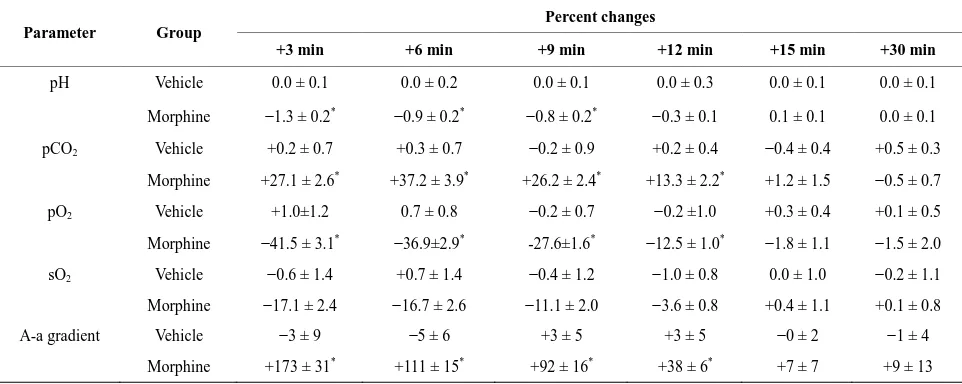
Table 1. Percent changes in arterial blood gas chemistry and Alveolar-arterial gradient elicited by injections of vehicle or morphine.
Parameter Group
Percent changes
+3 min +6 min +9 min +12 min +15 min +30 min
pH Vehicle 0.0 ± 0.1 0.0 ± 0.2 0.0 ± 0.1 0.0 ± 0.3 0.0 ± 0.1 0.0 ± 0.1
Morphine −1.3 ± 0.2* −0.9 ± 0.2* −0.8 ± 0.2* −0.3 ± 0.1 0.1 ± 0.1 0.0 ± 0.1
pCO2 Vehicle +0.2 ± 0.7 +0.3 ± 0.7 −0.2 ± 0.9 +0.2 ± 0.4 −0.4 ± 0.4 +0.5 ± 0.3
Morphine +27.1 ± 2.6* +37.2 ± 3.9* +26.2 ± 2.4* +13.3 ± 2.2* +1.2 ± 1.5 −0.5 ± 0.7
pO2 Vehicle +1.0±1.2 0.7 ± 0.8 −0.2 ± 0.7 −0.2 ±1.0 +0.3 ± 0.4 +0.1 ± 0.5
Morphine −41.5 ± 3.1* −36.9±2.9* -27.6±1.6* −12.5 ± 1.0* −1.8 ± 1.1 −1.5 ± 2.0
sO2 Vehicle −0.6 ± 1.4 +0.7 ± 1.4 −0.4 ± 1.2 −1.0 ± 0.8 0.0 ± 1.0 −0.2 ± 1.1
Morphine −17.1 ± 2.4 −16.7 ± 2.6 −11.1 ± 2.0 −3.6 ± 0.8 +0.4 ± 1.1 +0.1 ± 0.8
A-a gradient Vehicle −3 ± 9 −5 ± 6 +3 ± 5 +3 ± 5 −0 ± 2 −1 ± 4
Morphine +173 ± 31* +111 ± 15* +92 ± 16* +38 ± 6* +7 ± 7 +9 ± 13
All data are mean ± SEM. There were 8 rats in each group. A-a gradient, Alveolar-arterial gradient. *P < 0.05, significant percent change from Pre values. A-a gradient = PAO2− PaO2, where PAO2 is the partial pressure of alveolar O2 (calculated from the alveolar gas equation) and PaO2 is the partial pressure of O2 in
arterial blood. PAO2 is determined by the formula, PAO2=
(
FiO2×(
Patm−PH O2)
−(PaCO respiratory quotient 2 ))
, where FiO2 is the fraction of O2 inin-spired air; Patm is atmospheric pressure; PH O2 is the partial pressure of water in inspired air; and PaCO2 is the partial pressure of CO2 in arterial blood. We took
2.5. Body Temperature Measurements
Body temperatures (Bt) of male Sprague Dawley rats were measured via telemetry [32]. Rats were anesthe- tized (2% isoflurane) and a telemetry probe (ETA-F20, Data Sciences International, MN) was implanted into the peritoneal cavity. A jugular vein catheter was also im- planted. After 4 days, the rats were placed in plethys- mography chambers, under each of which was placed a telemetry receiver connected via a data exchange matrix to a computer. The Bt signals were sampled once a min- ute using internal hardware and Dataquest ART Gold3.1 software. Digitized signals were transferred to MATLAB (MathWorks, Natick, MA) for analysis.
2.6. Protocols for Analgesia Studies
Paw-withdraw latencies were determined in a group of 4 rats (290 ± 4 grams) before and 20, 40, 60 and 90 min after a bolus injection of morphine (5 mg/kg, i.v.). All studies used an injectable form of (+)-morphine sulfate (10 mg/ml) from Baxter Healthcare (Deerfield, IL, USA).
2.7. Protocols for Arterial Blood Gas and
Alveolar-Arterial Gradient Studies
Arterial blood sample (100 µL) were taken before (2 - 3 samples and values averaged) and 3, 6, 9, 12, 15 and 30 min after bolus injections of vehicle (saline, n = 8 rats; 293 ± 3 grams) or morphine (5 mg/kg, i.v., n = 8 rats; 291 ± 3 grams).
2.8. Protocols for Ventilation Studies
The rats were placed in the plethysmography chambers and allowed 45 - 60 min to acclimatize. Data was continuously recorded (i.e., breath by breath) throughout the acclimatization period (the last 15 min was used for analysis and graphing) and subsequent experimentation. The data were collected into 1 min bins for analysis and graphing. Two groups of rats were used in these studies. One group of rats (n = 12, 281 ± 1 grams) received an injection of vehicle (saline, i.v.) whereas another group (n = 12, 284 ± 3 grams) received an injection of morphine (10 mg/kg, i.v.). After 15 min, the rats were exposed to 3 consecutive 15 min episodes of hypoxia (10% O2, 90% N2), each of which was separated by a 15
min exposure to room air.
2.9. Protocols for Body Temperature Studies
Rats with jugular vein catheters and telemetry devices were placed in plethysmography chambers and allowed 45 - 60 min to acclimatize. One group (n = 6, 303 ± 2 g) received an i.v. injection of vehicle (saline) whereas another group (n = 6, 304 ± 3 g) received morphine (5
mg/kg, i.v.). After 15 min, the rats underwent the hypoxia-normoxia challenges as described above.
2.10. Statistics
Data recorded during the ventilatory studies (1 minbins) and derived parameters, namely, TV/Ti and cumulative response (cumulative arithmetic changes from pre-values), were taken for analyses. All data are presented as mean ± SEM and were analyzed by one-way or two-way
ANOVA followed by Student’s modified t test with
Bonferroni corrections for multiple comparisons between means [33]. P < 0.05 denoted significance.
3. RESULTS
3.1. Analgesia
Paw-withdraw latencies in rats prior to injection of morphine (5 mg/kg, i.v.) were 18.9 ± 0.5 sec. The paw withdrawal latencies at 20, 40, 60 and 90 min after morphine injection were 40 ± 0, 40 ± 0, 39.4 ± 0.6 and 38.9 ± 1.1 sec, respectively (P < 0.05, for all com- parisons to pre-injection values). Remembering that the cutoff-latency was 40 seconds, it was evident that of morphine elicited a sustained analgesia in these rats.
3.2. Arterial Blood Gas and Alveolar-Arterial
Gradient Studies
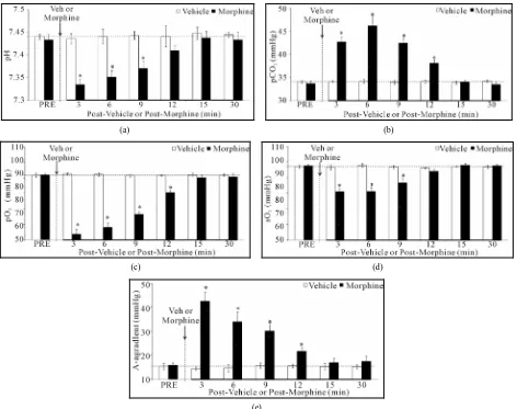
The effects of vehicle or morphine (5 mg/kg, i.v.) on
ABG chemistry and A-a gradient are summarized in Fig-
ure 1 and Table 1. Vehicle did not affect these para- meters whereas morphine elicited prompt decreases in arterial blood pH, pO2 and sO2 and increases in pCO2
and A-a gradient. All responses were resolved by 15 min and no changes were observed at 30 min.
3.3. Ventilatory Responses Elicited by Morphine
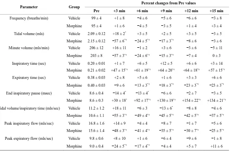
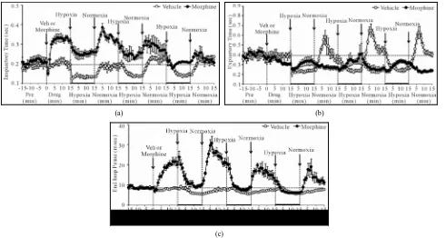
Morphine (5 mg/kg, i.v.) elicited transient increases in fr which were associated with decreases in Vt and Vm of 9 - 10 min in duration (Figure 2, Table 2). Despite the transient nature of the morphine-induced increase in fr, morphine elicited sustained increases in Ti and EIP but a sustained decrease in Te (Figure3, Table2). In addition, morphine elicited sustained decreases in Vt/Ti and PIF but relatively transient changes in PEF (Figure4, Table 2). Vehicle elicited minor responses (Figures2-4, Table
2). Taken together, vehicle elicited minor cumulative
responses whereas morphine elicited significant cumu- lative responses in all parameters except fr and PEF (Ta- ble3).
3.4. Ventilatory Responses during Hypoxic
Challenges and upon Return to Room-Air
(a) (b)
(c) (d)
[image:4.595.63.535.83.456.2](e)
Figure 1. Temporal changes in arterial blood gas chemistry values and Alveolar-arterial (A-a) gradient elicited by bolus injections of vehicle or morphine (5 mg/kg, i.v.) in conscious rats. There were 8 rats in each group. The data are mean ± SEM. *P < 0.05, significant change from Pre values.
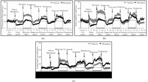
and sustained increases in fr, Vt and Vm in vehicle- treated (VEH) rats (Figure2, Table4). The increases in fr and Vm developed more slowly during H2 and H3. In morphine-treated (MOR) rats, H1-H3 transiently in- creased fr (Figure2, Table4). The increases in Vt dur- ing H1 were similar to those in VEH rats whereas the increases during H2 and H3 were greater. The increases in Vm during H1 in MOR rats were smaller than in VEH rats (due to lesser increases in fr) whereas the increases in Vm during H2 and H3 were similar in both groups. In VEH rats, fr returned to pre-levels during episode 1 of normoxia (N1) but fell below pre-values for several min during N2 and N3. Vt and Vm dipped below pre-levels during N1 - N3 but normalized within 5 - 10 min. In MOR rats, fr changed minimally during N1 whereas it rose and remained elevated during N2 and N3. Vt tended to return to pre-levels during N1 - N3 in MOR rats. Vm returned to pre-levels during N1 - N3 in MOR rats but remained above pre-levels during N2 and N3. fr and Vm
values in MOR rats were higher than in VEH rats at the end of N3 (Figure2).
(a) (b)
(c)
Figure 2. Changes in frequency of breathing (a), tidal volume (b) and Minute Volume (c) elicited by injections of vehicle or morphine (5 mg/kg, i.v.) and subsequent exposure to three 15 min episodes of hypoxia (10% O2, 90% N2) each of which was
[image:5.595.60.535.83.337.2]followed by a 15 min period of normoxia (room-air). The first episode of hypoxia began 15 min after injection of vehicle or morphine. There were 6 rats in each group. Data are mean ± SEM.
Table 2. Ventilatory responses elicited by vehicle or morphine.
Parameter Group Percent changes from Pre values
Pre +3 min +6 min +9 min +12 min +15 min
Frequency (breaths/min) Vehicle 99 ± 4 +1 ± 8 −4 ± 6 −5 ± 6 −6 ± 6 −3 ± 8
Morphine 95 ± 4 +1 ± 6 −4 ± 5 −3 ± 5 +1 ± 4 +3 ± 4
Tidal volume (mls) Vehicle 2.09 ± 0.12 +18 ± 2* +3 ± 5 +2 ± 5 +3 ± 5 −3 ± 5
Morphine 2.15 ± 0.12 −37 ± 6*† −24 ± 5*† −17 ± 3*† −8 ± 6 −3 ± 6
Minute volume (mls/min) Vehicle 206 ± 12 +16 ± 11 −1 ± 2 +3 ± 6 −3 ± 6 −5 ± 11
Morphine 203 ± 8 −37 ± 3*† −24 ± 6*† −15 ± 3*† −7 ± 4 0 ± 3
Inspiratory time (sec) Vehicle 0.20 ± 0.01 +1 ± 7 +6 ± 5 +12 ± 5 +6 ± 6 +3 ± 14
Morphine 0.21 ± 0.02 +47 ± 15*† +61 ± 19*† +64 ± 20*† +64 ± 18*† +57 ± 15*†
Expiratory time (sec) Vehicle 0.38 ± 0.03 +2 ± 8 +5 ± 6 +1 ± 6 +3 ± 3 +6 ± 6
Morphine 0.40 ± 0.03 −9 ± 6 −13 ± 3*† −18 ± 3*† −23 ± 3*† −23 ± 3*†
End inspiratory pause (msec) Vehicle 8.6 ± 0.4 −14 ± 4* −13 ± 4* −6 ± 6 −2 ± 7 −3 ± 5
Morphine 8.6 ± 0.3 +30 ± 18* +92 ± 17*† +130 ± 19*† +154 ± 22*† +134 ± 21*†
Tidal volume/inspiratory time (mls/sec) Vehicle 11.2 ± 1.2 +18 ± 11 −6 ± 3 −13 ± 6* −8 ± 8 −4 ± 6
Morphine 10.6 ± 1.1 −55 ± 3*† −49 ± 4*† −45 ± 5*† −42 ± 5*† −37 ± 5*†
Peak inspiratory flow (mls/sec) Vehicle 16.8 ± 1.6 +14 ± 9 −4 ± 4 −8 ± 7 −1 ± 7 −5 ± 6
Morphine 15.6 ± 1.4 −48 ± 3*† −41 ± 4*† −35 ± 5*† −30 ± 7*† −25 ± 5*†
Peak expiratory flow (mls/sec) Vehicle 9.8 ± 0.6 +8 ± 10 +1 ± 6 −6 ± 4 −9 ± 6 −1 ± 8
Morphine 9.0 ± 0.4 −24 ± 5*† −17 ± 4*† −4 ± 4 +5 ± 7 +11 ± 6
[image:5.595.69.540.416.726.2](a) (b)
[image:6.595.56.547.83.345.2](c)
Figure 3. Changes in inspiratory time (a), expiratory time (b) and end inspiratory pause (c) elicited by injections of vehicle or morphine (5 mg/kg, i.v.) and subsequent exposure to three 15 min episodes of hypoxia (10% O2, 90% N2) each of which was
followed by a 15 min period of normoxia (room-air). The first episode of hypoxia began 15 min after injection of vehicle or morphine. There were 6 rats in each group. Data are mean ± SEM.
Table 3. Cumulative responses elicited by vehicle or morphine.
Parameter Vehicle Morphine
Frequency (breaths/min) × min +24 ± 80 +37 ± 63
Tidal volume (mls) × min +1.3 ± 0.4* −5.5 ± 1.9*†
Minute volume (mls/min) × min +172 ± 103 −462 ± 94*†
Inspiratory time (sec) × min +0.1 ± 0. 2 +1.4 ± 0.3*†
Expiratory time (sec) × min +0.1 ± 0.3 −1.0 ± 0.2*†
End inspiratory pause (msec) × min −9.0 ± 6.8 +124.5 ± 11.5*†
Tidal volume/inspiratory time (mls/sec) × min −3.2 ± 8.0 −66.3 ± 11.9*†
Peak inspiratory flow (mls/sec) × min +4.8 ± 11.0 −71.6 ± 17.4*†
Peak expiratory flow (mls/sec) × min −1.0 ± 7.5 −1.0 ± 6.1
All data are mean ± SEM. There were 6 rats in each group. *P < 0.05, significant cumulative response. †P < 0.05, morphine versus vehicle.
similar to VEH rats at the end of N3 whereas Te was decreased (Figure3).
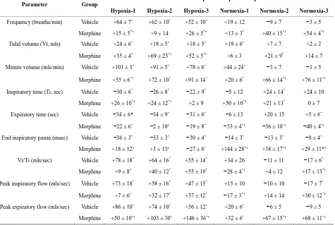
Vt/Ti, PIF and PEF: H1 - H3 elicited sustained in- creases in Vt/Ti in VEH and MOR rats (Figure4, Table 4). Vt/Ti values in MOR rats did not reach those of VEH rats during H1 but approached them in H2 and equaled them in H3. In VEH rats, Vt/Ti rapidly returned to base- line levels during N1 - N3. In MOR rats, Vt/Ti returned to the lower morphine-induced baseline during N1. The decreases in Vt/Ti during N2 and N3 were similar in both groups. H1 - H3 elicited sustained increases in PIF in
VEH and in MOR rats. The responses during H1 and H2 in MOR rats did not reach the maxima in VEH rats whe-reas the responses during H3 were equal to VEH rats. In VEH rats, PIF returned to pre-levels during N1 whereas it fell below pre-levels during N2 and N3. In MOR rats, PIF returned to post-morphine levels during N1 but re-turned to pre-morphine levels during N2 and N3. H1 - H3 elicited sustained increases in PEF in VEH and MOR
rats (Figure 4, Table 4). The responses during H1 in
[image:6.595.55.540.419.581.2](a) (b)
[image:7.595.56.541.84.352.2](c)
Figure 4. Changes in tidal volume/inspiratory time (a), peak inspiratory flow (b), and peak expiratory flow (c) elicited by injections of vehicle or morphine (5 mg/kg, i.v.) and subsequent exposure to three 15 min episodes of hypoxia (10% O2, 90% N2) each of
which was followed by a 15 min period of normoxia (room-air). The first episode of hypoxia began 15 min after injection of ve-hicle or morphine. There were 6 rats in each group. Data are mean ± SEM.
although higher values were reached in MOR rats during H3 because of higher pre-hypoxia values. In VEH rats, PEF returned to pre-levels during N1 - N3. In contrast, PEF in MOR rats remained elevated during N2 and N3. Vt/Ti values were similar in VEH and MOR rats at the end of N3, whereas PIF was lower in VEH rats because of the sustained post-hypoxic depression (Figure4). PEF was higher in MOR rats at the end of N3 because of the sustained effects of morphine (Figure4).
Cumulative responses: In VEH rats. H1 - H3 elicited similar cumulative responses in Vt, Ti, Te, and EIP whereas those in fr, Vm, Vt/Ti, PIF, and PEF gradually diminished with each episode (Table5). H1 - H3 elicited markedly different cumulative responses in MOR rats than in VEH rats, and the magnitude of these differences changed with each hypoxic episode (Table5). N1 - N3 elicited substantial cumulative responses in fr, Vm, Ti, EIP, PIF, and PEF but minimal responses in Vt, Ti, and Vt/Ti. Moreover, the magnitude and direction of these cumulative responses changed with each episode of nor- moxia. For example, N1 was associated with positive cumulative response for fr and Vm, whereas N2 and N3 were associated with negative responses. N1 - N3 also elicited substantial cumulative responses in MOR rats that changed markedly with each successive challenge (Table5). Although the cumulative responses during N1 for fr, Vt, and Vm were similar in VEH and MOR rats,
the cumulative responses in MOR rats greatly exceeded those in VEH rats during N2 and N3.
3.5. Body Temperature (Bt)
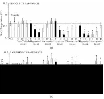
H1 - H3 elicited minor but sustained decreases in Bt in VEH rats (e.g., H1 at +15 min, −0.39˚C ± 0.04˚C, P < 0.05) (Figure 5). Bt returned to pre-hypoxia levels upon return to room-air (N1 - N3). Morphine elicited a minor increase in Bt, which was still evident at the time of exposure to H1 (+0.44˚C ± 0.08˚C, P < 0.05). H1 - H3 elicited minor decreases in Bt in MOR rats, although they never fell below pre-values. These decreases were still present at the end of each hypoxic challenge (e.g., H1 at +15 min, −0.55˚C ± 0.11˚C, P < 0.05). Bt returned to pre-hypoxia levels upon return to room air, such that the morphine-induced hyperthermia was sustained through- out the study.
4. DISCUSSION
normoxia.
Parameter Group
+15 min values expressed as percent of pre values
Hypoxia-1 Hypoxia-2 Hypoxia-3 Normoxia-1 Normoxia-2 Normoxia-3
Frequency (breaths/min) Vehicle +64 ± 7* +62 ± 10* +52 ± 10* +19 ± 12 −9 ± 7 −3 ± 5
Morphine +15 ± 5*† +9 ± 14 +26 ± 5*† +13 ± 3* +40 ± 15*† +54 ± 4*†
Tidal volume (Vt,mls) Vehicle +24 ± 6* +18 ± 5* +18 ± 5* +19 ± 6* +7 ± 7 +2 ± 2
Morphine +35 ± 4* +69 ± 23*† +52 ± 5*† +6 ± 3 +21 ± 9* +14 ± 7
Minute volume (mls/min) Vehicle +101 ± 3* +91 ± 5* +78 ± 6* +44 ± 24* −3 ± 7 −1 ± 5
Morphine +55 ± 6*† +72 ± 10* +91 ± 14* +20 ± 6* +66 ± 14*† +76 ± 11*†
Inspiratory time (Ti, sec) Vehicle −30 ± 6* −26 ± 8* −22 ± 9* −5 ± 12 +24 ± 14* +24 ± 10
Morphine +26 ± 10*† +24 ± 12*† +2 ± 9 +50 ± 10*† +21 ± 13* 0 ± 7
Expiratory time (sec) Vehicle −34 ± 6* −34 ± 9* −31 ± 6* −6 ± 13 +20 ± 15 +5 ± 6*
Morphine −22 ± 6* −2 ± 10† −19 ± 8* −33 ± 4*† −36 ± 10*† −40 ± 4*†
End inspiratory pause (msec) Vehicle −36 ± 3* −33 ± 3* −30 ± 4* −14 ± 3* −13 ± 3* −8 ± 4*
Morphine +18 ± 12† +3 ± 13† −27 ± 6* +144 ± 28*† +38 ± 17*† +29 ± 11*†
Vt/Ti (mls/sec) Vehicle +78 ± 18* +64 ± 16* +55 ± 14* +34 ± 26 −11 ± 11 −17 ± 6*
Morphine +9 ± 8† +40 ± 12* +55 ± 19* −28 ± 4*† +4 ± 12 +17 ± 13*†
Peak inspiratory flow (mls/sec) Vehicle +73 ± 18* +58 ± 16* +47 ± 15* +15 ± 10 −10 ± 10 −17 ± 7*
Morphine +7 ± 6† +32 ± 17* +37 ± 12* −17 ± 3*† +14 ± 14 +30 ± 12*†
Peak expiratory flow (mls/sec) Vehicle +86 ± 10* +74 ± 10* +56 ± 12* +20 ± 6* −6 ± 5 −9 ± 5
Morphine +50 ± 10*† +103 ± 30* +146 ± 36*† +32 ± 6* +67 ± 15*† +68 ± 11*†
All data are mean ± SEM. There were 6 rats in each group. *P < 0.05, significant response. †P < 0.05, morphine versus vehicle.
hypoxic challenge. These latent effects may involve direct effects on carotid body and/or brain mechanisms processing the hypoxic responses rather than changes in metabolism.
4.1. Analgesia and Body Temperature
The ability of 5 mg/kg morphine to elicit a long-lasting analgesia validated our choice of this dose for the ventilatory studies. Our finding that a 5 mg/kg dose of
morphine elicited a minor sustained hyperthermia of ≈
0.4˚C is consistent with findings from other laboratories [34,35]. Exposure of VEH rats to 15 min episodes of hypoxia (10% O2, 90% N2) elicited minor decreases in Bt
of 0.4˚C, consistent with evidence that exposure of rats to 10% - 12% O2 for 15 min elicits no effects [36] or a
decrease of ≈0.5˚C [37,38]. Since each episode of
hypoxia elicited very similar arithmetic falls in Bt in VEH and MOR rats, it appears that despite elevating Bt, morphine does not impair the metabolic processes that cause hypoxia-induced hypothermia. Taken together, the morphine-induced changes in BT are likely to have had minimal effects on the ventilatory responses to hypoxia.
4.2. Morphine-Induced Changes in ABG
Chemistry, A-Arterial Gradient and
Ventilation
The effects of morphine on ABG chemistry (i.e., de-
creases in pH, pO2 and sO2, increases in pCO2) are
con-sistent with hypoventilation (i.e., decreases in Vt) whe-reas the incwhe-reases in A-a gradient (mismatch of ventila-tion-perfusion) may have arisen via increases in pulmo-nary airways resistance [18,19], pulmopulmo-nary vasoconstric-tion reducing arterial blood flow to alveoli [20,39,40] and/or exacerbated hypoxic pulmonary vasoconstriction
via morphine-induced reductions in arterial pO2.
[image:8.595.65.540.124.443.2]Table 5. Cumulative responses during exposures to hypoxia and normoxia.
Parameter Group
Cumulative Responses during Episodes of Hypoxia and Normoxia
Hypoxia-1 Hypoxia-2 Hypoxia-3 Normoxia-1 Normoxia-2 Normoxia-3
Frequency (breaths/min)
× min Vehicle +1147 ± 143
*
+798 ± 146* +711 ± 126* +152 ± 70* −122 ± 47* −64 ± 36
Morphine +285 ± 88*† +283 ± 112*† +492 ± 109*† +111 ± 24* +364 ± 147*† +644 ± 99*†
Tidal volume (Vt, mls)
× min Vehicle +8.4 ± 1.2
* +6.6 ± 1.6* +6.6 ± 1.4* −0.5 ± 0.8 −1.3 ± 1.2 −1.5 ± 1.3
Morphine +11.4 ± 1.0* +18.7 ± 2.8*† +19.6 ± 1.8*† +0.7 ± 0.6 +5.9 ± 1.7*† +3.2 ± 0.7*†
Minute volume (mls/min)
× min Vehicle +3819 ± 241
* +2599 ± 102* +2399 ± 103* +291 ± 101* −374 ± 89* −251 ± 72*
Morphine +1876 ± 304*† +2536 ± 224* +3489 ± 320*† +329 ± 61* +1351 ± 250*† +1764 ± 236*†
Inspiratory time (Ti, sec)
× min Vehicle −0.9 ± 0.3
* −
0.6 ± 0.2* −0.6 ± 0.2* −0.2 ± 0.3 +0.3 ± 0.3 +0.3 ± 0.3
Morphine +0.7 ± 0.2*† +0.6 ± 0.2*† −0.1 ± 0.3 +1.8 ± 0.2*† +1.2 ± 0.3*† +0.3 ± 0.2
Expiratory time (sec)
× min Vehicle −2.3 ± 0.3
* −1.8 ± 0.3* −1.7 ± 0.4* 0.5 ± 0.6 +1.6 ± 0.7* +1.1 ± 0.4*
Morphine −1.5 ± 0.3*† −1.2 ± 0.4* −1.5 ± 0.6* −1.9 ± 0.3*† −2.0 ± 0.3* −2.3 ± 0.4*†
End inspiratory pause
(msec) × min Vehicle −25 ± 8
* −26 ± 5* −24 ± 5* −24 ± 5* −24 ± 6* −25 ± 7*
Morphine +62 ± 21*† +24 ± 10*† −22 ± 10* +219 ± 22*† +112 ± 21*† +67 ± 22*†
Vt/Ti (mls/sec) × min Vehicle +131 ± 17* +80 ± 10* +74 ± 15* +9 ± 14 −27 ± 17 −25 ± 12
Morphine +16 ± 11† +46 ± 8*† +97 ± 22* −56 ± 12*† −21 ± 14 0 ± 19
Peak inspiratory flow
(mls/sec) × min Vehicle +193 ± 35
* +104 ± 24* +95 ± 17* +15 ± 16 −39 ± 23 −40 ± 10*
Morphine +29 ± 15*† +62 ± 17* +117 ± 27* −51 ± 15*† +2 ± 19 +29 ± 15†
Peak expiratory flow
(mls/sec) × min Vehicle +140 ± 14
* +79 ± 8* +68 ± 8* +24 ± 7* −10 ± 9 −7 ± 5
Morphine +79 ± 15*† +121 ± 16*† +179 ± 24*† +39 ± 10* +88 ± 13*† +87 ± 11*†
All data are mean ± SEM. There were 6 rats in each group. *P < 0.05, significant cumulative response. †P < 0.05, morphine versus vehicle.
morphine-induced reductions in Vt/Ti had not resolved by 15 min, it appears that the negative influence on Res-piratory Drive was present despite the resolution of ABG chemistry, A-a gradient and Vm. Morphine also elicited sustained decreases in Te (i.e., expiration was faster), which may have been due to diminished Vt requiring less time to exhale. However, the decreased Te may have involved direct (presumably central) effects of morphine on pathways driving expiration.
4.3. Ventilatory Responses to Hypoxic
Challenges in Vehicle-Treated Rats
In VEH rats, the hypoxic challenges elicited prompt and sustained ventilatory responses including 1) increases in Vt/Ti and Vm (via increases in fr and Vt), 2) decreases in Ti and Te but minor decreases in EIP, and 3) increases in PIF and PEF. The rates of increase in fr, PIF, PEF, and
Vt/Ti were slower during H2 and H3 than H1. This sug-gests that the mechanisms responsible for these rapid responses had “adapted” to hypoxic challenge whereas those involved in maintaining the responses did not. Whether this adaptation occurred in response elements sensitive to reductions in blood pO2 and in the carotid
bodies [41-43], or within the brain is unknown. Ventila-tory drive can diminish during exposures to severe hy-poxia or to longer duration exposures to mild hyhy-poxia. This “ventilatory roll-off” involves neurochemical processes in the NTS under mild hypoxia [44,45], and the direct depressive effects of severe hypoxia on brain neurons regulating ventilation [45,46]. However, in the present study, none of the key ventilatory parameters (e.g., fr, Vt, Inspiratory Drive) displayed of roll-off indi-cating that 15 min exposures to 10% O2 did not engender
(a)
[image:10.595.130.468.79.399.2](b)
Figure 5. Changes in body temperature elicited by injections of vehicle (upper panel) or morphine (5 mg/kg, i.v., lower panel) and subsequent exposure to three 15 min episodes of hypoxia (10% O2, 90% N2) each of which was followed by 15 min periods of normoxia
(room-air). The first episode of hypoxia began 15 min after injection of vehicle or mor-phine. There were 8 rats in each group. Data are mean ± SEM.
4.4. Ventilatory Responses to Hypoxic
Challenges in Morphine-Treated Rats—
Episode H1
H1 elicited markedly smaller increases in fr in MOR rats although resting fr was not diminished. As such, mor- phine elicited latent effects on systems including those within the carotid bodies that drive the hypoxic res-ponses. Our data are consistent with evidence that the negative effects of opioids in humans may be latent since the morphine metabolite, M6G, does not affect resting ventilatory parameters whereas it substantially blunts the ventilatory response to hypercapnic challenge [5]. Since morphine did not markedly blunt the increase in Vt dur-ing H1, it is evident that it did not negatively affect neur-al drive to the chest muscles or diaphragm. This is sup-ported by the finding that Ti in MOR rats (elevated im-mediately prior to exposure to hypoxia) decreased sub-stantially during H1. Taken together, our data support the concept that morphine affected brainstem centers re-sponsible for generating breathing such as the respiratory pattern central generator, including the pre-Botzinger complex [47,48], rather than affecting peripheral motor/
skeletal muscle components driving breathing. Nonethe-less, it was evident that H1 elicited substantially smaller increases in PIF and PEF in MOR rats than in VEH rats thereby compromising the dynamics of active inspirato-ry/expiratory ventilation. It should be noted that resting PIF was still depressed in MOR rats (−24% ± 7%, P < 0.05) prior to H1 whereas resting PEF was not (+8% ± 6%, P > 0.05). In addition, Te was diminished prior to H1 in MOR rats (−23% ± 5%, P < 0.05) but actually rose appreciably during H1, suggesting that morphine inhi-bited the hypoxia-induced stimulation of active expira-tion.
4.5. Ventilatory Responses upon Reintroduction
of Room-Air in Vehicle-Treated Rats
re-turned to pre-values upon return to room-air whereas fr, Vt, Vm and PIF declined below pre-levels. Consistent with previous findings [52,53], this PHFD was associ- ated with an elongation in Te. The finding that the pattern of ventilatory responses was similar for each of the three normoxic episodes indicates that adaptations in carotid body or brainstem circuitry that may affect the normoxia responses did not occur during repeated exposure to hy- poxia.
4.6. Temporal Changes in Ventilatory Responses
to Hypoxia-Normoxia in
Morphine-Treated Rats
The pattern of ventilatory responses in MOR rats changed during the study. Although fr during each epi-sode of hypoxia was depressed in MOR rats, baseline fr gradually rose during N1 - N3 such that fr was higher than in VEH rats during N3. Since the PHFD observed in VEH rats was absent in MOR rats, morphine may have directly interfered with pontomedullary systems respon- sible for this phenomenon [52,53,55,56]. Although rest- ing Vt returned to baseline levels during N1-N3 in MOR rats, the responses during H1 - H3 changed in that the increases in Vt following H1 were slightly greater than those of VEH rats whereas they were greater than VEH rats during H2 and H3. As a result of the temporal pat- terns of changes in fr and TV, resting Vm in MOR rats was substantially above that of VEH rats during N2 and N3. Moreover, although the peak increases in Vm in MOR rats were less than in VEH rats during H1, the in- creases in Vm in MOR rats were equal to those in VEH rats during H2 and reached higher values (perhaps due to elevated starting baselines) during H3. Although the PHFD in Vm appeared qualitatively similar in both groups, PHFD in VEH rats was due to a decline in fr and to a lesser degree a decrease in Vt whereas it was due solely to a decrease in Vt in MOR rats. The prolongation of Ti by morphine was essentially resolved when N3 was applied. Although each episode of hypoxia decreased Ti in the MOR rats, none of the decreases were as pro- nounced as those in VEH rats. Moreover, Ti promptly returned to elevated levels during N1 and N2 but not N3. Although morphine elicited reductions in Vt/Ti and PIF, which had not resolved prior to H1 or H2, these chal- lenges still elicited robust increases in these parameters. In addition, although morphine elicited a sustained in- crease in EIP that was evident during N1 - N3, each epi- sode of hypoxia elicited robust decreases in EIP. As such, it appears that whereas morphine reduced EIP, Vt/Ti and PIF, it did not depress the mechanisms by which the ca- rotid bodies responded to hypoxia or central/efferent processing of this input. The findings that the PEF during N2 and N3 were elevated and that the post-hypoxia in- creases in Te in VEH rats (N1 - N3) were absent in MOR
rats are suggestive of facilitatory effects of morphine and/or metabolites on expiratory control.
4.7. Mechanisms Responsible for the Delayed
Effects of Morphine—Morphine Metabolites
The presence of (+)-morphine metabolites [57-60] may explain the delayed effects of the opioid on the ventila- tory responses to hypoxia. In rats, maximal plasma levels of (+)-morphine occur within 10 min and the elimination half-life is approximately 45 - 50 min. As such, pharma- cologically active plasma concentrations of morphine were likely present in our rats during H1 and N1 (15 to 45 min post-morphine injection), during H2 and N2 (46 - 75 min post-morphine) but perhaps less so during H3 and N3 (76 - 105 min post-morphine). It is feasible that mor- phine sequestered into relevant tissues may have effects when plasma levels are minimal. Obvious tissues would be 1) carotid bodies and neuromuscular elements of the chest wall [61], 2) brainstem nuclei receiving/processing carotid body chemoafferent input [62], and 3) brain structures devoid of blood-brain barriers [63]. It may also be possible that concentrations of morphine that are in- sufficient to affect central and/or peripheral drive may still be able to blunt the mechanisms responsible for sensing/expressing ventilatory responses to hypoxia. In humans, (+)-morphine is metabolized to M6G [57,60], which is relatively more selective for µ-opioid receptors (µ-ORs) than δ- or κ-ORs and elicits pronounced analge- sia and respiratory depression [58,64]).
In rats, (+)-morphine is metabolized primarily to M3G [57,59,60]. Maximal plasma levels of M3G occur within 30 min of morphine administration and the elimination half-life is over 200 min. Unlike M6G, M3G has a low affinity for ORs [65,66] and minimal analgesic or respi- ratory depressant activity [65,67]. However, M3G is not inert since 1) intracerebroventricular [68,69] or intrathe- cal [70,71] injections of M3G cause behavioral excita- tion, 2) rats develop hyperaesthesia and allodynia after intrathecal M3G [70], and 3) intracerebroventricular in- jections of M3G elicit electroencephalographic spiking, excessive grooming, and epiliptoform discharges [68,72]. Taken together, the latent effects of morphine on the ven- tilatory responses to hypoxia may be due to the actions of M3G in the brain and/or periphery via mechanisms other than the activation of ORs.
5. CONCLUSION
sleep apnea (OSA) are at risk for respiratory complica- tions to opioids given post-operatively [73]. Moreover, children undergoing adenotonsillectomy for relief of OSA symptoms have a higher sensitivity to the venti-
latory depressant effects of μ-OR agonists, and higher
post-operative difficulty in breathing compared to those without OSA [73,74]. There may be many patients with undiagnosed OSA [75], which are at risk for impaired breathing post-operatively [76-78]. The present study suggests that even normal patients may be under threat from opioids that blunt ventilatory responses to hypoxia even when monitoring suggests that the effects of opioids on ABG chemistry and Vm had subsided. Considering the ability of opioids to destabilize breathing during sleep, the latent deleterious actions of opioids on ventila- tory responses to hypoxia may be especially dangerous when the patient is asleep. Our studies suggest that M6G in humans and M3G in rats should be further investi- gated with respect to their potential ability (and mecha- nisms of action) to exert deleterious effects on the hy- poxic ventilatory response.
6. ACKNOWLEDGEMENTS
This work was supported by grants from Galleon Pharmaceuticals (S. J. L).
REFERENCES
[1] Cepeda, M.S., Farrar, J.T., Baumgarten, M., Boston, R., Carr, D.B. and Strom, B.L. (2003) Side effects of opioids during short-term administration: Effect of age, gender, and race. Clinical Pharmacology and Therapeutics, 74, 102-112.
[2] Cashman, J.N. and Dolin, S.J. (2004) Respiratory and haemodynamic effects of acute postoperative pain man- agement: Evidence from published data. British Journal of Anaesthesia, 93, 212-223.
[3] Taylor, S., Kirton, O.C., Staff, I. and Kozol, R.A. (2005) Postoperative day one: A high risk period for respiratory events. American Journal of Surgery, 190, 752-756.
[4] Lötsch, J., Dudziak, R., Freynhagen, R., Marschner, J. and Geisslinger, G. (2006) Fatal respiratory depression after multiple intravenous morphine injections. Clinical Pharmacokinetics, 45, 1051-1060.
[5] Peat, S.J., Hanna, M.H., Woodham, M., Knibb, A.A. and Ponte, J. (1991) Morphine-6-glucuronide: Effects on ven- tilation in normal volunteers. Pain, 45, 101-104.
[6] Berkenbosch, A., Teppema, L.J., Olievier, C.N. and Da- han, A. (1997) Influences of morphine on the ventilatory
1349.
[7] Dahan, A., Sarton, E., Teppema, L. and Olievier, C. (1998) Sex-related differences in the influence of mor- phine on ventilatory control in humans. Anesthesiology, 88, 903-913.
[8] Sarton, E., Teppema, L. and Dahan, A. (1999) Sex dif- ferences in morphine-induced ventilatory depression re- side within the peripheral chemoreflex loop. Anesthesi- ology, 90, 1329-1338.
[9] Fatemian, M., Nieuwenhuijs, D.J., Teppema, L.J., Mein- esz, S., van der Mey, A.G., Dahan, A., Robbins, P.A. (2003) The respiratory response to carbon dioxide in hu- mans with unilateral and bilateral resections of the carotid bodies. Journal of Physiology, 549, 965-973.
[10] Timmers, H.J., Wieling, W., Karemaker, J.M. and Lend- ers, J.W. (2003) Denervation of carotid baro- and chem- oreceptors in humans. Journal of Physiology, 553, 3-11.
[11] Shook, J.E., Watkins, W.D. and Camporesi, E.M. (1990) Differential roles of opioid receptors in respiration, res- piratory disease, and opiate-induced respiratory depres- sion. American Review of Respiratory Disease, 142, 895-
909.
[12] Satoh, M. and Minami, M. (1995) Molecular pharmacol- ogy of the opioid receptors. Pharmacology and Thera- peutics, 68, 343-364.
[13] Haji, A., Takeda, R. and Okazaki, M. (2000) Neurophar- macology of control of respiratory rhythm and pattern in mature mammals. Pharmacology and Therapeutics, 86, 277-304.
[14] Lewanowitsch, T. and Irvine, R.J. (2002) Naloxone me- thiodide reverses opioid-induced respiratory depression and analgesia without withdrawal. European Journal of Pharmacology, 445, 61-67.
[15] Lewanowitsch, T., Miller, J.H. and Irvine, R.J. (2006) Reversal of morphine, methadone and heroin induced ef- fects in mice by naloxone methiodide. Life Sciences, 78,
682-6
[16] Campbell, C., Weinger, M.B. and Quinn, M. (1995) Al- terations in diaphragm EMG activity during opiate-in- duced respiratory depression. Respiratory Physiology, 100, 107-117.
[17] Weinger, M.B. and Bednarczyk, J.M. (1994) Atipame- zole, an alpha 2 antagonist, augments opiate-induced muscle rigidity in the rat. Pharmacology, Biochemistry and Behavior, 49, 523-529.
of Pharmacology, 80, 57-63.
[19] Willette, R.N., Barcas, P.P., Krieger, A.J. and Sapru H.N. (1983) Pulmonary resistance and compliance changes evoked by pulmonary opiate receptor stimulation. Euro- pean Journal of Pharmacology, 91, 181-188.
[20] Schurig, J.E., Cavanagh, R.L. and Buyniski, J.P. (1978) Effect of butorphanol and morphine on pulmonary me- chanics, arterial blood pressure and venous plasma hista- mine in the anesthetized dog. Archives Internationales de Pharmacodynamie et de Thérapie, 233, 296-304.
[21] Zhang, Z., Xu, F., Zhang, C. and Liang, X. (2009) Opioid μ-receptors in medullary raphe region affect the hypoxic ventilation in anesthetized rats. Respiratory Physiology and Neurobiology, 168, 281-288.
[22] McQueen, D.S. and Ribeiro, J.A. (1980) Inhibitory ac- tions of methionine-enkephalin and morphine on the cat carotid chemoreceptors. British Journal of Pharmacology, 71, 297-305.
[23] Zimpfer, M., Beck, A., Mayer, N., Raberger, G. and Steinbereithner, K. (1983) Effects of morphine on the control of the cardiovascular system by the carotid-sinus- reflex and by the carotid chemoreflex. Anaesthesist, 32, 60-66.
[24] Kirby, G.C. and McQueen, D.S. (1986) Characterization of opioid receptors in the cat carotid body involved in chemosensory depression in vivo. British Journal of Pharmacology, 88, 889-898.
[25] Hargreaves, K., Dubner, R., Brown, F., Flores, C. and Joris, J. (1988) A new and sensitive method for measur- ing thermal nociception in cutaneous hyperalgesia. Pain, 32, 77-88.
[26] Rivat, C., Vera-Portocarrero, L.P., Ibrahim, M.M., Mata, H.P., Stagg, N.J., De Felice, M., Porreca, F. and Malan, T.P. (2009) Spinal NK-1 receptor-expressing neurons and descending pathways support fentanyl-induced pain hy- persensitivity in a rat model of postoperative pain. Euro- pean Journal of Neuroscience, 29, 727-737.
[27] Stein, P.D., Goldhaber, S.Z. and Henry, J.W. (1995) Al- veolar-arterial oxygen gradient in the assessment of acute pulmonary embolism. Chest, 107, 139-143.
[28] Story, D.A. (1996) Alveolar oxygen partial pressure, alveolar carbon dioxide partial pressure, and the alveolar gas equation. Anesthesiology, 84, 1011.
[29] Kanbar, R., Stornetta, R.L., Cash, D.R., Lewis, S.J. and Guyenet, P.G. (2010) Photostimulation of Phox2b medul- lary neurons activates cardiorespiratory function in con- scious rats. American Journal of Respiratory Critical Care Medicine, 182, 1184-1194.
[30] Young, A.P., Gruber, R.B., Discala, J.F., May, W.J.,
Palmer, L.A. and Lewis, S.J. (2013) Co-activation of μ- and δ-opioid receptors elicits tolerance to morphine-in- duced ventilatory depression via generation of peroxyni- trite. Respiratory Physiology and Neurobiology, 186,
255-264
[31] Laferriere, A., Colin-Durand, J. and Moss I.R. (2005) Ontogeny of respiratory sensitivity and tolerance to the mu-opioid agonist fentanyl in rat. Developmental Brain Research, 156, 210-217.
[32] Fairchild, K.D., Saucerman, J.J., Raynor, L.L., Sivak, J.A., Xiao, Y., Lake, D.E. and Moorman, J.R. (2009) Endotoxin depresses heart rate variability in mice: Cyto- kine and steroid effects. American Journal of Physiol- ogy—Regulatory, Integrative and Comparative Physiol- ogy, 297, R1019-R1027.
[33] Wallenstein, S., Zucker, C.L. and Fleiss, J.L. (1980) Some statistical methods useful in circulation research. Circulation Research, 47, 1-9.
[34] Quock, R.M., Vaughn, L.K., Barlament, J. and Wojce-chowskyj, J.A. (1985) Sex and strain differences in mor-phine-induced temperature effects in WKYs and SHRs. Brain Research Bulletin, 14, 323-326.
[35] Jorenby, D.E., Keesey, R.E. and Baker, T.B. (1988) Cracterization of morphine’s excitatory effects. Be- ha-vioral Neuroscience, 102, 975-985.
[36] Haouzi, P., Bell, H.J., Notet, V. and Bihain, B. (2009) Comparison of the metabolic and ventilatory response to hypoxia and H2S in unsedated mice and rats. Respiratory
Physiology and Neurobiology, 167, 316-322.
[37] Mortola, J.P. and Saiki, C. (1996) Ventilatory response to hypoxia in rats: Gender differences. Respiratory Physi- ology, 106, 21-34.
[38] Baig, M.S. and Joseph, V. (2008) Age specific effect of MK-801 on hypoxic body temperature regulation in rats. Respiratory Physiology and Neurobiology, 160, 181-186.
[39] Popio, K.A., Jackson, D.H., Ross, A.M., Schreiner, B.F. and Yu, P.N. (1978) Hemodynamic and respiratory ef- fects of morphine and butorphanol. Clinical Pharmacol- ogy and Therapeutics, 23, 281-287.
[40] Mitaka, C., Sakanishi, N., Tsunoda, Y. and Mishima, Y. (1985) Comparison of hemodynamic effects of morphine, butorphanol, buprenorphine and pentazocine on ICU pa- tients. The Bulletin of Tokyo Medical and Dental Univer- sity, 32, 31-39.
[41] Lahiri, S., Roy A., Baby, S.M., Hoshi, T., Semenza, G.L. and Prabhakar, N.R. (2006) Oxygen sensing in the body. Progress in Biophysics and Molecular Biology, 91, 249-
286.
response to hypoxia in mammals: Mechanisms, meas- urement, and analysis. Physiology Reviews, 90, 675-754.
[44] Gozal, D., Gozal, E. and Simakajornboon, N. (2000) Sig- naling pathways of the acute hypoxic ventilatory response in the nucleus tractus solitarius. Respiratory Physiology, 121, 209-221.
[45] Marczak, M., Kolesnikova, E.E. and Pokorski M. (2004) Hypoxic ventilatory profile in the anesthetized rat. Jour- nal of Physiology and Pharmacology, 55, 89-94.
[46] Kongo, M., Yamamoto, R., Kobayashi, M. and Nosaka, S. (1999) Hypoxia inhibits baroreflex vagal bradycardia via a central action in anaesthetized rats. Experimental Phy- siology, 84, 47-56.
[47] Spyer, K.M. and Gourine, A.V. (2009) Chemosensory pathways in the brainstem controlling cardiorespiratory activity. Philosophical Transactions of the Royal Phio- sophical Society, Series B: Biological Sciences, 364,
2603-26
[48] Smith, J.C., Abdala, A.P., Rybak, I.A. and Paton, J.F. (2009) Structural and functional architecture of respira- tory networks in the mammalian brainstem. Philosophical Transactions of the Royal Phiosophical Society, Series B: Biological Sciences, 364, 2577-2587.
[49] Badr, M.S., Skatrud, J.B. and Dempsey, J.A. (1992) De- terminants of poststimulus potentiation in humans during NREM sleep. Journal of Applied Physiology, 73, 1958- 1971.
[50] Badr, M.S., Skatrud, J.B. and Dempsey, J.A. (1994) Ef- fect of chemoreceptor stimulation and inhibition on total pulmonary resistance in humans during NREM sleep. Journal of Applied Physiology, 76, 1682-1692.
[51] Hayashi, F., Coles, S.K., Bach, K.B., Mitchell, G.S. and McCrimmon, D.R. (1993). Time-dependent phrenic nerve responses to carotid afferent activation: intact vs. decree- bellate rats. American Journal of Physiology—Regula- tory, Integrative and Comparative Physiology, 265, R811-R819.
[52] Coles, S.K. and Dick, T.E. (1996) Neurones in the ven- trolateral pons are required for post-hypoxic frequency decline in rats. Journal of Physiology, 497, 79-94. [53] Schlenker, E.H. and Prestbo, A. (2003) Elimination of the
post-hypoxic frequency decline in conscious rats lesioned in pontine A5 region. Respiratory Physiology Neurobi- ology, 138, 179-191.
[54] Hayashi, F. and Fukuda, Y. (2000) Neuronal mechanisms mediating the integration of respiratory responses to hy- poxia. Japanese Journal of Physiology, 50, 15-24.
[55] Jodkowski, J.S., Coles, S.K. and Dick, T.E. (1997) Pro- longation in expiration evoked from ventrolateral pons of adult rats. Journal of Applied Physiology, 82, 377-381.
[56] Dick, T.E. and Coles, S.K. (2000) Ventrolateral pons me- diates short-term depression of respiratory frequency af-
[57] Stain, F., Barjavel, M.J., Sandouk, P., Plotkine, M., Scherrmann, J.M. and Bhargava, H.N. (1995) Analgesic response and plasma and brain extracellular fluid phar- macokinetics of morphine and morphine-6-beta-D-glu- curonide in the rat. Journal of Pharmacology and Expe- rimental Therapeutics, 274, 852-857.
[58] Christrup, L.L. (1997) Morphine metabolites. Acta An- aesthesiologica Scandinavica, 41, 116-122.
[59] Zheng, M., McErlane, K.M. and Ong, M.C. (1998) High- performance liquid chromatography-mass spectrometry- mass spectrometry analysis of morphine and morphine metabolites and its application to a pharmacokinetic study in male Sprague-Dawley rats. Journal of Pharmaceutical and Biomedical Analysis, 16, 971-980.
[60] Hasegawa, Y., Kishimoto, S., Shibatani, N., Nomura, H., Ishii, Y., Onishi, M., Inotsume, N., Takeuchi, Y. and Fu-kushima, S. (2010) The pharmacokinetics of morphine and its glucuronide conjugate in a rat model of strepto- zotocin-induced diabetes and the expression of MRP2, MRP3 and UGT2B1 in the liver. Journal of Pharmacy and Pharmacology, 62, 310-314.
[61] Fitzgerald, R.S., Eyzaguirre, C. and Zapata, P. (2009) Fifty years of progress in carotid body physiology—In- vited article. Advances in Experimental Medicine and Bi- ology, 648, 19-28.
[62] Guyenet, P.G., Stornetta, R.L., Abbott, S.B., Depuy, S.D., Fortuna, M.G. and Kanbar, R. (2010) Central CO2-chem-
oreception and integrated neural mechanisms of cardio- vascular and respiratory control. Journal of Applied Phy- siology, 108, 995-1002.
[63] Johnson, A.K. and Gross, P.M. (1993) Sensory circum- ventricular organs and brain homeostatic pathways. FA-SEB Journal, 7, 678-686.
[64] Kilpatrick, G.J. and Smith, T.W. (2005) Morphine-6-glu- curonide: Actions and mechanisms. Medicinal Research Reviews, 25, 521-544.
[65] Christensen, C.B. and Jargensen, L.N. (1987) Morphine- 6-glucuronide has high affinity for the opioid receptor. Pharmacology and Toxicology, 60, 75-76.
[66] Oguri, K., Yamada-Mori, I., Shigezane, J., Hirano, T. and Yoshimura, H. (1987) Enhanced binding of morphine and nalorphine to opioid delta receptor by glucuronate and sulfate conjugations at the 6-position. Life Sciences, 41, 1457-1464.
[68] Labella, F.S., Pinsky, C. and Havlicek, V. (1979) Mor-
phine derivatives with diminished opiate receptor potency show enhanced central excitatory activity. Brain Re- search, 174, 263-271.
[69] Smith, M.T., Watt, J.A. and Cramond, T. (1990) Mor- phine-3-glucuronide—A potent antagonist of morphine analgesia. Life Sciences, 47, 579-585.
[70] Yaksh, T.L., Harty, G.J., and Onofrio, B.M. (1986) High doses of spinal morphine produce a nonopiate receptor mediated hyperesthesia: Clinical and theoretic implica- tions. Anesthesiology, 64, 590-597.
[71] Yaksh, T.L. and Harty, G.J. (1987) Pharmacology of the allodynia in rats evoked by high dose intrathecal mor- phine. Journal of Pharmacology and Experimental Ther- apeutics, 244, 501-507.
[72] Bartlett, S.E., Cramond, T. and Smith, M.T. (1994) The excitatory effects of morphine-3-glucuronide are attenu- ated by LY274614, a competitive NMDA receptor an- tagonist and by midazolam, an antagonist at the benzodi- azepine site on the GABAA receptor complex. Life Sci- ences, 54, 687-694.
[73] Kryger, M.H. (2000) Management of obstructive sleep apnea-hypopnea syndrome: Overview. In Kryger, M.H., Roth, T., Dement, W.C. and Saunders, W.B. (Eds.), Prin- ciples and Practice of Sleep Medicine, 3rd Edition, Phil-adelphia, 940-954.
[74] Lerman, J. (2006) Unraveling the mysteries of sleep- disordered breathing in children. Anesthesiology, 105, 645-647.
[75] Young, T., Peppard, P.E. and Gottlieb, D.J. (2002) Epi- demiology of obstructive sleep apnea: a population health perspective. American Journal of Critical Care Medicine, 165, 1217-1239.
[76] Dahan, A. (2007). Novel data on opioid effect on breath- ing and analgesia. Seminars in Anesthesia Perioperative Medicine and Pain, 26, 58-64.
[77] Dahan, A. (2007) Respiratory depression with opioids. Journal of Pain and Palliative Care Pharmacotherapy,
21, 63-66
[78] Wang, D. and Teichtahl, H. (2007) Opioids, sleep archi-tecture and sleep-disordered breathing. Sleep Medicine Reviews, 11, 35-46.
![Crystal structure of aqua(2 {[2 ({2 [bis(carboxylato κO methyl)amino κN]ethyl}(carboxylato κO methyl)amino κN)ethyl](carboxymethyl)azaniumyl}acetato)gallium(III) trihydrate](data:image/gif;base64,R0lGODlhAQABAIAAAP///wAAACH5BAEAAAAALAAAAAABAAEAAAICRAEAOw==)