Georgia State University
Georgia State University
ScholarWorks @ Georgia State University
ScholarWorks @ Georgia State University
Chemistry Dissertations
Department of Chemistry
Winter 12-11-2012
Biophysical Characterization of Synthetic Imidazole and Pyrrole
Biophysical Characterization of Synthetic Imidazole and Pyrrole
Containing Analogues of Netropsin and Distamycin that Target
Containing Analogues of Netropsin and Distamycin that Target
Specific DNA Sequences for the Treatment of Various Diseases
Specific DNA Sequences for the Treatment of Various Diseases
Joseph P. Ramos
Georgia State UniversityFollow this and additional works at: https://scholarworks.gsu.edu/chemistry_diss
Recommended Citation
Recommended Citation
Ramos, Joseph P., "Biophysical Characterization of Synthetic Imidazole and Pyrrole Containing Analogues of Netropsin and Distamycin that Target Specific DNA Sequences for the Treatment of Various Diseases." Dissertation, Georgia State University, 2012.
https://scholarworks.gsu.edu/chemistry_diss/75
BIOPHYSICAL CHARACTERIZATION OF SYNTHETIC IMIDAZOLE AND PYRROLE CONTAINING ANALOGUES OF NETROPSIN AND DISTAMYCIN THAT TARGET
SPECIFIC DNA SEQUENCES FOR THE TREATMENT OF VARIOUS DISEASES
by
JOSEPH PAUL RAMOS
Under the Direction of Dr. W. David Wilson
ABSTRACT
The development of small-molecules which target nucleic acids, more specifically the minor groove of DNA, in a sequence specific manner and control gene expression are currently being investigated as potential therapeutic compounds for the treatment of various diseases, including cancer, as well as viral and bacterial infections. The naturally occurring compounds netropsin and distamycin have been shown to demonstrate antitumor and antibacterial properties. Currently, there are synthetic efforts to create pyrrole and imidazole-containing polyamide derivatives of netropsin and distamycin that show potential as medicinal agents. Synthetic pyrrole and imidazole-containing polyamides are potentially useful for targeting and modulating the expression of genes, including those associated with cancer cell growth.
high affinity. As part of a systematic study within the authors’ laboratory, our goal is to develop polyamides which can be synthesized readily yet possess excellent sequence specificity, stronger binding affinity, high solubility in biological media and enhanced cell penetration and nuclear localization properties.
There is a need to develop a library of modified polyamides which target DNA and exhibit improved biological properties. The present study is a systematic examination of the binding properties of various modified synthetic polyamide compounds. The synthetic polyamide derivatives presented have more potential as therapeutic candidates over other synthetic polyamides because of their increased water solubility, smaller molecular weights, and molecular design, thus, allowing them to penetrate into cells and localize in the nucleus.
BIOPHYSICAL CHARACTERIZATION OF SYNTHETIC IMIDAZOLE AND PYRROLE CONTAINING ANALOGUES OF NETROPSIN AND DISTAMYCIN THAT TARGET
SPECIFIC DNA SEQUENCES FOR THE TREATMENT OF VARIOUS DISEASES
by
JOSEPH PAUL RAMOS
A Dissertation Submitted in Partial Fulfillment of the Requirements for the Degree of
Doctor of Philosophy
in the College of Arts and Sciences
Georgia State University
Copyright By Joseph Paul Ramos
BIOPHYSICAL CHARACTERIZATION OF SYNTHETIC IMIDAZOLE AND PYRROLE CONTAINING ANALOGUES OF NETROPSIN AND DISTAMYCIN THAT TARGET
SPECIFIC DNA SEQUENCES FOR THE TREATMENT OF VARIOUS DISEASES
by
JOSEPH PAUL RAMOS
Committee Chair: Dr. W. David Wilson Committee: Dr. Markus W. Germann Dr. Dabney W. Dixon
Electronic Version Approved:
DEDICATIONS
…This work is dedicated to my family, most importantly, my parents Karla and Barney, whose unwavering love and support have made this all possible! You two are the greatest parents a person could have! I love you both with every fiber of my being!
ACKNOWLEDGEMENTS
It would be truly impossible to list all the wonderful people who have helped and/or influenced me in various ways throughout the complex process of obtaining my doctoral degree, never the less, I must acknowledge a certain few.
First and foremost, I feel it is imperative, an absolute must, and of the utmost importance to recognize one of the most humble, kindhearted, genuine, intelligent, and enthusiastic persons I have ever had the privilege of knowing, Dr. W. David Wilson. Without Dr. Wilson’s
unwavering support, guidance, understanding, and belief in my abilities, I would not have had the opportunity to finish the very daunting task of completing my doctoral degree. Dr. Wilson’s ever-lasting love and enthusiasm for science is without a doubt, absolutely unrivaled! Not only has he been an outstanding advisor and mentor, but he has also become a very well-respected and life-long friend, someone whom I admire greatly! Along with all the science he has taught me, even more importantly he has taught me to be a man, a good friend and colleague, and a good human-being. I have the utmost gratitude and am forever in debt to Dr. Wilson for everything he has done for me!
I would also like to thank all the members of the Wilson Laboratory, both past and present. Without their support and them laying the foundation for my work, none of this would have been possible. I would like to specifically thank Drs. Yang Liu, Rupesh Nanjunda, and Manoj Munde for sharing their knowledge of SPR with me and for taking the time, as well as having the patience, to teach me this brilliant technique! Drs. Liu and Munde were always more than willing to take the time to answer my questions, show me how to operate and take care of the SPR instruments, and engage in discussions regarding our work together, and they did this without hesitation! For that I am eternally grateful.
Along with members of the Wilson laboratory, this research could not have been completed without the collaboration of Dr. Moses Lee and Dr. James Baskin. Dr. Moses Lee was gracious enough to provide the majority of the compounds studied in this research. His youthful attitude and love of chemistry is only matched by that of Dr. Wilson. Dr. Lee and his group’s synthetic efforts and contributions to the field of polyamides are without comparison! Without them, this work would not have been possible!
Without funding, the work presented in this dissertation would not have been possible. Therefore, I would like to thank NIH and NSF for funding the projects presented here. Also, I would like to thank the Georgia State University Department of Chemistry for providing four years of tuition waivers, as well as my first year stipend, and the Molecular Basis for Disease for funding my stipend the last three years of my doctoral research.
TABLE OF CONTENTS
ACKNOWLEDGMENTS………...vii
LIST OF TABLES……… …………...xv
LIST OF FIGURES………...xvii
LIST OF SCHEMES………...xx
LIST OF ABBREVIATIONS………xxi
CHAPTER 1: AN INTRODUCTION TO AND HISTORY OF POLYAMIDES: NETROPSIN AND DISTAMYCIN, NATURALLY OCCURING POLYAMIDES THAT TARGET THE MINOR GROOVE OF DNA IN A SEQUENCE SPECIFIC MANNER AND DEMONSTRATE BIOLOGICAL ACTIVITY………1
1.1 Discovery of netropsin and distamycin...………..……...2
1.2 Polyamides as minor groove binders..………..…..…3
1.3 Netropsin and distamycin interactions with the minor groove of DNA: a look at the energetics..……….…...……8
1.4 Synthetic derivatives of netropsin and distamycin: Dervan’s binding rules………..…..9
1.5 Polyamides potential as therapeutics to treat various disease…...11
1.6 Objectives of this dissertation………13
1.7 References...………14
CHAPTER 2: DNA RECOGNITION: DESIGN, SYNTHESIS AND BIOPHYSICAL CHARACTERISTICS OF PYRROLE (H) BASED POLYAMIDES…...16
2.2 Introduction…….……….………..…19
2.3 Results and Discussion ……….…...…20
2.3.1 Polyamide and DNA sequence design………...……..…..20
2.3.2 Synthesis………..21
2.3.3 Thermal Melting Studies………...22
2.3.4 Circular Dichroism Studies………...22
2.3.5 DNase I footprinting Studies……….…23
2.3.6 Surface Plasmon Resonance Studies………....24
2.4 Conclusions………..………..….25
2.4.1 Experimental………...………...….25
2.4.2 Synthesis…..………...………..…...26
2.4.3 Biophysical……..…..………...………..….29
2.4.4 Circular Dichroism……...………...…………..….30
2.4.5 Thermal Denaturation (∆Tm)…….………..….31
2.4.6 Surface Plasmon Resonance...………..….31
2.4.7 DNase I Footprinting……...……….….….33
2.5 Acknowledgments...……….………..….34
2.6 References………...……….……….…..35
CHAPTER 3:
SYNTHESIS AND DNA BINDING PROPERTIES OF 1-(3-AMINOPROPYL)IMIDAZOLE-CONTAINING TRIAMIDE f-Im*PyIm: A NOVEL DIAMINO POLYAMIDE DESIGNED TO TARGET 5’-ACGCGT-3’………44
3.1 Abstract…………...……….……...46
3.3 Experimental………...50
3.3.1 Synthesis………..…50
3.3.2 DNase I Footprinting………..………...50
3.3.3 Thermal Denaturation……….………..51
3.3.4 Circular Dichroism……….…51
3.3.5 Surface Plasmon Resonance……….….52
3.3.6 Isothermal Titration Calorimetry……….…55
3.4 Results and discussion………55
3.5 Conclusions………...57
3.6 Acknowledgments………..……….…....60
3.7 References and notes....………..……….…...60
CHAPTER 4: NOVEL DIAMINO IMIDAZOLE AND PYRROLE –CONTAINING POLYAMIDES: SYNTHESIS AND DNA BINDING STUDIES OF MONO- AND DIAMINO-PHENYL f-ImPy*Im DESIGNED TO TARGET 5’-ACGCGT-3’……….………..………62
4.1 Abstract…………...……….……...64
4.2 Introduction...…...……….…...65
4.3 Results and discussion………68
4.3.1 Synthesis………..68
4.3.2 DNase I Footprinting………..………68
4.3.3 Thermal Denaturation……….………..69
4.3.4 Circular Dichroism……….70
4.3.5 Surface Plasmon Resonance………..70
4.4 Conclusions……….73
4.5 Experimental………...74
4.6 Acknowledgments………..……….……82
4.7 References and notes....………..……….…...82
CHAPTER 5: AFFINITY AND KINETIC MODULATION OF NOVEL SYNTHETIC POLYAMIDE DERIVATIVES………..……….………….94
5.1 Abstract………..…….96
5.2 Introduction……….…….…..97
5.3 Experimental……….….98
5.3.1 Surface Plasmon Resonance……….…98
5.3.2 Isothermal Titration Calorimetry ……….100
5.3.3 DNase I Footprinting………..……….100
5.3.4 Thermal Denaturation……….………....101
5.3.5 Circular Dichroism……….……….101
5.3.6 Synthesis……….………...101
5.4 Results………....105
5.5 Discussion………..109
5.6 Acknowledgments………...112
5.7 References……….…………...……….112
6.1 Abstract……….………...……….125
6.2 Introduction………..126
6.3 Materials and Methods………...……….128
6.3.1 Synthesis……….…………..………….128
6.3.2 Fluorescence Spectroscopy…..………….………...128
6.3.3 Competition Fluorescence Spectroscopy…….…………..………….129
6.3.4 Surface Plasmon Resonance....………….………...129
6.3.5 Quantitative DNase I Footprinting by Capillary Electrophoresis...130
6.4 Results………..…..………….………...132
6.4.1 Polyamide and DNA Targeting Design..…….…………..……….…132
6.4.2 Fluorescence Spectroscopy…..………….………...133
6.4.3 Surface Plasmon Resonance....………….………...133
6.4.4 Quantitative DNase I Footprinting……….134
6.5 Acknowledgments……….136
LIST OF TABLES
CHAPTER 2: DNA RECOGNITION: DESIGN, SYNTHESIS AND BIOPHYSICAL CHARACTERISTICS OF PYRROLE (H) BASED POLYAMIDES
Table 2.1 Thermal denaturation data and binding constants from data of Figure 5 and scheme 1……….43
CHAPTER 3:SYNTHESIS AND DNA BINDING PROPERTIES OF
1-(3-AMINOPROPYL)IMIDAZOLE-CONTAINING TRIAMIDE f-Im*PyIm: A NOVEL DIAMINO POLYAMIDE DESIGNED TO TARGET 5’-ACGCGT-3’
Table 3.1 Results from DNase I footprinting, thermal denaturation, SPR, and ITC experiments………..……….58
CHAPTER 4: NOVEL DIAMINO IMIDAZOLE AND PYRROLE –CONTAINING POLYAMIDES: SYNTHESIS AND DNA BINDING STUDIES OF MONO- AND DIAMINO-PHENYL f-ImPy*Im DESIGNED TO TARGET 5’-ACGCGT-3’
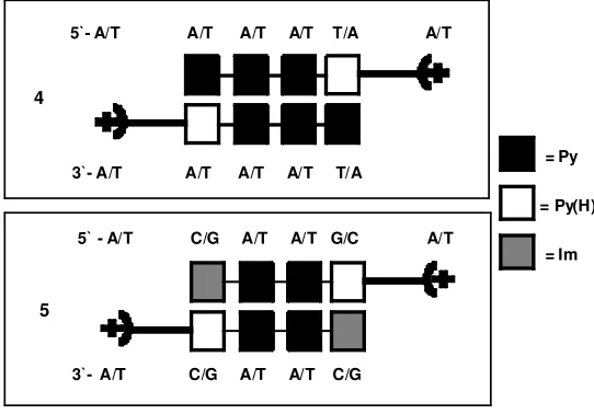
Table 4.1List of thermal denaturation data and SPR binding constants……..….92 Table 4.2 Binding constants of polyamides 3, 5 and 1 to three differing DNA
sequences………..…….93
CHAPTER 5: AFFINITY AND KINETIC MODULATION OF NOVEL SYNTHETIC POLYAMIDE DERIVATIVES
Table 5.1Results for ITC and SPR experiments………...121 Table 5.2 Results for thermal denaturation and DNase I footprinting
experiments………...………..122 Table 5.3 Estimated half-lives for the rate of dissociation of polyamides……..123
CHAPTER 6: PROMOTER SCANNING OF THE HUMAN COX-2 GENE WITH 8-RING POLYAMIDES: UNEXPECTED WEAKENING OF POLYAMIDE-DNA BINDING AND SELECTIVITY BY REPLACING AN INTERNAL N-Me-PYRROLE WITH β-ALANINE
LIST OF FIGURES
CHAPTER 1: AN INTRODUCTION TO AND HISTORY OF POLYAMIDES: NETROPSIN AND DISTAMYCIN, NATURALLY OCCURING
POLYAMIDES THAT TARGET THE MINOR GROOVE OF DNA IN A SEQUENCE SPECIFIC MANNER AND DEMONSTRATE BIOLOGICAL ACTIVITY
Figure 1.1Chemical structure of the polyamide netropsin (A) and distamycin (B)………..3 Figure 1.2 X-ray crystallographic structure of 1:1 netropsin-DNA complex……..5 Figure 1.3 NMR structure of 2:1 distamycin-DNA complex………..7 Figure 1.4Schematic representation of G-C recognition of Im-Py………...10
CHAPTER 2: DNA RECOGNITION: DESIGN, SYNTHESIS AND BIOPHYSICAL CHARACTERISTICS OF PYRROLE (H) BASED POLYAMIDES
Figure 2.1 Structures of Distamycin 1, synthesized polyamides 2 and 3………..37 Figure 2.2 Binding of 2 and 3 to DNA in a 2:1 motif………...38 Figure 2.3 Circular dichroism data for 2 and 3………..39 Figure 2.4 DNase I footprinting data of polyamides 2 (A) and 3 (B)………40 Figure 2.5 SPR sensorgrams of 3 with 3 different DNA sequences...…………...41
CHAPTER 3:SYNTHESIS AND DNA BINDING PROPERTIES OF
1-(3-AMINOPROPYL)IMIDAZOLE-CONTAINING TRIAMIDE f-Im*PyIm: A NOVEL DIAMINO POLYAMIDE DESIGNED TO TARGET 5’-ACGCGT-3’
CHAPTER 4: NOVEL DIAMINO IMIDAZOLE AND PYRROLE –CONTAINING POLYAMIDES: SYNTHESIS AND DNA BINDING STUDIES OF MONO- AND DIAMINO-PHENYL f-ImPy*Im DESIGNED TO TARGET 5’-ACGCGT-3’
Figure 4.1 Structures of f-ImPyIm and ph-Im-Py-Im………...………….84
Figure 4.2 Schematic of phenyl containing polyamides bound to DNA………...85
Figure 4.3 DNase I footprinting of ph-ImPyIm mono and dication………..86
Figure 4.4 CD data for ph-ImPyIm and the dication...………..87
Figure 4.5 SPR sensorgrams for mono and dicationic ph-ImPyIm………...88
Figure 4.6 Raw ITC and integrated heat data………...……….89
CHAPTER 5: AFFINITY AND KINETIC MODULATION OF NOVEL SYNTHETIC POLYAMIDE DERIVATIVES Figure 5.1 Structures of f-ImPyIm derivatives………116
Figure 5.2 SPR sensorgrams for f-ImPy(Gly)Im……….117
Figure 5.3 Raw ITC and integrated heat data for f-ImPy(Gly)Im ………..118
Figure 5.4 DNase I footprinting of f-ImPy(Gly)Im ...……….119
Figure 5.5 CD data for f-ImPy(Gly)Im ..………...120
CHAPTER 6: PROMOTER SCANNING OF THE HUMAN COX-2 GENE WITH 8-RING POLYAMIDES: UNEXPECTED WEAKENING OF POLYAMIDE-DNA BINDING AND SELECTIVITY BY REPLACING AN INTERNAL N-Me-PYRROLE WITH β-ALANINE Figure 6.1 Structures of compounds PA1-PA4………...140
Figure 6.2Map of predicted polyamide-DNA interactions………141
Figure 6.3Polyamide-DNA binding as observed via fluorescence spectroscopy………142
LIST OF SCHEMES
CHAPTER 2: DNA RECOGNITION: DESIGN, SYNTHESIS AND BIOPHYSICAL CHARACTERISTICS OF PYRROLE (H) BASED POLYAMIDES
Scheme 21 Synthesis of 2 and 3………...42
CHAPTER 3:SYNTHESIS AND DNA BINDING PROPERTIES OF
1-(3-AMINOPROPYL)IMIDAZOLE-CONTAINING TRIAMIDE f-Im*PyIm: A NOVEL DIAMINO POLYAMIDE DESIGNED TO TARGET 5’-ACGCGT-3’
Scheme 3.1 Reagents and conditions………49 CHAPTER 4: NOVEL DIAMINO IMIDAZOLE AND PYRROLE –CONTAINING
POLYAMIDES: SYNTHESIS AND DNA BINDING STUDIES OF MONO- AND DIAMINO-PHENYL f-ImPy*Im DESIGNED TO TARGET 5’-ACGCGT-3’
LIST OF ABBREVIATIONS
Im N-methylimidazole
Py N-methylpyrrole
f formamido
Hp 3’-hydroxy-1 H-pyrrole
DNA deoxyribonucleic acid
A adenine
T thymine
G guanine
C cytosine
MG minor groove
Oligo oligonucleotide
nt nucleotide
ss single-stranded
ds double-stranded
HP DNA hairpin deoxyribonucleic acid
ITC isothermal titration calorimetry
SPR surface plasmon resonance
RU response units
CD circular dichroism
UV ultraviolet
Vis visible
ppm parts per million
CCL cacodylic acid
HEPES N-[2-Hydroxyethyl]piperazine-N´-[2-enthanesulfonic acid]
EDTA ethylenediaminetetraacetic acid
NIH National Institutes of Health
CHAPTER 1:
AN INTRODUCTION TO AND HISTORY OF POLYAMIDES: NETROPSIN AND DISTAMYCIN, NATURALLY OCCURING POLYAMIDES THAT TARGET THE
1.1 Discovery of netropsin and distamycin
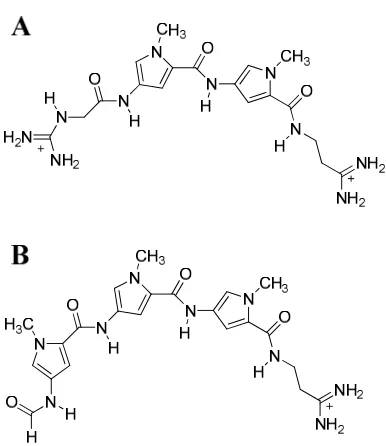
Netropsin is a naturally occurring oligopeptide first discovered and isolated by Finlay et al. in 1951 (1). This oligopeptide has been of significant interest over the last sixty years because of its biological activity. Netropsin has shown both antibacterial and antiviral activity and is classified as a pyrrole (Py)-containing amide antibiotic, although it is not used clinically because of toxicity. It was isolated from the actinobacterium Streptomyces netropsis and given the name “netropsin” (1). Netropsin small molecules consist of two pyrrole rings and two terminal amidine groups linked together by amide (peptide) bonds, as shown in Figure 1A.
Distamycin is also a naturally occurring oligopeptide discovered in 1958 by Arcamone et al. and isolated from an actinomycete bacterium, Streptomyces distallcius, of the same
Figure 1. Chemical structure of the polyamide netropsin (A) and distamycin (B).
1.2 Polyamides as DNA minor groove binders
distamycin, preferentially bind to the minor groove of DNA in a sequence specific manner. It has been shown that netropsin and distamycin preferentially bind to AT-rich sequences. The narrower minor groove enhances electrostatic interactions between the small molecule and the minor groove.
Netropsin and distamycin are comprised of multiple simple aromatic ring systems, linked together by bonds which have torsional freedom. The torsional freedom of these bonds allows them to adopt a crescent shape which is a typical, yet a key structural property of minor groove binding (2). The torsional freedom and crescent shape adopted by the small molecule allows it to twist in such a way that it becomes isohelical with the DNA minor groove. Because the polyamide and DNA are isohelical, contacts between the two are optimized (2).
that of the minor groove of GC-rich DNA sequences, therefore, the favorable binding to AT-rich sequences by positively charged small molecules, such as netropsin and distamycin, is favorable. The ability of the exocyclic amine to become a hydrogen bond donor can be exploited for sequence recognition and enhanced binding affinity by modified polyamides. This concept will be addressed in section 1.4.
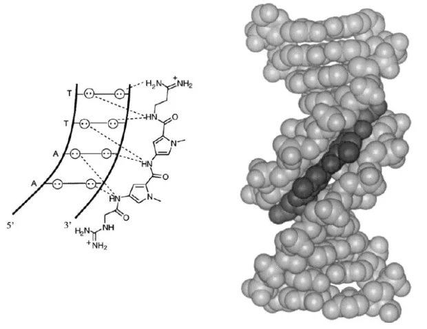
The first crystal structure of a netropsin-DNA complexwith 5’-(dCGCGAATTCGCG)2-3’ was solved by Dickerson et al. in 1985 (5). The complex showed a single netropsin molecule bound to the AATT-site, as was expected. The three amide –NH groups point towards the floor of the minor groove forming bifurcated hydrogen bonds with the O2 of thymine and N3 of adenine, as shown in Figure 2. van der Waals contacts are formed between the tightly bound polyamide and the atoms which comprise the sugar-phosphate walls of the groove, creating a parallel plane between the pyrrole rings of netropsin and the walls of the groove. The interaction of the two cationic amidine groups and the N3 of the 5’ adenine is favorable and a key
component of complex formation. The methyl groups at the N5 position of the pyrrole rings point into the solution, resulting in an ability for the compound to penetrate into the minor groove.
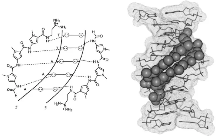
The crystal structure for distamycin has several key features very similar to that of netropsin. However, an NMR study by Wemmer et al. in 1990 showed distamycin bound as a stacked homodimer to the sequence 5’-CGCGAAATTTCGCG-3’(Figure 3) (3, 6-8). The additional two base pairs (compared to the AATT site) in the central recognition site allowed for the “longer” distamycin molecule and could accommodate the stacked dimer even in the narrower AT-rich minor groove. Data from this study showed that the 2:1 distamycin-DNA complex was favored for binding sites six base pairs or longer, and the 1:1 complex for a binding site of four base pairs (5’-AATT-3’).
are at opposite ends (antiparallel), thus maximizing the distance between the charges and reducing the electrostatic repulsion. Because netropsin has two positive charges on the terminal amidine groups, one on each end, the electrostatic repulsion would be too great for netropsin to bind as a homodimer and this explains why the observed stoichiometry of netropsin-DNA complexation is 1:1 and not 2:1. In order to accommodate the two distamycin molecules, the minor groove must widen. The structural change as a result of 2:1 distamycin-DNA
complexation causes a positive, unfavorable overall free energy contribution. Although the widening of the groove is unfavorable, it is compensated by a highly favorable hydrophobic interaction that is a direct result of stacking and binding two ligands within one binding site of the minor groove.
[image:30.612.79.437.396.621.2]
1.3 Netropsin and distamycin interactions: A look at energetics
The thermodynamic and binding properties of both netropsin and distamycin to DNA have been studied extensively. Because of advancements in instrumentation over the last twenty years, enthalpic and entropic contributions to the overall free energy have been measured, giving a more complete thermodynamic profile and defining the driving force behind complexation. The 2:1 binding of distamycin to 5’-AAATT-3’ compared to the 1:1 complex formation of netropsin and 5’-AAATT-3’ have been examined and compared (9). Both netropsin and distamycin have favorable binding enthalpy contributions and are exothermic processes. Drug binding is most often associated with a favorable enthalpic binding process (2, 3). The enthalpy for netropsin binding to 5’-AAATT-3’ is ~-31 kJ/mol. The enthalpy for binding two distamycin molecules to the same DNA sequence are ~-52 kJ/mol for the first molecule and ~79 kJ/mol for the second (2). Keq for netropsin is ~108 M-1 (10) and K1 and K2 for distamycin are ~107 M-1 and
106 M-1, respectively. Because K2 is ~10 times less than K1, the binding of two distamycin
the overall Gibbs free energy are opposite for the two polyamides. Netropsin has a favorable entropy whereas distamycin has an unfavorable entropy. The differences in entropy are attributed to changes in hydration; water is released or taken up upon binding, and the release of counter ions.
1.4 Synthetic derivatives of netropsin and distamycin
Since their discovery sixty years ago, netropsin and distamycin have served as templates for the design of synthetic polyamides. In the mid-late 1980’s, Lown (11) and Dickerson et al.
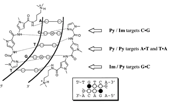
(12), developed synthetic polyamides, lexitropsins, which could target G-C and C-G sequences (13). This was accomplished by substituting an N-methyl pyrrole (Py) with an N-methyl imidazole (Im). The additional nitrogen in the imidazole ring acts as a hydrogen bond acceptor and can hydrogen bond with the exocyclic amino group of guanine. Targeting of G-C and C-G sequences through the use of Im rings developed by Lown and Dickerson were exploited by Dervan, who eventually developed a set of binding rules which are outlined in Figure 4 (14-18). Figure 4 shows a schematic view of binding the synthetic polyamide Im-Py-Py to the sequence 5’-TGTCA-3’ (4). In theory, this concept allowed for the programmable targeting of any specific DNA sequence. The idea of programmably targeting specific DNA sequences led to an explosion in synthetic efforts of numerous laboratories throughout the scientific community.
The two differences between netropsin and distamycin are: 1) netropsin has two pyrrole rings and distamycin has three, and 2) netropsin has two positively charged termini groups and distamycin has one cationic amidine terminus and a formamido (f) group at the other terminus. Most synthetic efforts have left off the terminal formamido group because of synthetic
addressed and studied by Lacy et al. in 2001 (20). The addition of a terminal formamido led to a staggered, antiparallel stacking formation and a 10-100 fold increase in binding affinity
[image:33.612.60.399.302.504.2]depending on the synthetic polyamide. This systematic study revealed the importance of the terminal formamido group and its influence on the mode of stacking, which directly impacts the binding affinity and kinetics of complexation (20). The study also showed the presence of a terminal formamido group had no advantage in terms of sequence specificity.
From the development of Dervan’s pairing rules, numerous different types of polyamides have been developed to optimize both binding affinity and sequence specificity. Upon the discovery of the 2:1 complex formation of distamycin with 5’-AAATTT-3’ through Wemmer’s NMR structure, synthetic developments have been made to covalently link the two dimers. This led to the development of hairpin polyamides, where two polyamides are linked from the 3’ terminus (end) of one to the 5’ terminus (tail) of the other, H-pin polyamides, where two polyamides are linked via N5 position of both central Py or Im rings, and cyclic polyamides, where two polyamides are linked from both the 5’ and 3’ ends to the opposing 3’ and 5’ ends of the second polyamide molecule. The lengths of the linking moieties have been well studied for all three polyamide derivatives, as have the addition of functional groups on the linkers (21-24).
1.5 Polyamides control gene expression: potential as therapeutics to treat various diseases
Since their discovery and isolation, netropsin and distamycin have been known to have biological activity as antibiotic, antiviral and antitumor therapeutics, leading to the hypothesis that these two molecules are synthesized naturally by streptomycete strains as a potential defense mechanism (3). Though too toxic for clinical use, these compounds have been used to elucidate the origins of sequence specificity and biological activity. These molecules are thought to interfere with topoisomerase in some fashion, although their true mechanism is not completely understood. Excitingly, synthetic Py- and Im-containing polyamides have not shown the same levels of toxicity as netropsin and distamycin.
the (CH2)3 alkyl linker which links the terminal secondary amine and the last pyrrole ring. This
synthetic hairpin polyamide binds to the sequence 5’-TAGTACT-3’, which is found within the promoter site of the TFIIA gene, and causes inhibition of the 5S RNA through direct
competition. In vivo studies showed the presence of the hairpin polyamide led to a reduction in 5S RNA by interfering with the transcriptional process of the TFIIA gene. Although a debate remains about the viability of larger hairpin polyamides and their ability to permeate into the cells and localize in the nucleus, Gottesfeld et al. presented a strong first argument for the use of polyamides as therapeutics showing direct evidence addressing these issues (3, 25).
Modulation of gene expression, specifically the inverted 5’-CCAAT-3’ of the
1.6 Objectives of this Dissertation
References
1. Finlay, A. C., Hochstein, F. A., Sobina, B. A., and Murphy, F. X. (1951) Netropsin, a new antibiotic produced by a Streptomyces, J. Am. Chem. Soc.73, 341-343.
2. Blackburn, G. M., and Gait, M. J. (1997) Nucleic Acids in Chemistry and Biology, 2nd ed., Oxford University Press, New York.
3. Wemmer, D. E. (2001) Ligands recognizing the minor groove of DNA: Development and applications, Biopolymers52, 197-211.
4. Dervan, P. B. (2001) Molecular recognition of DNA by small molecules, Bioorg. Med. Chem.9, 2215-2235.
5. Kopka, M. L., Yoon, C., Goodsell, D., Pjura, P., and Dickerson, R. E. (1985) Binding of an antitumor drug to DNA, netropsin and C-G-C-G-A-A-T-T-BrC-G-C-G, J. Mol. Biol. 183, 553-563.
6. Pelton, J. G., and Wemmer, D. E. (1990) Binding modes of distamycin A with
d(CGCAAATTTGCG)2 determined by two-dimensional NMR, J. Am. Chem. Soc.112, 1393-1399.
7. Coll, M., Aymami, J., van der Marel, G. A., van Boom, J. H., Rich, A., and Wang, A. H. (1989) Molecular structure of the netropsin-d(CGCGATATCGCG) complex: DNA conformation in an alternating AT segment, Biochemistry28, 310-320.
8. Coll, M., Fredrick, C. A., Wang, A. H.-J., Rich, A. P., and Natl Acad Sci USA 1987. (1987) A bifurcated hydrogen-bonded conformation in the d(A.T) base pairs of the DNA dodecamer d(CGCAAATTTGCG) and its complex with distamycin., Natl Acad Sci USA 84, 8385–8389.
9. Rentzeperis, D., Marky, L. A., Dwyer, T. J., Geierstanger, B. H., Pelton, J. G., and Wemmer, D. E. (1995) Interaction of minor groove ligands to an AAATT/AATTT site: Correlation of thermodynamic characterization and solution structure, Biochemistry34, 2937-2945.
10. Nguyen, B., Tanious, F., and Wilson, W. D. (2006) Biosensor-surface plasmon resonance: Quantitative analysis of small molecule–nucleic acid interactions, Methods 42, 150-161.
11. Lown, J. W., Krowicki, K., Bhat, U. G., Skorobogaty, A., Ward, B., and Dabrowiak, J. C. (1986) Molecular recognition between oligopeptides and nucleic acids: novel imidazole-containing oligopeptides related to netropsin that exhibit altered DNA sequence specificity, Biochemistry25, 7408–7416.
12. Kopka, M. L., Yoon, C., Goodsell, D., Pjura, P., and Dickerson, R. E. (1985) The molecular origin of DNA-drug specificity in netropsin and distamycin, Proc. Natl. Acad. Sci. U S A82, 1376-1380.
13. Lee, M., Hartley, J. A., Pon, R. T., Krowicki, K., and Lown, J. W. (1988) Sequence spcific molecular recognition by a monocationic lexitropsin of the DNA
d(CATGGCCATG): structural and dynamic aspects deduced from high field proton NMR studies, Nucleic Acids Res.16, 665-684.
15. Geierstanger, B. H., Jacobsen, J. P., Mrksich, M., Dervan, P. B., and Wemmer, D. E. (1994) Structural and dynamic characterization of the heterodimeric and homodimeric complexes of distamycin and 1-methylimidazole-2-carboxamide-netropsin bound to the minor groove of DNA, Biochemistry33, 3055-3062.
16. Geierstanger, B. H., Mrksich, M., Dervan, P. B., and Wemmer, D. E. (1994) Design of a G.C-specific DNA minor groove-binding peptide, Science266, 646-650.
17. Kielkopf, C. L., Baird, E. E., Dervan, P. B., and Rees, D. C. (1998) Structural basis for G.C recognition in the DNA minor groove, Nat. Struct. Biol.5, 104-109.
18. Kielkopf, C. L., White, S., Szewczyk, J. W., Turner, J. M., Baird, E. E., Dervan, P. B., and Rees, D. C. (1998) A structural basis for recognition of A.T and T.A base pairs in the minor groove of B-DNA, Science282, 111-115.
19. Hawkins, C. A., Clairac, R. P. d., Dominey, R. N., Baird, E. E., White, S., Dervan, P. B., and Wemmer, D. E. (2000) Controlling binding orientation in hairpin polyamide DNA complexes, J. Am. Chem. Soc.122, 5235-5243.
20. Lacy, E. R., Le, N. M., Price, C. A., Lee, M., and Wilson, W. D. (2002) Influence of a terminal formamido group on the sequence recognition of DNA by polyamides, J. Am. Chem. Soc.124, 2153-2163.
21. O'Hare, C. C., Mack, D., Tandon, M., Sharma, S. K., Lown, J. W., Kopka, M. L., Dickerson, R. E., and Hartley, J. A. (2002) DNA sequence recognition in the minor groove by crosslinked polyamides: The effect of N-terminal head group and linker length on binding affinity and specificity, Proc Natl Acad Sci U S A99, 72-77.
22. Chenoweth, D. M., and Dervan, P. B. (2009) Allosteric modulation of DNA by small molecules., Proc. Natl. Acad. Sci.106, 13175-13179.
23. Mrksich, M., Parks, M. E., and Dervan, P. B. (1994) Hairpin peptide motif: A new class of oligopeptides for sequence specific recognition in the minor groove of double helical DNA, J. Am. Chem. Soc.116, 7983-7988.
24. Mrksich, M., and Dervan, P. B. (1993) Enhanced sequence specific recognition in the minor groove of DNA by covalent peptide dimers:
bis(pyridine-2-carboxamideonetropsin)(CH2)3-6, J. Am. Chem. Soc.115, 9892-9899.
25. Gottesfeld, J. M., Neely, L., Trauger, J. W., Baird, E. E., and Dervan, P. B. (1997) Regulation of gene expression by small molecules, Nature387, 202-205.
26. Le, N. M., Sielaff, A., Cooper, A. J., Mackay, H., Brown, T., Kotecha, M., O'Hare, C., Hochhauser, D., Lee, M., and Hartley, J. A. (2006) Binding of f-PIP, a pyrrole- and imidazole-containing triamide, to the inverted CCATT box-2 of the topoisomerase IIα
promoter and modulation of gene expression in cells Bioorg Med Chem Letters16, 6161-6164.
27. Fortune, J. M., Velea, L., Graves, D. E., Utsugi, T., Yamada, Y., and Osheroff, N. (1999) DNA topoisomerases as targets for the anticancer drug TAS-103: DNA interactions and topoisomerase catalytic inhibition, Biochemistry38, 15580-15586.
28. Fortune, J. M., and Osheroff, N. (2000) Topoisomerase II as a target for anticancer drugs: When enzymes stop being nice, Prog. Nucleic Acid Res. Mol. Biol. 64, 221-253.
29. Isaacs, R. J., Harris, A. L., and Hickson, I. D. (1996) Regulation of the human
CHAPTER 2:
DNA RECOGNITION: DESIGN, SYNTHESIS AND BIOPHYSICAL CHARACTERISTICS OF PYRROLE (H) BASED POLYAMIDES
DNA Recognition: Design, Synthesis and Biophysical
Characteristics of Pyrrole(H) based Polyamides.
Sameer Chavdaa, Keith Muldera, Toni Browna, Hilary Mackaya, Balaji Babua, Laura Westratea, Amanda Fergusona, Shicai Linc, Konstantinos Kiakosc, Joseph P. Ramosb, Manoj Mundeb, W. David Wilsonb, John A. Hartleyc and Moses Leea,
a
Department of Chemistry and the Division of Natural and Applied Sciences, Hope College 49423, USA
bDepartment of Chemistry, Georgia State University Atlanta, GA, 30303, USA
2.1 Abstract
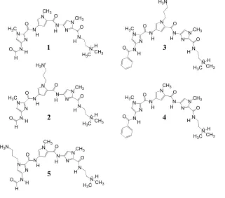
N-Methyl imidazole (Im) and N-methyl pyrrole (Py)-containing polyamides that can form stacked dimers can be programmed to target specific DNA sequences in the minor groove of DNA and control gene expression. Polyamides are being investigated as potential medicinal agents for treating diseases including cancer. The naturally occurring polyamide distamycin binds as a dimer in the minor groove of DNA and recognizes sequences rich in A/T and T/A base pairs indiscriminately. Synthetic analogs of distamycin that incorporate N-methylimidazole into the heterocyclic core have been shown to bind to G/C rich sequences with a high degree of specificity. The purpose of this study is to investigate the behavior of polyamides containing the 2, 5-linked N-methylpyrrole-2-carboxamide or pyrrole(H) [Py(H)] moiety upon binding to DNA. The synthesis and biophysical characteristics of two polyamides PyPyPyPy(H) 2 and
ImPyPyPy(H) 3 designed to test the binding preference of a Py/Pyrrole(H) pairing [Py/Py(H)] and a [Im/Py(H)] is described. Studies utilizing circular dichroism, thermal denaturation (∆TM),
biosensor-surface plasmon resonance and DNase I footprinting show that an [Im/Py(H), 3] pairing prefers a G/C or C/G pairing whilst a [Py/Py(H), 2] pairing tolerates A/T or T/A base pairs and avoids a G/C base pair.
2.2 Introduction
The development of polyamide analogs of distamycin 1 (Figure 2.1) that can target specific DNA sequences is a fascinating and well documented area of research. Such compounds exhibit potential for use as gene regulatory agents by inhibiting the binding of native
transcription factors at the target promoter site, and are being investigated as potential medicinal agents for treating diseases including cancer [1]. The pyrrole (Py) heterocyclic moieties in distamycin stack as a 2:1 antiparallel dimer in the DNA minor groove and can bind indiscriminately to an A/T or a T/A base pair [2a-d]. Alternatively, an imidazole (Im)
heterocycle moiety can be paired opposite a pyrrole to selectively target a G/C base pair [2d]. In addition, 3-hydroxy-1H-pyrrole (Hp) has been paired opposite Py and shown to selectively target T/A base pairs but with a significant loss in binding affinity. This could possibly attribute to a handful of factors ranging from sterics to chemical instability [3a-c]. The bulky hydroxyl group on an Hp containing polyamide (which forms an additional hydrogen bond to the O-2 group of thymine), makes the 2:1 complex formed in the minor groove sterically encumbered leading to unfavorable binding. Accordingly the additional hydrogen bonding to thymine O-2 could be critical for distinguishing A/T from T/A. We envisaged that a polyamide containing a reversed conformation Py moiety [Py(H) or 2,5-linked N-methylpyrrole-2-carboxamide] stacked over a polyamide containing Py [Py(H)/Py] may recognize a T/A base pair. In addition, it has been reported that the N-H group on this Py(H) moiety should form a more favorable van der Waals distance with thymine thus resulting in enhanced binding affinity [4]. In an attempt to test this hypothesis, we chose to synthesize two polyamides PyPyPyPy(H)-N
shown that incorporation of multiple 2,5-linked [3a] or 2,4-linked Py(H) [4a] units may result in a loss in sequence specificity, thus allowing for an increased tolerance of G/C base recognition [4a]. Even though polyamides bind DNA effectively through the 2:1 stacked motif, they are also known to bind in a 1:1 fashion within the minor groove of DNA [4b].
2.3 Results and Discussion
2.3.1 Polyamide and DNA sequence design
2.3.2 Synthesis
The synthetic approach towards obtaining polyamides 2 and 3 is outlined in Scheme 2.1. Nitration of 4 furnished 4-nitro-2-trichloroacetylpyrrole 5 in good yield, which in turn, was coupled with commercially available N,N-dimethyl-3-aminopropylamine in the presence triethylamine to furnish the monoheterocyclic component 6 [8]. This was followed by
subsequent reduction of 6 using standard palladium catalyzed hydrogenation [9]to the resulting amino derivative (not shown) followed by immediate coupling to 4-nitro-1-methylpyrrole-2-carbonyl chloride 7 (formed from the corresponding carboxylic acid). The resulting diamide (O2N-PyPy(H)-N-dimethyl-3-aminopropylamine, 8), formed in 22% yield was, in turn, coupled
to another molecule of 4-nitro-1-methylpyrrole-2-carbonyl chloride 7 using the protocol described, to furnish the corresponding triamide (O2N-PyPyPy(H)-tail 9 in 39% yield (Scheme
2.3.3 Thermal melting studies
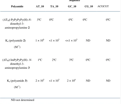
Thermal melting studieswere conducted to determine whether polyamides 2 and 3
stabilize duplex DNA at elevated temperatures. The data shown in Table 1 indicates that polyamide 2 can only produce a ∆TM value of 3°C for the AT_10 sequence. TA_10, CG_10 and
GC_10 did not increase the melting temperature in the presence of tetraamide 2. On the other hand polyamide 3 exhibits some stabilization on sequences AT_10, TA_10 and GC_10 giving
∆TM values of 2, 2 and 3°C, respectively but did not induce a ∆TM for CG_10.
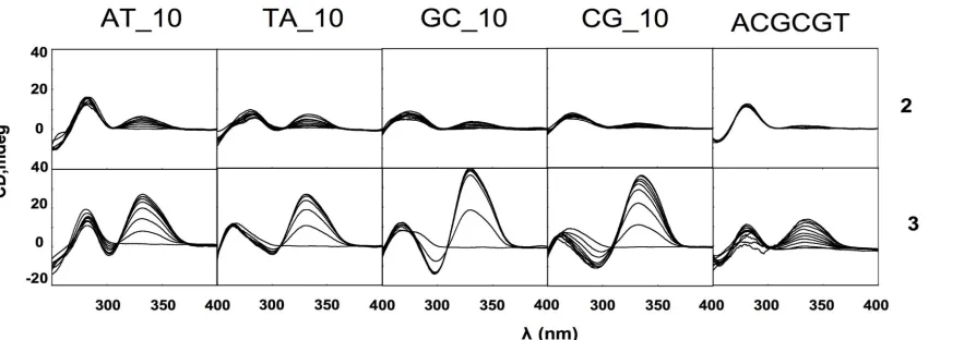
2.3.4 Circular dichroism studies
Circular dichroismstudies (the ability of chiral molecules to absorb circularly polarized light: the DNA/polyamide complex is chiral due to the DNA’s inherent chirality) were performed on PyPyPyPy(H)-N-dimethyl-3-aminopropylamine 2 and 3 ImPyPyPy(H)-N -dimethyl-3-aminopropylamineon the same DNA sequences used in the thermal denaturation studies (Figure 3). The results shown in Figure 2.3 for 3 confirm that it binds within the floor of the minor groove of the DNAs tested due to the appearance of an induced band at 330 nm [12]. The results also indicate that polyamide 2 gaveweakerinduced CD bands than 3 when titrated into solutions of the DNAs mentioned. Except for AT_10, the results were in agreement with the thermal denaturation data shown in Table 2.1. However, as depicted in Figure 3 polyamide 2
produced clear DNA induced ligand bands when titrated into solutions of AT_10 and TA_10 suggesting a limited preference of Py/Py(H) for an AT or TA base pair. The most impressive result was achieved when polyamide 3 was titrated into a solution of GC_10 with near-saturation being achieved after addition of two equivalents of 3, further corroborated by a ∆TM value of 3
occurring via a single motif characteristic of a typical DNA minor groove binder. It is interesting to note that, though a ∆TM value of 0°C was observed for polyamide 3 when titrated into a
solution of CG_10 (Table 2.1), a significant amount of binding is observed in the CD study (Figure 3) in turn, suggesting binding of 3 to this DNA sequence is possibly thermodynamically less favorable than when binding to GC_10.
2.3.5 DNase I footprinting
DNase I footprinting experiments were performed on 2 and 3 using a 131 bp 5`-[32 P]-radiolabelled DNA fragment containing the following sequences: (a) 5`-AAATTT-`3; AT_10 (b) ATATAT-`3; TA_10 (c) ACATGT-`3; CG_10 (d) AGATCT-`3; GC_10 and (d) 5`-AGAGCT-`3. Autoradiogram A (Figure 2.4) shows that polyamide 2 exhibits good sequence specificity mostly for TA_10 DNA. A clear footprint appears at 20 µM, which is consistent with CD and ∆TM measurements (Table 2.1 and Figure 2.3). Polyamide 2 also displays negligible
binding to GC_10 as shown by the appearance of a weak footprint at 100 µM. In comparison, 3
seems to show better binding affinity towards the sequences tested but poorer sequence
2.3.6 Surface Plasmon Resonance
To obtain a more accurate measure of binding affinity, selectivity and to probe the stoichiometry of binding, SPR-biosensor experiments were performed. As described in the Methods Section, the biotin-labeled GC_10, AT_10 and TA_10 DNA hairpin duplexes were immobilized by streptavidin capture in three of the four flow cells. The fourth was left blank and used as a control. Compounds 2 and 3 were then injected over the DNA and blank flow cells at a range of concentrations. From the sensorgrams in Figure 2.5, it can be observed that 3 shows good selectivity for GC_10. The kinetics are relatively fast and all of the sensorgrams reach a steady state plateau below 200 s. Binding constants were determined by plotting the steady state RU values, determined by averaging over a 10 s time period in the steady state region, versus the compound concentration and fitting with either a one or two site binding model (Methods Section). Compound 3 binds strongly to GC_10 (K= 2 × 10⁶ M-1, Table 2.1) with 2:1
stoichiometry but shows weaker binding towards AT_10 by 10-fold (K= 2 × 10⁵ M-1) with the data best fitting a 1:1 stoichiometry. Very weak binding was observed for 3 with TA_10 and the stoichiometry could not be determined. Binding constants were determined by the same
2.4 Conclusion
Overall, this study has shown that, polyamide 2 containing the Py/Py(H) pairing, displays weaker binding than expected to its cognate sequences. The results do show sequence selectivity (towards AT_10), in agreement with its predicted specificity, but with a lack of marked binding affinity. However, it prefers to avoid a G/C base pair, as ascertained by SPR, DNase I
footprinting and thermal melting techniques. On the other hand, 3, which contains an Im/Py(H) pairing, prefers a G/C or C/G base pair. For these reasons, we believe that additional polyamides related to 2 and 3 are worthy of further investigation.
2.4.1 Experimental
Solvents and organic reagents were purchased from Aldrich or Fisher, and in most cases were used without further purification. DCM (P2O5), and DMF (BaO) were distilled prior to use.
Melting points (mp) were performed using a Mel-temp instrument and are uncorrected. Infrared (IR) spectra were recorded using a FT-IR instrument as films on NaCl discs. 1H-NMR spectra were obtained using a Varian Unity Inova 400 instrument. Chemical shifts (δ) are reported at 20
°C in parts per million (ppm) downfield using tetramethylsilane (Me4Si) as an internal standard.
High-resolution mass spectra (HRMS) and low-resolution mass spectra (LRMS) were provided by the Mass Spectrometry Laboratory, University of South Carolina, Columbia. Reaction progress was assessed by thin-layer chromatography (TLC) using Merck silica gel (60 F254) on
2.4.2 Synthesis
2-Trichloroacetylpyrrole 4 and 5-nitro-2-trichloroacetylpyrrole 5
These precursor compounds were synthesized according to a literature protocol and were spectroscopically identical to those reported [3].
O2N-Py(H) N,N-dimethyl-3-aminopropylamine 6
O2N-Py(H)-CCl3 (500 mg, 1.95 mmol) was dissolved in dry CH2Cl2 (60 mL) and triethylamine
(0.41 mL, 2.93 mmol), N,N-dimethyl-3-aminopropylamine (0.37 mL, 2.93 mmol) was added and the reaction mixture was heated at reflux for ~18 h. The reaction was quenched with water (50 ml) and the resulting aqueous basified to pH 10 was and the organic layer was collected. The aqueous was extracted with CHCl2 (2 × 40 mL). The combined organic layers were dried over
Na2SO4 and evaporated to dryness. The crude product was purified using flash chromatography
on silica gel eluting with to yield 6 as an orange/yellow solid (300 mg, 68%); RF 0.46 (silica gel,
40:39.5:0.5 MeOH:CHCl3:NH4OH); mp 154-160 ºC; υ (cm-1) 1692 (C=O); δH (400 MHz;
CDCl3) 7.65 (1 H, d, J 1.6 Hz); 7.15 (1 H, d, J 1.6), 3.37 (2 H, t, J 6.8), 2.49 (2 H, t, J 6.8); 2.34
(6 H, s) and 1.76 (2 H, quintet, J 6.8); LRMS: (direct probe) m/z (rel. intensity) 240 ([M+], 5%), 139 (4%), 93 (4%), 72 (5%).
O2N-Py-Py(H) N,N-dimethyl-3-aminopropylamine 8
A round bottom flask purged with argon, containing O2N-Py(H)-N
-dimethyl-3-aminopropylamine, 6 (186 mg, 0.82 mmol), 5% Pd/C (93 mg, 50% by wt.) cold MeOH (60 ml) was cautiously added and the resulting solution was stirred under H2 for 12 h. The Pd/C catalyst
yellow/brown oil which was stored under high vacuum out of light. NO2-Py-COOH (0.168 g,
0.987 mmol) was dissolved in SOCl2 (3 mL) and refluxed for 15 minutes. The resulting solution
was co-evaporated with CHCl2 (3 × 15 mL) to give the acid chloride 7 as an oil which was
diluted with dry CH2Cl2 (5 mL) and added dropwise to a stirring solution of the reduced form of 6 and dry triethylamine (0.14 ml, 0.99 mmol) in dry CHCl2 (10 mL) cold (0 ºC, ice/water). The
reaction mixture was stirred at room temperature overnight. A basic aqueous work-up was performed and the organic layers collected, dried over (Na2SO4) and evaporated to dryness. The
crude product was purified using flash-column chromatography to yield 8 as a yellow solid (67 mg, 22%), mp. decomposition at 200 ºC; RF 0.20 (silica gel, 80:19.5:0.5 % v/v;
CHCl3:MeOH:NH4OH); υ (CH2Cl2, cm-1) 1640 (C=O) 1636 (C=O); δH (400 MHz, CDCl3) 10.08
(1 H, s (br), 9.24 (1 H, s (br)), 7.74 (1 H, s (br)), 7.59 ( 1 H, d, J 1.6), 7.37 (1 H, s), 7.36 (1 H, d,
J 1.6), 6.70 (1 H, s), 4.02 (3 H, s), 3.42 (2 H, t, J 6.4), 2.44 (2 H, t, J 6.4), 2.30 (6 H, s) and 1.76 (2 H, quintet, J 6.4); MS: (ES+) m/z (rel. intensity) 363 ([M+H] 100%), 255 (5%).
O2N-Py-Py-Py(H) N,N-dimethyl-3-aminopropylamine 9
In the same way as for 8,O2N-Py-Py(H)-N-dimethyl-3-aminopropylamine 8 (67 mg, 0.184
mmol), 5% Pd/C (35 mg, 50% by wt.) and cold MeOH (40 mL) gave the corresponding amine as a yellow oil. Also in the same way as 8, NO2-Py-COCl 7 (from NO2-Py-COOH, 37 mg, 0.22
mmol and 3) and dry triethylamine (0.03 ml, 0.22 mmol) gave crude 9 which was purified using flash chromatography eluting in to yield 9 as a yellow/brown solid (35 mg, 39%); mp
decomposition at 200 ºC: RF 0.3 (silica gel, 60:39.5:0.5 CHCl3:MeOH:NH4OH); υ (CH2Cl2, cm -1) 1640 (C=O) 1636 (C=O), 1638 (C=O); δ
H (400 MHz, CDCl3) 7.57 (1 H, d, J 2.0); 7.35 (1 H,
H, s), 3.90 (3 H, s), 3.39 ( 2 H, t, J 6.4), 2.63 (2 H, t, J 7.2); 2.45 (6 H, s) and 1.83 (2 H, quintet, J
7.2); LRMS: (ES+) m/z (rel. intensity) 485 ([M+H], 100%), 377 (20%).
Py-Py-Py-Py(H) N,N-dimethyl-3-aminopropylamine 2
In the same way as for the method stated in for the reduction of 8 and coupling of the resultant amine, O2N-Py-Py-Py(H)-N-dimethyl-3-aminopropylamine 9 (75 mg, 0.155 mmol), 5% Pd/C
(40 mg, 50% by wt) and cold MeOH (8 mL) gave the resultant amine as a orange/yellow oil. The amine from 11, Py-COCl 10 (from 39 mg, 0.309 mmol) pyrrole-2-carboxylic acid and dry triethylamine (0.04 mL, 0.30 mmol) gave a crude product was purified using flash
chromatography to yield 2 as an orange solid (25 mg, 28%), mp decomposition at 210 ºC; RF
0.30 (silica gel, 60:39.5:0.5 CHCl3/MeOH/NH4OH); 1640 (C=O) 1636 (C=O), 1638 (C=O) and
1637 (C=O); δH (400 MHz, CDCl3) 7.77 (1 H, d, J 2.0); 7.52 (1 H, d, J 1.6), 7.33 (1 H, d, J 1.6);
7.23 ( 1 H, d, J 1.6), 7.12 (1 H, s), 6.77 (2 H, d, J 1.6), 6.10 (1 H, d, J =1.6), 6.09 (1 H, d, J 1.6), 5.55 (2 H, 2 × s (br)), 5.15 (2 H, 2 × s (br)), 3.93 (3 H, s), 3.90 (3 H, s), 3.89 (3 H, s), 3.35 ( 2 H, t, J 7.2), 2.63 (2 H, t, J 7.2); 2.55 (6 H, s) and 1.85 (2 H, quintet, J 7.2); LRMS: 562 ([M+H], 100%), 282 (80%), 145 (90%); HRMS: C28H36N9O4 requires 562.2890; found 562.2901.
Im-Py-Py-Py(H) N,N-dimethyl-3-aminopropylamine 3
The reduction of 9 was achieved by following the same protocol as stated in for 8 using the following: O2N-Py-Py-Py(H)-tail 9 (75 mg, 0.155 mmol), 5%Pd/C (40 mg, 50% by wt.) and cold
(similar to that of described previously and the organic layer collected, dried over (Na2SO4) and
evaporated to dryness. The crude product was purified using flash-column chromatography to yield 3 as a yellow solid (15 mg, 17%), mp. decomposition at 210 ºC; RF 0.40 (silica gel,
60:39.5:0.5 CHCl3:MeOH:NH4OH); υ (neat, cm-1) 1640 (C=O) 1636 (C=O), 1638 (C=O) and
1635 (C=O); δH (400 MHz, CDCl3); 10.32 (1 H, s (br)), 9.45 (1 H, s (br)), 8.48 (1 H, s (br), 8.27
(1 H, s (br)) 7.89 (1 H, s (br)), 7.34 (1 H, d, J 2.4), 7.21 (1 H, d, J 2.4), 7.00 (1 H, t, J 4.8), 6.94 (1 H, s), 6.79 (1 H, s), 6.68 (1 H, d, J 6.5), 6.65 (1 H, d, J 6.5), 4.03 (3 H, s), 3.88 (3 H, s,), 3.38 (3 H, s), 3.40 (2 H, t, J 7.2), 2.45 (2 H, t, J 7.2), 2.21 (6 H, 2 × s) and 1.71-1.63 (2 H, quintet, J
7.2); LRMS: (ES+) m/z (rel. intensity) 563 ([M+H], 15%), 297 (45%), 282 (100%), 145 (20%); HRMS, C27H35N10O4 requires 563.2842; found C27H35N10O4 563.2847.
2.4.3 Biophysical
All buffer reagents were obtained from Sigma-Aldrich or Fisher, and used without further purification unless stated otherwise. The synthetic oligonucleotides were obtained from Operon (Huntsville, AL): ACGCGT (5'-ACGCGT-3'): 5'-GA ACGCGT CG CTCT CGACGCGTTC-3';
A3T3_10 (5'-A3T3-3'): 5'-CG AAATTT CC CTCT GG AAATTT CG-3'; AT_10 (AT-3'):
5'-CC AAATTT 5'-CC CTCTGG AAATTT GG; TA_10 (5'-TA-3'): 5'-CC ATATAT CC CTCTGG ATATAT GG; CG_10 (5'-CG-3'); 5'-CC ACATGT CC CTCTGG ACATGT GG and GC_10
(5'-GC-3'); 5'-CC AGATCT CC CTCTGG AGATCT GG were used in the thermal denaturation and circular dichroism experiments without further purification. DNA used in the footprinting and SPR experiments are described below. CD data was obtained using an Olis DSM20 instrument. ∆TM data were obtained using a Varian-Cary 100 Biomelt UV–visible
pH 6.4) was used for CD and Tm experiments. All data were analyzed using KaleidaGraph
(Snergy Software, Reading PA), unless otherwise stated. Ligand stock solutions were prepared in double distilled water at a concentration of 0.5 mM, unless otherwise specified.
2.4.4 Circular dichroism (CD)
CD studies were performed using previously reported procedures [14b,f] and were conducted at ambient temperature in a 1-mm path length quartz cell using PO4 buffer. Buffer
and stock DNA were added to the cuvette to give a final DNA concentration of 9 µM. Each polyamide (in 500µL in double distilled) H2O was titrated in 1 molar equivalents into the
relevant DNA (160 µL of 9 µM DNA) until saturation was achieved. Each run was performed over 400-220 nm. The CD response at the λmax of the induced peak was plotted against the mole
ratio of ligand:DNA.
2.4.5 Thermal denaturation (∆TM)
Thermal denaturation studies were performed using published procedures [3d].
Experiments for polyamides 2 and 3 were performed at a concentration of 3 µM ligand and 1 µM DNA. All experiments were run in PO4 buffer. Oligonucleotide samples were reannealed prior to
denaturation studies by heating at 70 ºC for 1 min then cooling to RT. Heating runs were typically performed between 25 and 95 ºC, with a heating rate of 0.5 ºC min–1, while
continuously monitoring the absorbance at 260 nm (digitally sampled at 200 ms intervals). All melts were performed in 10-mm path length quartz cells. Tm values were determined as the
2.4.6 Surface plasmon resonance (SPR)
Buffers: a 0.01 M cacodylic acid (CCA) solution pH 6.25 contained 0.001 M EDTA (disodium ethylenediamine tetraacetate), 0.1 M NaCl and 0.005% (by vol.) surfactant P20 which was (Biacore AB) used to reduce the nonspecific binding of polyamides to the fluidics and sensor chip surface. In addition, a 0.01 M HEPES (N-[2-Hydroxyethyl]piperazine-N’-[2-ethanesulfonic acid]) solution pH 7.4 containing 0.15 M NaCl, 0.003 M EDTA and 0.005% (by volume) surfactant P20 was used during the DNA immobilization process. All buffers were degassed and filtered prior to experiments. DNA sequences were obtained from Integrated DNA Technologies (San Diego, CA) with HPLC purification and were used without further purifying. The lyophilized 5’ biotin-labeled DNA hairpin constructs were dissolved in the appropriate amount of DI H2O to create 1.0 mM DNA stock solutions. Further dilutions were made using
0.01 M HEPES buffer during the DNA immobilization step. DNA concentrations were determined spectrophotometrically using extinction coefficients calculated for each individual DNA sequence by using the nearest neighboring for singled stranded DNA method. Stock solutions of 4 and 5 were prepared by dissolving the solid compound in the necessary amount of distilled H2O to create a stock solution with a concentration of approximately 1.0 x 10⁻³ M. Stock
solutions were kept frozen at 4 ˚C until experimental use to minimize any degradation of the compound. Samples for SPR experiments were prepared by a series of dilutions from the stock solution using a 0.01 M cacodylic acid buffer solution.
The surface plasmon resonance (SPR) experiments were conducted to analyze real time interactions between the polyamide compounds and specific DNA sequences. All SPR
streptavidin-coated sensor chip (BIAsensorSA) as previously reported [14]. The 5’-biotin-labeled DNA hairpins were immobilized on three of the four flow cells. The fourth was left blank and used as a control. All SPR experiments were performed at 25 ˚C and used 0.01 M CCA as the running buffer. The amount of DNA immobilized was approximately 400 response units (RU). This was achieved by continuously injecting ~ 20 µL of an approximately 50 nM DNA solution at a rate of 2 µL/min onto the sensor chip surface until a relative response of 400 units was reached. Binding data was obtained by injecting known concentrations and were analyzed with one or two site binding models as previously described [14].
2.4.7 DNase I Footprinting Studies
The designed DNA fragment was amplified by using PCR as follows. The forward primer 5’- GTCGGTTAGGAGAGCTCCACTTG-3’ (4 pmol) was 5’ 32P end-labeled with [-32P] ATP using T4 kinase (Invitrogen) following standard protocols. PCR amplification was performed in a reaction mixture of 50 µL containing the 32P labeled primer, 4 pmol of reverse primer 5’-CTCCAGAAAGCCGGCACTCAG-3’, 2 µL of 25 mM MgCl2, 2.5 µL of 2.5 mM
dNTPs (Promega, UK), 1U Flexi GoTaq polymerase (Promega,UK), the supplied 5x GoTaq buffer, and 0.125 pmol of each of the templates
5’ATGCTCCAGAAAGCCGGCACTCAGTCTACAAAAATTTCATCTTGATCATATATGTT CACAGAGCTCTCTCTAGATCTAGATCTAACTCTAGTACATGTCTTCAAGCAAGTGGA GCTCTCCTAACCGACTTT-3′ and
5′-AAAGTCGGTTAGGAGAGCTCCACTTGCTTGAAGACATGTACTAGAGTTA
PCR cycling conditions consisted of an initial denaturation step for 3 min at 95 °C, and 1 min at 94 °C, 1 min at 63 °C, and 1 min at 72 °C for a total of 35 cycles. The amplified fragment was isolated on a 2% agarose gel electrophoresis and purified with the Mermaid Kit (Q-biogene) according to the manufacturer’s instructions.
DNase I digestions were conducted in a total volume of 8 µL. The labeled DNA fragment (2 µL, 200 counts s-1) was incubated for 30min at room temperature in 4 µL TN binding buffer (10 mM Tris Base, 10 mM NaCl, pH 7) containing the required drug concentration. Cleavage by DNase I was initiated by addition of 2 µL of DNase I solution (20 mM NaCl, 2 mM MgCl2, 2
mM MnCl2, DNase I 0.02U, pH 8.0) and stopped after 3 min by snap freezing the samples on dry
ice. The samples were subsequently lyophilized to dryness and resuspended in 5 µL of
formamide loading dye (95% formamide, 20 mM EDTA, 0.05% bromophenol blue, and 0.05% cyanol blue). Following heat denaturation for 5 min at 90oC, the samples were loaded on a denaturing polyacrylamide (10%) gel (Sequagel, National Diagnostics, UK) containing urea (7.5 mM). Electrophoresis was carried out for 2 hours at 1650 V (~70W, 50°C) in 1X TBE buffer. The gel was then transferred onto Whatman 3MM and dried under vacuum at 80°C for 2 h. The gel was exposed overnight to medical X-Ray film (Super RX, Fuji) and developed on a Konica Medical Film Processor SRX-101A,
2.5 Acknowledgements
2.6 References and notes
1. (a) Melander, C; Burnett, R; Gottesfeld, J. M. Regulation of Gene Expression with Pyrrole-Imidazole Polyamides J. Biotechnol. 2004, 112, 195-220; (b) Dervan, P. B.; Edelson, B. S. Recognition of the DNA minor groove by pyrrole-imidazole polyamides. Curr. Opin. Struct.
Biol. 2003, 13, 284-299; (c) Reddy, B. S.; Sharma, S. K.; Lown, J. W. Recent developments in sequence selective minor groove DNA effectors. Curr. Med. Chem.2001, 8, 475-508. 2. (a) Wade, W. S.; Mrksich, M; Dervan, P. B. Design of peptides that bind in the minor groove
of DNA at 5’-(A,T)G(A,T)C(A,T)-3’ sequences by a dimeric side by side motif. J. Am.
Chem. Soc. 1992, 114, 8783-8794; (b) Pelton, J. G.; Wemmer, D. E. Binding modes of distamycin A with d(CGCAAATTTGCG)2. J. Am. Chem. Soc. 1990, 112, 1393-1399; (c) Wade, W. S.; Mrksich, M; Dervan, P. B. Binding Affinities of Synthetic Peptides, Pyridine-2-carboxamide-netropsin and 1-Methylimidazole-Pyridine-2-carboxamide-netropsin, that Form 2:1 Complexes in the Minor Groove of Double-Helical DNA. Biochemistry1993, 32, 11385-11389; (d) Kopka, M. L.; Yoon, C.; Goodsell, D.; Pjura, P.; Dickerson, R. E. The molecular origin of DNA-drug specificity in netropsin and distamycin. Proc. Natl. Acad. Sci. U.S.A.
1985, 89, 1376-1399.
3. (a) Marques, M. A.; Doss, R. M.; Urbach, A. R.; Dervan, P.B. Toward an Understanding of the Chemical Etiology for DNA Minor-Groove Recognition by Polyamides. Helv. Chim.
Acta. 2002, 85, 4485-4517; (b) Kielkopf, C. L.; Bremer, R. E.; White, S.; Szewcyk, J. W.; Turner, J. M.; Baird, E. E.; Dervan; P. B. Structural Effects of DNA Sequence on T.A Recognition by Hydroxypyrrole/Pyrrole Pairs in the Minor Groove. J. Mol. Biol.2000, 295, 557-567; (c) Kielkopf, C. L.; White, S.; Szewcyk, J. W.; Turner, J. M.; Baird, E. E.; Dervan, P.B., Rees, D. C. A Structural Basis for Recognition of A… and T… Base Pairs in the Minor Groove of B-DNA. Science, (Washington D.C.)1998, 282, 111-121; (d) Buchmueller, K. L.; Staples, A. M.; Howard, C. M.; Horrick, S. M.; Uthe, P. B.; Minh Le, N.; Cox, K. K.; Nguyen, B.; Pacheco, K. A.O.; Wilson, W. D.; C.; Lee, M. Extending the Language of DNA Molecular Recognition by Polyamides: Unexpected Influence of Imidazole and Pyrrole Arrangement on Binding Affinity and Specificity. J. Am. Chem. Soc., 2005, 127, 742-750. 4. (a) Bremer, R. E.; Szewcyk, J. W.; Baird, E. E.; Dervan; P. B. Recognition of the DNA Minor Groove by Pyrrole-Imidazole Polyamides: Comparison of Desmethy- and N-Methylpyrrole. Bioorg. Med. Chem. 2000, 8, 1947-1955; (b) Urbach, A. R.; Dervan, P. B. Toward rules for 1:1 polyamide:DNA recognition. Proc Natl Acad Sci U S A. 2001, 98, 4343-4348.
5. Verma, R.; Patapoutian, A.; Gordon, C.B.; Campbell, J. L. Identification and purification of a factor that binds to the Mlu I cell cycle box of yeast DNA replication genes. Proc. Natl. Acad.Sci. U.S.A.1991, 88, 7155-7159.
6. (a) Wu. X.; Lee, H. Human Dbf4/ASK promoter is activated through the Sp1 and MluI cell-cycle box (MCB) transcription elements. Oncogene2002, 21, 7786-7796; (b) Yamada, M.; Sato, N.; Taniyama, C.; Ohtani, K.; Arai, K.; Masai, H. J. Biol. Chem. 2002, 277, 27668-27681.
8. Jaramillo, D.; Liu.; Aldrich-Wright, J.; Tor, Y. Synthesis of Methylpyrrole and N-Methylimidazole Amino Acids Suitable for Solid-Phase Synthesis. J. Org. Chem.,2004, 69, 8151-8153.
9. Buchmueller, K. L.; Bailey, S. L.; Matthews, D. A.; Taherbhai, Z. T.; Register, J. K.; Davis, Z. S.; Bruce, C. D.; O’Hare, C.; Hartley, J. A.; Lee, M. Physical and Structural basis for the Strong Interactions of the -ImPy- Central Pairing Motif in the Polyamide f-ImPyIm.
Biochemistry, 2006, 45, 13551-13565.
10.Heckel, A.; Dervan, P. B. U-pin Polyamide Motif for Recognition of the DNA Minor Groove. Chem. Eur. J.2003, 9, 3353-3366.
11.Yields for tri and diamides synthesized reported previously in the author’s laboratory have typically ranged from 20-40% despite the development of specific handling techniques in an attempt to minimize the losses at each stage in the construction of each polyamide, see Brown, T.; Mackay, H.; Turlington, M.; Sutterfield, A.; Smith, T.; Sielaff, A.; Westrate, L.; Bruce, C.; Kluza, J.; O’Hare, C.; Nguyen, B.; Wilson, D.W.; Hartley, J. A.; Lee, M. Modifying the N-terminus of polyamides: PyImPyIm has improved sequence specificity over f-ImPyIm. Bioorg. Med. Chem. 2008, 16, 5266-5276.
12.(a) Lyng, R.; Rodger, A.; Nordén. B. The CD of ligand-DNA systems. 2. Poly(dA-dT) B-DNA.Biopolymers 1992, 32, 1201-1214; (b) Lyng, R.; Rodger, A.; Nordén. B. The CD of ligand-DNA systems. I. Poly(dG-dC) B-DNA. Biopolymers1991, 31, 1709-1720.
13.(a) Lown, W. J.; Krowicki, K,; Bhat. U. G.; Skorobogarty, A.; Ward, B.; Dabrowiak, J. C. Molecular recognition oligopeptides and nucleic acids: novel imidazole-containing oligopeptides related to netropsin that exhibit altered DNA sequence specificity. Biochemistry 1986, 25, 7408-7416; (b) Lown, W. J.; Sondhi, S.M.; Ong, C. W. Skorobogarty, A.; Kishikawa, H.; Dabrowiak, J. C. Deoxyribonucleic acid cleavage specificity of a series of acridine and acodazole –iron porphyrins as functional bleomycin models. Biochemistry 1986, 25, 5111-5117.
14. (a) Lacy, E. R., Nguyen, B.; Minh Le, N.; Cox, K. K.; O’ Hare, C.; Hartley, J. A.; Lee, M.; Wilson, D. Energetic basis for selective recognition of T*G mismatched in DNA by imidazole-rich polyamides. Nucleic Acids Research, 2004, 32, 2000-2007; (b) Lacy, E. R.; Minh Le, N.; Price, C. A.; Lee, M.; Wilson, D. Influence of the Terminal Formamido Group on the Sequence Recognition of DNA by polyamides. J. Am. Chem. Soc. 2002, 124, 2153-2163; (c) Nguyen, B.; Tanious, F.; Wilson. Biosensor Surface Plasmon Resonance: Quantitative Analysis of Small-Molecule Nucleic Acid Interactions. Methods. 2007, 42, 150-161; (d) Munde, M.; Ismail, M. A.; Arafa, R.; Peixoto, P.; Collar, C. J.; Liu, Y.; Hu, L.; David-Cordonnier, M-H.; Lansiaux, A.; Bailly, C.; Boykin, D. W.; Wilson, D. W.; Design of Minor Groove Binding Diamidines That Recognize GC Base Pair Sequences: A Dimeric Hinge Interaction Motif. J. Am. Chem. Soc. 2007, 129, 13732-13743; (e) Buchmueller, K.L.; Staples, A.M.; Howard, C.M.; Horick, S.M.; Uthe, P.B.; Le, N.M.; Cox, K.K.; Nguyen, B.; Pacheco, K.A.O.; Wilson, W.D.; Lee, M. Nucleic Acids Research, 2005, 33, 912-921; (f) Lacy, E. R., Nguyen, B.; Minh Le, N.; Cox, K. K.; O’ Hare, C.; Hartley, J. A.; Lee, M.; Wilson, D. Recognition of T*G Mismatched Base Pairs in DNA by Stacked Imidazole-Containing Polyamides- Surface Plasmon Resonance and Circular Dichroism Studies.