The Staphylococcus aureus Map protein is an
immunomodulator that interferes with T cell–
mediated responses
Lawrence Y. Lee, … , Damien McDevitt, Eric L. Brown
J Clin Invest.
2002;
110(10)
:1461-1471.
https://doi.org/10.1172/JCI16318
.
Staphylococcus aureus
(SA) is an opportunistic pathogen that affects a variety of organ
systems and is responsible for many diseases worldwide. SA express an MHC class II
analog protein (Map), which may potentiate SA survival by modulating host immunity. We
tested this hypothesis in mice by generating Map-deficient SA (Map
–SA) and comparing
disease outcome to wild-type Map
+SA–infected mice. Map
–SA–infected mice presented
with significantly reduced levels of arthritis, osteomyelitis, and abscess formation compared
with control animals. Furthermore, Map
–SA–infected nude mice developed arthritis and
osteomyelitis to a severity similar to Map
+SA–infected controls, suggesting that T cells can
affect disease outcome following SA infection and Map may attenuate cellular immunity
against SA. The capacity of Map to alter T cell function was tested more specifically in vitro
and in vivo using native and recombinant forms of Map. T cells or mice treated with
recombinant Map had reduced T cell proliferative responses and a significantly reduced
delayed-type hypersensitivity response to challenge antigen, respectively. These data
suggest a role for Map as an immunomodulatory protein that may play a role in persistent
SA infections by affecting protective cellular immunity.
Article
Infectious disease
Find the latest version:
Introduction
Staphylococcus aureus(SA) is an opportunistic pathogen that colonizes the skin (primarily the anterior nasal vestibule) of approximately 20% of the population with-out causing clinical symptoms (1, 2). If, however, the skin is damaged by trauma, inoculation by needles, or direct implantation of medical devices, SA can gain entry into
the host and colonize and infect a variety of tissues (3). SA can cause lethal infections often associated with abscess formation such as endocarditis and pneumonia. Due to a developing resistance to multiple antibiotics, SA is rec-ognized worldwide as a major health threat (2, 4–7).
Adherence of SA to host tissues is mediated by a family of adhesins termed MSCRAMMs (microbial surface com-ponents recognizing adhesive matrix molecules), and expression of MSCRAMMs appears to be necessary for colonization of different tissues (8). The SA Map (MHC class II analog protein) protein, also referred to as EAP and p70, is a secreted protein that can bind to a variety of ECM components, including fibronectin, fibrinogen, vi-tronectin, bone sialoprotein, and thrombospondin (9–18). Map-deficient SA, however, is not impaired in its adhesion to different ECM components, suggesting that other adhesins can mediate SA adherence to the ECM and that Map may serve functions other than that of an adhesin (19, 20). Map contains six repeated domains of 110 amino acids; each containing a 30–amino acid sub-domain with similarity to a sequence in the peptide-bind-ing groove of the MHC class II βchain of various mam-mals (21). This striking similarity between Map and MHC II molecules suggested the possibility that Map may posses the ability to affect T cell function.
Map has been demonstrated to induce immunoglob-ulin synthesis and the proliferation of blood mononu-clear cells in addition to shifting T cell responses in a Th2 direction (12, 13, 20, 21). The effects of Map on T cell responses may play a critical role in SA survival since
The Staphylococcus aureus Map protein is an
immunomodulator that interferes
with T cell–mediated responses
Lawrence Y. Lee,
1Yuko J. Miyamoto,
2Bradley W. McIntyre,
2Magnus Höök,
1Kirk W. McCrea,
1Damien McDevitt,
1and Eric L. Brown
11The Center for Extracellular Matrix Biology, Texas A&M University System Health Science Center,
Albert B. Alkek Institute of Biosciences and Technology, Houston, Texas, USA
2Department of Immunology, The University of Texas M.D. Anderson Cancer Center, Houston, Texas, USA
Staphylococcus aureus(SA) is an opportunistic pathogen that affects a variety of organ systems and is responsible for many diseases worldwide. SA express an MHC class II analog protein (Map), which may potentiate SA survival by modulating host immunity. We tested this hypothesis in mice by gen-erating Map-deficient SA (Map–SA) and comparing disease outcome to wild-type Map+SA–infected
mice. Map–SA–infected mice presented with significantly reduced levels of arthritis, osteomyelitis, and
abscess formation compared with control animals. Furthermore, Map–SA–infected nude mice
devel-oped arthritis and osteomyelitis to a severity similar to Map+SA–infected controls, suggesting that T
cells can affect disease outcome following SA infection and Map may attenuate cellular immunity against SA. The capacity of Map to alter T cell function was tested more specifically in vitro and in vivo using native and recombinant forms of Map. T cells or mice treated with recombinant Map had reduced T cell proliferative responses and a significantly reduced delayed-type hypersensitivity response to challenge antigen, respectively. These data suggest a role for Map as an immunomodula-tory protein that may play a role in persistent SA infections by affecting protective cellular immunity.
J. Clin. Invest.110:1461–1471 (2002). doi:10.1172/JCI200216318.
Received for publication July 1, 2002, and accepted in revised form September 23, 2002.
Address correspondence to:Eric L. Brown, The Center for Extracellular Matrix Biology, Texas A&M University Health Science Center, Albert B. Alkek Institute of Biosciences and Technology, 2121 West Holcombe Boulevard, Suite 603, Houston, Texas 77030-7552, USA. Phone: (713) 677-7572; Fax: (713) 677-7576; E-mail: [email protected]. Yuko J. Miyamoto’s present address is: Department of
Pharmacology, School of Medicine, University of North Carolina, Chapel Hill, North Carolina, USA.
Kirk W. McCrea’s present address is: University of Michigan, School of Public Health, Ann Arbor, Michigan, USA. Damien McDevitt’s present address is: Microbiology Department, Glaxo-Smith-Kline Pharmaceuticals, Collegeville, Pennsylvania, USA.
Conflict of interest:The authors have declared that no conflict of interest exists.
the induction of Th1 (cellular immunity) responses dur-ing the course of SA infections have been associated with bacterial clearance in mice (22). The role of T cells in pro-tection against SA infections in humans is less well-defined. SA infections do not usually result in protective immunity, and individuals can be subjected to persistent and repeated staphylococcal infections (23–26). While SA infections affecting the skin appear to be exacerbat-ed by strong Th1-type response, it is clear that cellular immunity is critical in orchestrating the clearance of sys-temic SA infections and in preventing reinfection with the same or similar pathogen(s) (27–31). The capacity of Map to inhibit or shift the T cell response in a Th2 direc-tion may serve as an addidirec-tional mechanism facilitating SA survival and persistence.
Recently, Map has been described as an anti-inflam-matory agent with the capacity to interfere with the innate host defense systems by preventing neutrophil recruitment, primarily by interacting with ICAM-1 on endothelial cells (20). In the studies reported here, we demonstrate that Map can also interfere with acquired immunity in a chronic murine SA-infection model (32). We demonstrated that BALB/c mice infected with SA genetically manipulated to be deficient in Map (Map–SA) had significantly reduced levels of arthritis and abscess formation (heart and kidneys) following reinfection with wild-type SA (Map+SA) compared with mice infected and reinfected with Map+SA or mice receiving a single inocu-lum of Map+SA. Furthermore, when T cell–deficient nude mice (nu/nu) were used in SA infection studies, Map–SA was only marginally less effective in causing arthritis and osteomyelitis compared with Map+SA– infected nude mice. Conversely, infections of the geno-type control mice (nu/+) with Map+SA or Map–SA, respectively, resulted in disease presentation similar to that observed in SA-infected BALB/c mice. Since Th1 responses appear to be important in the control of SA infections (22), we examined the ability of isolated native or recombinant Map to modulate the Th1-mediated delayed-type hypersensitivity (DTH) response in mice and the effects of recombinant Map on the proliferation of a Th1 cell line in vitro. These data suggest that Map is a virulence factor that may play a role in SA persistence and survival by altering T cell function in vivo; however, it is unclear if and how Map’s homology to MHC II relates to its T cell inhibitory properties.
Methods
Mice. Specific pathogen-free (MTV–) BALB/c,
C3H/Hen, nu/nu(nude), and nu/+ mice were pur-chased from Harlan Sprague Dawley (Indianapolis, Indiana, USA). The animals were maintained in facilities approved by the American Association for Accreditation of Laboratory Animal Care in accordance with current regulations and standards of the United States Department of Agriculture, Department of Health and Human Services, and NIH. All animal procedures were approved by the In-stitutional Animal Care and Use Committee. In the
experiments reported here we used female mice that were 8–10 weeks old at the start of each experiment. Construction of a SA strain Newman Map deletion mutant. An allelic-replacement mutagenesis strategy was adopt-ed to replace the native map locus with a gene that encodes drug resistance in SA strain Newman. The map gene and its up- and downstream flanking sequences were cloned on two overlapping plasmids: pMap5 har-boring 1.9 kb of the mapgene and 3.3 kb of downstream sequence and pMap6 harboring 1 kb of the mapgene and 1 kb of upstream sequence. These are pBluescript SK(+) plasmids containing gene fragments from the original lambda gt 11 library of SA strain FDA 574 (21).
The 3.3-kb downstream sequence of map was PCR
amplified using pMap5 as template and primers R1 (NcoI/HincII), 5′ -TGACCATGGCGTCGACATTTGTAGA-GATGT-3′(2174–2202 bp) and F1 (T7 primer from pBluescript), 5′-AATACGACTCACTATAGG-3′. The PCR product was purified, digested with KpnI/NcoI and the 1.7-kb digestion product was gel purified and ligated into pMap6, which had been digested with KpnI/NcoI to remove the map sequence. The resulting plasmid pMap7 contained both flanking sequences and only 50 nucleotides of the mapgene.
Plasmid pRN6680 was digested with HincII/SmaI and the tmn-TcRmarker was blunt-end ligated into Hin cII-digested pMap7 to generate plasmid pMap8 (33). To construct a shuttle plasmid that would replicate in SA but had a temperature sensitive origin of replication (ts ori), plasmid pMap8 was ligated with the SA ts orifrom pE194. This ts oriis on a 3.7-kb PstI fragment that had been cloned from pRN5101 into the PstI site in pBlue-script. The PstI sites of this ts oriwere filled in and ligat-ed into the SmaI site of pMap8. The resulting plasmid was called pMap9. Plasmid pMap9 was introduced into SA strain RN4220 by electroporation and then plated on TSA containing 10 µg/ml tetracycline and incubat-ed at 30°C (34, 35). The temperature sensitivity of plas-mid pMap9 in RN4220 was confirmed. The growth of a culture at 40°C in the presence of 10 µg/ml of tetra-cycline selected for plasmid pMap9 loss and isolation of mutants where allelic-replacement had occurred. The Dmap:tmn mutation was then transferred from SA strain RN4220 into SA strain Newman by phage 85 transduction (36). The correct chromosomal mutations in both strains RN4220 and Newman were confirmed by Southern hybridization analysis using gene probes for both the mapand tmn genes (data not shown). West-ern immunoblotting of proteins released from the cell surface of Newman wild-type and Newman Dmap:tmn cells with 1 M LiCl also confirmed that the mutant failed to express the Map protein (data not shown).
resulting proteins contain an N-terminal His tag that allowed for purification using metal ion-chelating chromatography. Map19 contains residues 50–237 encompassing the first and second MHC II analog sub-domains and is derived from strain FDA574 (21). The amino acid sequence is 84% identical and 95% similar to that of Newman Map in this region (21). E. coliwere grown at 37°C in Lennox broth (LB) containing the appropriate antibiotics until they reached an A600of 0.6 (42). Isopropyl-β-D-thiogalactopyranoside (Life
Tech-nologies Inc., Gaithersburg, Maryland, USA) was added to a final concentration of 0.2 mM, and the cells were incubated at 37°C for an additional 4 hours. Cells from a 1-l culture were harvested by centrifugation, resus-pended in 10 ml binding buffer (BB) (20 mM Tris HCl, 0.5 M NaCl, 15 mM imidazole, pH 8.0), and lysed in a French pressure cell at 11,000 pounds/inch2(41). The lysate was centrifuged at 40,000gfor 15 minutes and the supernatant filtered through a 0.45-µm filter. A 1-ml iminodiacetic acid Sepharose column (Sigma-Aldrich, St. Louis, Missouri, USA) was charged with 75 mM NiCl2.6H2O and equilibrated with BB. The filtered supernatant was applied to the column and washed with 10 volumes of BB, then 10 volumes of BB con-taining 60 mM imidazole. The bound proteins were eluted with BB containing 200 mM imidazole, dialyzed against PBS containing 10 mM EDTA, then dialyzed against PBS (41). Protein concentrations were deter-mined by the bicinchoninic acid protein assay (Pierce Chemical Co., Rockford, Illinois, USA), and proteins were stored at –20°C until use.
Quantitation of SA and intravenous injections. Map+SA and Map–SA (strain Newman) were grown overnight in LB (Difco Laboratories, Detroit, Michigan, USA) media at 37°C with shaking and were used in all infection experi-ments. Five hundred microliters of this culture was used to inoculate 50 ml of fresh LB in a 250-ml Erlenmeyer flask. The new cultures were grown as above until the optical density reached 0.5 at 600 nm using a 1-cm quartz cuvette. Aliquots of each culture were quantified for colony-forming units. The remainder of each culture was washed three times in sterile PBS. The cultures, based on prior growth curve determinations, were diluted to approximately 2 ×10⁷CFU/ml. Mice were injected intra-venously with 107SA in 0.5 ml PBS and monitored for up to 8 weeks. At the conclusion of the experiment, mice were sacrificed and the joints were examined histo-logically for osteomyelitis and arthritis as described
previously (43, 44). Briefly, histopathological examina-tion of hematoxylin- and eosin-stained formalin-fixed hind tibiotarsal joints were scored according to the level of cellular infiltrate (primarily neutrophils) as follows: 0, no arthritis/osteomyelitis; 1, minimal or rare (<10% tis-sue involvement); 2, mild (10–20%); 3, frequent (20–50%); 4, severe (>50%) (43, 44). All sections were examined with-out knowledge of the infection status of the mouse.
Quantification of SA in tissues. Mice (four mice/group) were infected with Map–SA or Map+SA as described above. Mice were sacrificed at various time points after infection (0.5, 1, 2, and 4 days), and their blood, heart, kidneys, and tibiotarsal joints (devoid of skin) were collected. Blood was collected aseptically into sterile Vacutainer tubes containing sodium heparin (Becton Dickinson and Co., Franklin Lakes, New Jer-sey, USA) as described (43, 44). The heart and both kidneys were collected aseptically and homogenized under sterile conditions using a mortar and pestle. Both hind tibiotarsal joints were homogenized using a Brinkman PT 10/35 homogenizer under sterile con-ditions. All homogenized tissues were serially dilut-ed in sterile PBS and inoculatdilut-ed onto 5% sheep blood agar plates to estimate the bacterial density for each tissue. Plates were incubated at 37°C, and the num-ber of colonies were counted 24–48 hours later. No
Table 1
Recombinant proteins
Protein Relevant characteristics Reference
Map19 30 kDa; recombinant His tag protein contains the first and second MHC II analog domains (12–13, 20–21) from S. aureusstrain FDA574, potential MSCRAMM, and anti-inflammatory agent
Ace40 40 kDA; recombinant His tag protein contains the minimal collagen-binding domain from E. faecalis, MSCRAMM (39–40) Ace19 19 kDa; recombinant His tag protein (40), Truncated form of Ace40 with no collagen-binding activity, MSCRAMM
[image:4.576.319.524.503.656.2]DbpA 20 kDa; recombinant His tag protein, Decorin-binding protein A from B. burdorferi, MSCRAMM (41)
Figure 1
bacterial colonies were detected in tissues harvested and cultured from uninfected mice.
Extraction of Map from SA. Map+SA and Map–SA were grown overnight as described above. Bacteria were pellet-ed by centrifugation and resuspendpellet-ed in 1 M LiCl (one-tenth of the original media volume). The suspension was incubated at 42°C with shaking for 2 hours. The bacte-ria were pelleted and the supernatant containing nonco-valently attached surface proteins was removed and quantified for protein by determining the A280using 1 M LiCl as a blank. Extracted proteins were diluted to 0.2 mg/ml in PBS and passed through a 0.22-µm filter for sterilization prior to intraperitoneal injection (45).
DTH assay. Mice were immunized with 20 µg of DbpA in CFA (Sigma-Aldrich) (day 0) (44). Seven days after immunization, mice were challenged in the hind foot-pads with 2.5 µg DbpA (50 µl PBS) (44). At the time of immunization, days 2, 4, and 6 after immunization, mice were injected intraperitoneally with 100 µg of native Map extracted from Map+SA, supernatant from Map–SA, or with various concentrations (1–500 µg) of the recombinant proteins Map19, Ace19, or Ace40 in 500 µl of PBS (38, 39, 41, 46–48). The footpads were measured before challenge and 24 hours later, using a spring-loaded micrometer (Mitutoyo, Tokyo, Japan). Mice were anesthetized with Isoflurane during footpad measurements (49).
Adoptive T cell transfer. BALB/c mice (five mice/group) were immunized with DbpA and were treated with
[image:5.576.61.383.56.270.2]recombinant Map19 or recombinant Ace40 as described above. The day after the last Map19 or Ace40 treatment, mice were sacrificed, and the spleens from each treatment group were enriched for T cells by pas-sage over nylon wool columns as described previously (50). Twenty-four hours after intraperitoneal injection
Figure 2
The number of bacteria isolated from various tissues. CFUs from blood, heart, kidneys, and joints were deter-mined from mice infected with either Map–SA (open squares) or Map+SA
(filled squares) at various times after infection. Data are expressed as the mean CFU ± SE of the mean of 4 mice/group at each time point. There was no statistical differences between infection groups at any of the time points examined (Student ttest). *The bacterial density in the joints harvest-ed from Map–SA–infected mice 4 days
after infection was 5,750 ± 2,453.
Figure 3
Hematoxylin- and eosin-stained tissue sections from Map–SA– or
Map+SA–infected mice. (a) Severe osteomyelitis (BALB/c mouse infected with Map+SA). (b) No arthritis or osteomyelitis (BALB/c mouse infected with Map–SA). (c–d) Severe arthritis (nu/nuand nu/+
mice infected with either Map–SA or Map+SA, respectively). (e–f) Kid-ney sections (BALB/c mice infected with Map–SA or Map+SA, respec-tively) and (g–h) heart sections (BALB/c mice infected with Map–SA
[image:5.576.314.518.386.737.2]of T cells (107nylon wool–enriched T cells/mouse in 500 µl of complete T lymphocyte media [CTL]), mice were challenged in the hind footpads with DbpA, and the DTH response was assessed as described above. CTL medium: RPMI-1640 containing 2 mM L
-gluta-mine, 100 U/ml penicillin, 100 µg/ml streptomycin, 50
µg/ml gentamicin, 0.2 mM nonessential amino acids, 11 µg/ml sodium pyruvate, 0.02 M N -2-hydrox-yethylpiperaxine-N′-2ethanesulfonic acid, and 5 ×10⁻⁵ N 2-mercaptoethanol plus 10% heat-inactivated FBS. In vitro proliferation of BAT2.2 T cells. The Borrelia burgdorferi–specific mouse Th1 cell line BAT2.2 was stimulated with whole, inactive B. burgdorferi (iBb) and syngeneic (C3H/Hen) antigen-presenting cells (APCs), as described previously (43, 50). Briefly, 105BAT2.2 T cells were cultured in 96-well flat-bottom plates (Corn-ing-Costar Corp., Cambridge, Massachusetts, USA) along with 3 ×10⁵mitomycin-treated APCs in CTL medium andBorrelia(2 µg) in the presence of various proteins. Each treatment group was done in triplicate in a final volume of 200 µl complete medium. Each pro-tein (10, 50, and 100 µg) was added to each well, and the T cells were allowed to proliferate for 24–48 hours at 37°C. Four hours before the end of the proliferation
period, 20 µl/well of 3-{4,5-Dimethylthizol-2-y}-2,5diphenyl-tetraxolium bromide (MTT) (5 mg/ml; Sigma-Aldrich) was added to each well. After 4 hours of incubation at 37°C, 100 µl of solubilization buffer (0.04 N HCL in isopropanol) was added to each well and absorbance measured at 590 nm. Data are expressed as mean plus or minus SE of the mean of triplicate wells. Assessment of Map-induced apoptosis of BAT2.2 cells. BAT2.2 T cells (106/well; 5 U IL-2/ml) were incubated in the pres-ence of Map19 or Ace19 in a final volume of 1.5 ml com-plete media and examined for apoptosis using an Apop-totic DNA Ladder Kit (Roche Molecular Biochemicals, Indianapolis, Indiana, USA), according to the manufac-turer’s instructions. Two hundred micrograms of each protein was used and apoptosis measured after a 24-hour incubation at 37°C. Extracted DNA was treated with 10
[image:6.576.58.535.81.170.2]µg/ml RNase (DNase free) for 1 hour at room tempera-ture before examination by agarose gel electrophoresis. Flow cytometry. T cells isolated for adoptive transfer experiments were nylon wool–enriched as described pre-viously and examined by flow cytometric analysis to ver-ify purity (50). Cells were washed in PBS containing 3% FBS (FACS buffer) and stained with the following mAb’s: FITC-conjugated anti-mouse CD8a (Ly2) and
Table 2
Histological examination of joints and abscess formation assessment in heart and kidneys harvested from Map–SA– and Map+SA–infected mice
Infecting strains Mean arthritis score Arthritis frequency(%) Mean osteomyelitis score Osteomyelitis frequency (%)
Map+A 1.93 ± 0.38B 13/14 (93%)C 2.86 ± 0.40D 12/14 (86%)E
Map–A 0.93 ± 0.28 8/16 (50%) 0.06 ± 0.06 1/16 (6%)
Abscess formation (tissue examined)
Infecting strains Heart Kidneys
Map+A 8/14 (57%)F 21/28 (75%)F
Map–A 0/16 (0%) 1/32 (3%)
ABALB/c mice were infected intravenously with either 107Map+SA or Map–SA strain Newman. Eight weeks later hind tibiotarsal joints were examined histo-logically for arthritis and hearts and kidneys were examined grossly and histohisto-logically for abscess formation. BP < 0.05 versus Map–/–8-week group; Student t test. CP < 0.05 versus Map–/–8-week group; Fisher’s exact test. DP < 0.0001 versus Map–/–8-week group; Student ttest. EP < 0.0001 versus Map–/–8-week group; Fisher’s exact test. FP < 0.0005 versus Map–/–8-week group; Fisher’s exact test.
Table 3
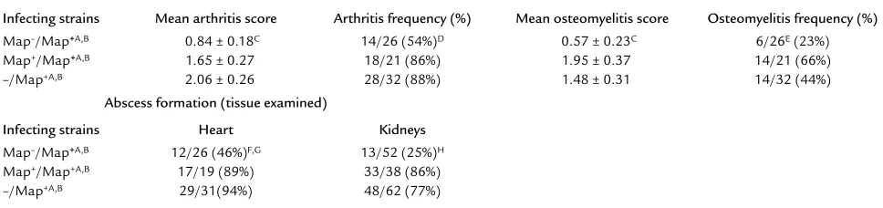
Histological examination of joints and abscess formation assessment in heart and kidneys harvested from Map–SA– and Map+SA–infected mice reinfected with Map+SA
Infecting strains Mean arthritis score Arthritis frequency (%) Mean osteomyelitis score Osteomyelitis frequency (%)
Map–/Map+A,B 0.84 ± 0.18C 14/26 (54%)D 0.57 ± 0.23C 6/26E(23%)
Map+/Map+A,B 1.65 ± 0.27 18/21 (86%) 1.95 ± 0.37 14/21 (66%)
–/Map+A,B 2.06 ± 0.26 28/32 (88%) 1.48 ± 0.31 14/32 (44%)
Abscess formation (tissue examined)
Infecting strains Heart Kidneys
Map–/Map+A,B 12/26 (46%)F,G 13/52 (25%)H
Map+/Map+A,B 17/19 (89%) 33/38 (86%)
–/Map+A,B 29/31(94%) 48/62 (77%)
[image:6.576.56.540.563.677.2]phycoerythrin-conjugated anti-mouse CD4 (L3T4) (PharMingen, San Diego, California, USA). The cells were incubated with the directly conjugated Ab’s for 1 hour at 4°C and then washed with cold FACS buffer prior to FACS analysis. Annexin V-FITC (PharMingen) and propidium iodide (PI) (Sigma-Aldrich) staining of 106BAT2.2 cells (5 U IL-2/ml) was performed 24 hours after incubation with either Map19 or DbpA (0.2 µg/µl) or left untreated in CTL media (2 ml), according to man-ufacturer’s instructions. Cells from all groups were washed in cold FACS buffer and then analyzed on a Coulter EpicProfile flow cytometer (Coulter Corp., Miami, Florida, USA).
Results
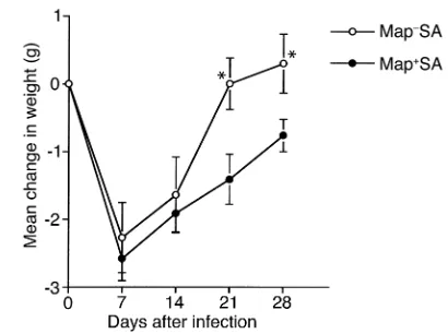
Experimental SA infection. Mice challenged with a sub-lethal dose of virulent SA can develop severe arthritis and osteomyelitis in addition to abscesses in the heart and kidneys (32). To explore the role that Map might play in the pathogenesis of SA infections, BALB/c mice were infected with either 107Map–SA or Map+SA strain Newman. During the initial stages of the infection, the groups of animals inoculated with the two strains were similarly affected, as indicated by recorded weight loss-es (Figure 1). The mean weight loss recorded for Map–SA– and Map+SA–infected mice 7 days after infec-tion was –2.27 ± 0.52 and –2.58 ± 0.32 g, respectively; however, by day 21 after infection, the weight of Map–SA–infected mice returned to postinfection levels compared with Map+SA–infected mice, which were an average of 1.41 ± 0.37 g below preinfection weight read-ings (Figure 1). Furthermore, bacterial densities between infection groups were indistinguishable during the early stages of infection (Figure 2), suggesting that there were no differences in the capacity of Map–SA to colonize tis-sues when compared with Map+SA colonization.
Eight weeks after infection, 12 of 14 (86%) Map+SA–infected mice developed osteomyelitis, but only 1 of 16 (6%) Map–SA–infected mice was diagnosed with a bone infection (Figure 3, a and b, and Table 2). Arthri-tis frequency and severity were also significantly greater in Map+SA–infected mice compared with Map–SA– infected counterparts (Table 2). Furthermore, 8 of 14 (57%) Map+SA–infected mice developed heart abscesses and 21 of 28 (75%) kidneys examined were similarly
affected (Figure 3, f and h, and Table 2). Conversely, none of the 16 Map–SA–infected mice developed heart abscesses and only 1 of 32 (3%) kidneys presented with an abscess (Figure 3, e and g, and Table 2). Taken togeth-er, these results demonstrated that the Map–SA strain was significantly less virulent in our animal model when compared with the parental Map+SA strain, suggesting that Map may be a virulence factor in chronic SA infec-tions. Based on the sequence similarities with MHC class II molecules, we speculated that Map might act as an immunomodulator and that the observed differences between the Map+SA and Map–SA strains were due to suppressed host defenses in Map+SA–infected mice.
[image:7.576.58.539.82.144.2]This hypothesis was tested in several experiments. First, mice were infected with either 107Map+SA, Map–SA, or left untreated. Four weeks later, mice from all groups were reinfected with 107Map+SA organisms. After an additional 4 weeks, mice from all groups were sacrificed and examined as described above. The results from these experiments suggested that mice initially infected with Map–SA were partially protected against the subsequent Map+SA challenge infection. These mice presented with
Figure 4
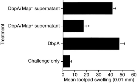
Native Map-mediated inhibition of DTH. DbpA-immunized mice were treated with native Map on the day of immunization (day 0) and on days 2, 4, and 6 after immunization. On day 7, mice were challenged with DbpA, and footpads were measured 0 and 24 hours after challenge. Mice treated with supernatant from Map+SA had a significantly reduced DTH response compared with immunized and challenged mice (*P< 0.0001; Student ttest). Data are expressed as the mean ± SE of five mice.
Table 4
Histological examination of joints harvested from Map–SA– or Map+SA–infected nude miceA
Infecting strains Mean arthritis score Arthritis frequency(%) Mean osteomyelitis score Osteomyelitis frequency (%)
nu/+/Map+SA 3.00 ± 0.24B 16/16 (100%) 3.19 ± 0.36D,E 14/16 (88%) nu/+/Map–SA 1.78 ± 0.21 21/23 (91%) 0.34 ± 0.15 5/23 (22%)G nu/nu/Map+SA 2.58 ± 0.25 12/12 (100%) 3.00 ± 0.32 12/12 (100%) nu/nu/Map–SA 2.50 ± 0.26C 21/24 (88%) 1.58 ± 0.20F 15/24 (63%)
[image:7.576.307.537.494.634.2]significantly fewer heart (46%) and kidney (25%) abscess-es following Map+SA challenge compared with more than 75% abscess formation in hearts and kidneys from Map+SA–infected and challenged mice or mice infected with a single dose of Map+SA (i.e., –/Map+) (Table 3). Arthritis and osteomyelitis incidence and severity were also significantly reduced in Map–SA–infected mice chal-lenged with Map+SA compared with control mice (Table 3). One possible explanation for these data is that Map–SA–infected mice were capable of clearing the infec-tion since their cellular responses were not impaired by Map while concomitantly establishing a memory response capable of clearing the second infection of Map+SA. If Map functioned to impair T cell–mediated immunity, then mice initially infected with Map+SA never fully cleared the infection and were likely not to have been able to develop a memory response capable of recogniz-ing a second SA infection.
SA-infected nude mice. T cell–deficient nude (nu/nu) and genotype control (nu/+) mice were used to further assess the connection between SA virulence, Map, and T cell immunity. If Map is an immunomodulator with T cell inhibitory activities that facilitate SA survival and persistence, then Map–SA–infected nu/numice should present with similar disease as Map+ SA–infect-ed nu/+ mice. Map–SA-infected nu/numice had signif-icantly higher arthritis and osteomyelitis scores com-pared with nu/+Map–SA–infected mice. Furthermore, the arthritis scores between nu/nu/Map–SA– and
nu/+/Map+SA–infected mice were similar (Figure 3, c and d, and Table 4), even though nu/+/Map+ SA–infect-ed mice presentSA–infect-ed with significantly higher osteomye-litis scores (Table 4). Abscess formation was observed in all Map+SA-infected mice but not in mice infected with Map–SA, regardless of genotype. These data
suggested that (a) the presence of Map on SA can affect the severity of arthritis and osteomyelitis through a T cell–mediated mechanism, and (b) Map plays a role in abscess formation through a T cell–inde-pendent mechanism(s).
Map-mediated inhibition of DTH. A direct in vivo assay for cellular immunity was used to assess the ability of Map to interfere with DTH. DTH responses are initiated and mediated by Th1 CD4+T cells in response to recall anti-gens, and these responses result in specific, measurable inflammation at the site of challenge. Mice immunized
with a recombinant form of the B. burgdorferi
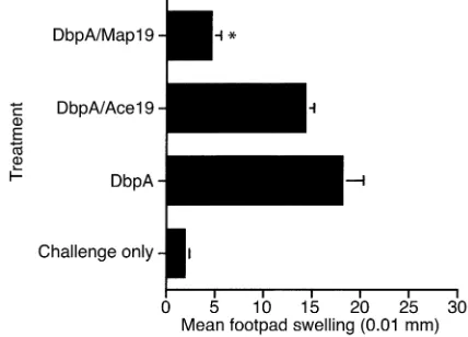
MSCRAMM, DbpA, emulsified in CFA developed a sig-nificant DTH response to DbpA as measured by footpad swelling 7 days after immunization (Figure 4 and 5) (41, 44). Mice treated with native Map (Map+SA super-natant), however, on the day of immunization (day 0) and days 2, 4, and 6 after immunization had a signifi-cantly reduced DTH response to DbpA challenge com-pared with untreated but immunized control mice and to mice treated with supernatants from Map–SA (Figure 4). Similarly, mice treated with a recombinant form of Map (Map19) had a significantly reduced DTH response following DbpA challenge compared with untreated but immunized controls or the recombinant control protein Ace19-treated mice (Figure 5). Recombinant Map19 inhibited the DTH response in both BALB/c and C3H/Hen mice following immunization and challenge, demonstrating that Map has inhibitory affects not spe-cific to particular mouse strains (Figure 5 and data not shown, respectively). The effects of Map19 were largely dose dependent with concentrations of 25 µg/mouse exhibiting significant inhibitory effects (Figure 6).
[image:8.576.66.280.57.211.2]Adoptively transferred T cells from Map19-treated mice. Since adoptively transferred immune T cells confer immunity in syngeneic recipient mice, a cell transfer model was used to further dissect the cellular events underlying Map-mediated inhibition of DTH. Mice
Figure 5
[image:8.576.310.537.519.653.2]Recombinant Map19-mediated inhibition of DTH. DbpA-immu-nized mice were treated with recombinant Map19 on the day of immunization (day 0) and on days 2, 4, and 6 after immunization. On day 7, mice were challenged with DbpA and footpads were meas-ured 0 and 24 hours after challenge. Mice treated with recombinant Map19 had a significantly reduced DTH response compared with immunized and challenged mice (*P< 0.0001; Student ttest). Data are expressed as the mean ± SE of five mice.
Figure 6
immunized with DbpA were either left untreated or injected intraperitoneally with either Map19 or the recombinant control protein Ace40 on the day of immu-nization (day 0) and on days 2, 4, and 6 after immuniza-tion. On day 7, mice were sacrificed, and single cell sus-pensions from whole spleens were prepared and enriched for T cells by passage over nylon wool columns (50). Adoptive transfer of nylon wool–purified T cells from Map19-treated mice did not elicit a DTH response to DbpA in naive recipients compared with mice adop-tively transferred with enriched T cells from Ace40-treat-ed mice or untreatAce40-treat-ed immune T cell control groups (Fig-ure 7). Flow cytometric analysis of nylon wool–collected cells revealed a profile that was 46.83% ± 0.92% CD4+, 31.63% ± 0.96% CD8+, 1.2% ± 0.26% CD4+CD8+, and 20.4% ± 1.33% CD4–CD8–. These data are expressed as the mean percentage of positive cells plus or minus SE for the three groups examined.
Inhibition of T cell proliferation and apoptosis assessment. Recombinant Map19 and Ace19 were tested for their abil-ity to inhibit the proliferation of the Borrelia-specific T cell line BAT 2.2 (43, 50). T cell proliferation was meas-ured 48 hours after plating in the presence of mitomycin C–treated syngeneic APCs and iBb (50). Proliferation was measured as a function of tetrazolium blue production following a 4-hour incubation in the presence of MTT. Map19-treated BAT 2.2 cells were inhibited significantly from proliferating (Figure 8). Map19 did not inhibit BAT2.2 proliferation if the APCs were preincubated in the presence of either Map19 or Ace19 for 6 hours and
then washed prior to the addition of iBb and BAT2.2 cells, suggesting Map had its inhibitory effects on T cells (data not shown). Furthermore, APCs preincubated with Map19 and iBb for 6 hours induced BAT2.2 prolifera-tion, suggesting that Map did not affect the ability of the APCs to process antigen (data not shown).
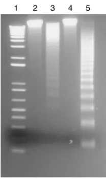
Microscopic analysis of BAT2.2 cells cultured in the presence of Map19 revealed significant morphological changes (e.g., cell shrinkage) compared with untreated cells or cells cultured in the presence of control proteins (data not shown), suggesting that apoptosis or necrosis but not anergy were likely induced by Map. FACS analy-sis of BAT2.2 cells stained with annexin V-FITC and PI revealed that 76% of BAT2.2 Map19-treated cells (Figure 9c) were both annexin V-FITC/PI positive compared with almost 11% double-positive cells for both untreated and DbpA-treated cells (Figure 9, a and b, respectively). FACS analysis at earlier time points did not reveal significant differences between treatment groups (data not shown). Since cells that have already undergone an apoptotic death will eventually also stain positive for PI, it was not possible to distinguish if Map-treated cells in this assay were in a state of late apoptosis or necrosis (51). To dif-ferentiate between these forms of cell death, DNA extract-ed from BAT2.2 T cells incubatextract-ed in the presence of Map19 was examined for fragmentation by gel elec-trophoresis (Figure 10). DNA extracted from Map19-treated cells (lane 3) was fragmented comparable to the apoptotic-positive control DNA (lane 5) but not DNA extracted from untreated (lane 2) or Ace40-treated (lane 4) T cells (Figure 10), suggesting that Map can, at least in vitro, prevent T cell function by induction of apoptosis.
Discussion
[image:9.576.66.281.57.210.2]Mechanisms of immune escape are used by many infec-tious agents affecting humans. The nature of these strate-gies significantly vary and can be classified into various categories: the generation of physical barriers (biofilm
Figure 7
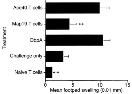
[image:9.576.312.527.522.655.2]DTH following adoptive T cell transfer. DbpA-immunized mice were treated with either Map19 or Ace40, as described above. On day 7, mice were sacrificed and spleens were harvested and enriched for T cells by nylon wool purification. Cells (5 × 10⁷) were injected intraperitoneally into syngeneic recipients. Twenty-four hours later, recipient mice were challenged with DbpA, and the DTH response was assessed as described above. T cells from DbpA-immunized Ace40-treated or T cells from DbpA-immunized (untreated) mice elicited a significant DTH response compared with unimmunized but challenged mice or to naive T cell–recipient mice (*P< 0.04; Student ttest). T cells from DbpA-immunized, Map19-treated mice did not elicit a significant DTH response compared with positive control treatment groups (**P< 0.001; Student ttest). Data are expressed as the mean ± SE of five mice.
Figure 8
formation) that protect bacteria against immune attack (52, 53); the secretion of immunoregulatory cytokine analogs or the induction of host cytokines that potenti-ate nonprotective immunity, resulting in pathogen sur-vival (54–58); antigenic variation, which can prevent the generation of a protective immune response (59–61); antigenic mimicry, which can result in either immune modulation (e.g., MHC I analogs) or sequestration of the pathogen from the immune system (62, 63); toxin and protease production, which can destroy different cell types, including immune cells (2, 52); and finally, direct modulation of host cell functions, which can result in the induction or inhibition of apoptosis, respectively, depending on the pathogen (64–69).
Reinfection of humans with SA is one of the hallmarks of diseases caused by this pathogen, and a difficulty in clearing these types of infections is not only a function of disseminated disease but of SA immune escape mecha-nisms (23–26). One possible reason for recurring infec-tions is that leukocytes from patients with chronic or recurrent SA infections have impaired chemotactic, phagocytic, and bactericidal functions (25, 30, 31). Whether these defects in lymphocyte function were a result of the bacterial infection or a preexisting condition in these individuals is not known (25, 30, 31). More specifically, superantigens and protein A produced by SA during an infection serve immune-evasion functions that may potentiate ongoing and recurrent infections (2).
Here we describe a third SA MIM (microbial immunomodulatory molecule) with potential T cell– limiting properties. The first observation suggesting Map acted in this capacity stemmed from double infec-tion studies in which a primary infecinfec-tion with Map–SA conferred significant protection against reinfection with Map+SA. This contrasted significantly with the patholo-gies observed in mice receiving primary and secondary Map+SA infections. One explanation for these data is that T cell–mediated responses in Map+SA–infected mice were abrogated by the presence of Map. In contrast, Map–SA–infected mice developed cell-mediated immu-nity over the course of infection, resulting in bacterial clearance and in a memory response capable of control-ling a secondary Map+SA infection. That a primary Map–SA infection conferred significant but incomplete protection against Map+SA challenge suggested that the balance or competition between an anamnestic response and Map-mediated immunomodulation could be affect-ed by the challenge dose. Although these data suggestaffect-ed
[image:10.576.61.343.58.151.2]that Map acted as an immunoregulatory molecule, we could not exclude the following two possibilities: first, if Map is a critical adhesin required during the infection process, its removal may have resulted in an attenuated SA strain that served to “vaccinate” mice against the sec-ond Map+SA infection. This is not a likely scenario since Map-deficient SA still efficiently adhere to individual ECM molecules (19, 20). Furthermore, the mean weight loss between Map–SA– and Map+SA–infected mice was similar during the first weeks after infection, and the bacterial densities in various tissues were indistinguish-able between infection groups, suggesting that bacterial adhesion, a critical early step during infection, was not compromised in Map–SA. This weight-loss pattern is sig-nificantly different from that recorded for mice infected with SA deficient in the collagen-binding protein CNA (CNA-SA). Weight loss differences in these animals, com-pared with CNA+SA–infected controls diverged from the start of the infection process (E.L. Brown et al., unpub-lished observations). Second, a diminished disease state following Map–SA infection may have been a result of polar effects caused by mapinactivation following trans-formation resulting in the inactivation of other genes required for SA survival or virulence. Although a map -complemented Map–SA strain Newman is not yet avail-able, the significant effects of both isolated native Map
Figure 9
The effect of Map19 on annexin V/PI staining. BAT2.2 cells (106) (5 U IL-2/ml) were incubated in media alone (a) or in the presence of either 400 µg of either Ace40 (b) or Map19 (c) for 24 hours and analyzed by FACS following incubations with annex-in V-FITC and PI.
Figure 10
[image:10.576.366.470.500.675.2]and of recombinant Map19 on cellular immunity strongly suggested that the lack of virulence observed for Map–SA was a function of the bacteria’s inability to affect host immunity and not a result of the map inacti-vation process. In addition, nu/numice infected with Map–SA presented with more severe arthritis compared with nu/+ mice infected with Map–SA. Interestingly, abscess formation was absent in nu/nu and nu/+ Map-SA–infected mice but not in Map+SA–infected mice of either genotype. These data suggested a potential role for T cells in controlling joint pathology but not in pro-tection against abscess formation. These experiments somewhat contradict findings in a rat model that sug-gested that T cells are critical for abscess formation and with recent work describing Map as an anti-inflamma-tory agent in a mouse peritonitis model (20, 70). How-ever, both of these studies examined acute SA infections in contrast to the assessment of chronic disease described here. Furthermore, the infection routes (intraperitoneal) and the parameters used to assess dis-ease were significantly different in both studies (20, 70). These differences make it difficult to reconcile the anti-inflammatory effects of Map described in a thioglycol-late-induced peritonitis model with the abscesses and severe joint disease caused by a chronic SA infection (20). Additional evidence suggesting that Map affected T cell function stemmed from experiments demonstrating that Map inhibited DTH responses and induced T cell death in vitro most likely by apoptosis. The effects of Map on T cell function appeared only to affect mature T cells stim-ulated in the context of antigen and MHC II. It is inter-esting to speculate that this effect of Map on T cells is a function of the homology between Map and MHC class II molecules since the proliferation of naive T cells fol-lowing incubation with concanavalin A or by Ab cross-linking of the T cell receptor was not inhibited by Map19 (McIntyre and Miyamoto, unpublished observations). Interference of Map with the interactions between acti-vated T cells and MHC class II–bearing APCs may explain why Map only affected T cell proliferation in the context of antigen and APCs. Furthermore, a human T cell line cultured in the presence of plate-bound fibronectin and
αCD3 proliferated in the presence of Map19 (B.W. McIn-tyre and Y.J. Miyamoto, unpublished observations). These data suggested that activated T cells, but not naive T cells, were susceptible to inhibition of proliferation by Map and that T cell lines induced to proliferate via “non-classical” pathways bypassed Map-mediated mechanisms that resulted in the loss of T cell function.
Sequence analysis of the SA genome revealed four open-reading frames (ORFs) encoding Map-like pro-teins in meticillin-resistant SA strain N315 (38). The ORF designated SA1751 encodes a protein with a 69% identity to Map from strain FDA574 (21, 38). The SA1751 sequence is truncated by a stop codon immedi-ately after an adenine-rich region, and expansion of this region by one adenine would remove the stop codon; the adjacent MHC II homologous region (SA1750) would be encoded (38). This suggested that SA can
express longer or shorter forms of Map by modulating the length of the alanine repeat region (38). The remain-ing ORFs (SA2006 and SA0841) encode proteins with 34 and 44% identity, respectively, to the FDA574 Map protein. The role of these Map homologues in SA infec-tion is unknown at this time. Perhaps the potential to encode a variety of MHC II–like proteins can serve to potentiate survival of various SA strains in mammals of varied genetic backgrounds. Extensive searches for Map homologues using the available public databases revealed no similar proteins in other bacteria.
SA uses various survival strategies that allow it to per-sist in mammalian hosts. Furthermore, perper-sistent and recurrent SA infections point to mechanisms that obstruct the development of protective immunity. One of the functions of the Map protein suggested that it may function to impede the development and mainte-nance of memory responses. Apoptotic modulation by MIMs is just one mechanism of immune evasion used by human pathogens. Elucidation of the structure of this class of molecules, their functions, and mecha-nisms of action may be used to design strategies to pre-vent or treat disease.
Acknowledgments
This work was supported by the Department of Health and Human Services, grants from the Center for Disease Control (CCU618387) to E. Brown, by NIH grant AI-20624 to M. Höök and grant CA-62596 to B. McIntyre, and Cancer Immunobiology Training Program Grant CA09598 to Y. Miyamoto. We thank Andrew Spicer for critical review of the manuscript and Karen Ramirez for flow cytometry operation and technical assistance.
1. Kissane, J.M. 1997. Staphylococcal infections. In Pathology of Infectious Dis-eases.Vol. I. D.H. Connor, F.W. Chandler, H.J. Schwartz, H.J. Manz, and E.E. Lack, editors. Appleton and Lange. Stamford, Connecticut, USA. 805–816.
2. Lowy, F.D. 1998. Staphylococcus aureus infections. N. Engl. J. Med. 339:520–532.
3. Rupp, M.E. 1997. Infections of intravascular catheters and vascular devices. In The staphylococci in human disease. K.B. Crossley and G.L. Archer, editors. Churchill Liningstone. New York, New York, USA. 379–399. 4. Tacconelli, E., Tumbarello, M., and Cauda, R. 1998. Staphylococcus aureus
infections. N. Engl. J. Med. 339:2026–2027.
5. Petros, S., et al. 1998. Severe community acquired pneumonia due to
Staphylococcus aureus. Intensive Care Med. 24:189.
6. Kim, H.S., Weilbaecher, D.G., Lie, J.T., and Titus, J.L. 1978. Myocardial abscesses. Am. J. Clin. Pathol. 70:18–23.
7. Hiramatsu, K., Cui, L., Kuroda, M., and Ito, T 2001. The emergence and evolution of methicillin-resistant Staphylococcus aureus. Trends Microbiol.
9:486–493.
8. Patti, J.M., Allen, B.L., McGavin, M.J., and Höök, M. 1994. MSCRAMM-mediated adherence of microorganisms to host tissues. Annu. Rev. Micro-biol. 48:585–617.
9. Herrmann, M., Suchard, S.J., Boxer, L.A., Waldvogel, F.A., and Lew, P.D. 1991. Thrombospondin binds to Staphylococcus aureusand promotes staphylococcal adherence to surfaces. Infect. Immun. 59:279–288. 10. Fujigaki, Y., et al. 1998. Glomerular injury induced by cationic 70-kD
staphylococcal protein; specific immune response is not involved in early phase in rats. J. Pathol. 184:436–445.
11. Chhatwal, G.S., Preissner, K.T., Muller-Berghaus, G., and Blobel, H. 1987. Specific binding of the human S protein (vitronectin) to streptococci,
IL-2 secretion, proliferation and immunoglobulin synthesis in periph-eral blood mononuclear cells (PBMC) of both healthy controls and patients with common variable immunodeficiency (CVID). Clin. Exp. Immunol. 100:406–411.
14. Hudson, M.C., Ramp, W.K., and Frankenburg, K.P. 1999. Staphylococcus aureusadhesion to bone matrix and bone-associated biomaterials. FEMS Microbiol. Lett. 173:279–284.
15. Lopes, J.D., dos Reis, M., and Brentani, R.R. 1985. Presence of laminin receptors in Staphylococcus aureus. Science. 229:275–277.
16. O’Connell, D.P., et al. 1998. The fibrinogen-binding MSCRAMM (clumping factor) of Staphylococcus aureushas a Ca2+-dependent
inhibito-ry site. J. Biol. Chem. 273:6821–6829.
17. Vercellotti, G.M., et al. 1985. Extracellular matrix proteins (fibronectin, laminin, and type IV collagen) bind and aggregate bacteria. Am. J. Pathol.
120:13–21.
18. Wann, E.R., Gurusiddappa, S., and Höök, M. 2000. The fibronectin-binding MSCRAMM FnbpA of Staphylococcus aureusis a bifunctional pro-tein that also binds to fibrinogen. J. Biol. Chem. 275:13863–13871. 19. Kreikemeyer, B., McDevitt, D., and Podbielski, A. 2002. The role of the
Map protein in S. aureusmatrix protein and eukaryotic cell adherence.
Int. J. Med. Microbiol.292:283–295.
20. Chavakis, T., et al. 2002. Staphylococcus aureusextracellular adherence pro-tein serves as anti- inflammatory factor by inhibiting the recruitment of host leukocytes. Nat. Med. 8:687–693.
21. Jonsson, K., McDevitt, D., McGavin, M.H., Patti, J.M., and Höök, M. 1995. Staphylococcus aureusexpresses a major histocompatibility complex class II analog. J. Biol. Chem. 270:21457–21460.
22. Guillen, C., et al. 2002. Enhanced Th1 response to Staphylococcus aureusinfection in human lactoferrin-transgenic mice. J. Immunol.
168:3950–3957.
23. Chang, H.R., et al. 2000. Use of pulsed-field gel electrophoresis in the analysis of recurrent Staphylococcus aureusinfections in patients on con-tinuous ambulatory peritoneal dialysis. Am. J. Nephrol. 20:463–467. 24. Hartstein, A.I., Mulligan, M.E., Morthland, V.H., and Kwok, R.Y. 1992.
Recurrent Staphylococcus aureusbacteremia. J. Clin. Microbiol. 30:670–674. 25. Monteil, M., Hobbs, J., and Citron, K. 1987. Selective
immunodeficien-cy affecting staphylococcal response. Lancet. 2:880–883.
26. Shayegani, M., De Courcy, S.J., Jr., and Mudd, S. 1973. Cell-mediated immunity in mice infected with S. aureusand elicited with specific bac-terial antigens. J. Reticuloendothel. Soc. 14:44–51.
27. Easmon, C.S., and Glynn, A.A. 1975. Cell-mediated immune responses in Staphylococcus aureusinfections in mice. Immunology. 29:75–85. 28. Ficker, L., Seal, D., and Wright, P. 1989. Staphylococcal infection and the
limbus: study of the cell-mediated immune response. Eye. 3:190–193. 29. Sarai, Y., et al. 1977. Immunological properties in staphylococcal toxic
epidermal necrolysis. Dermatologica. 155:315–318.
30. Verbrugh, H.A., et al. 1980. Phagocytic and chemotactic function of poly-morphonuclear and mononuclear leucocytes in patients with recurrent staphylococcal infections. Scand. J. Infect. Dis. 12:111–116.
31. Valmin, K., Hallberg, T., and Hedstrom, S.A. 1982. Recurrent Staphylo-coccal furunculosis: lymphocyte subsets and plasma immunoglobulins.
Scand. J. Infect. Dis. 14:153–154.
32. Bremell, T., Lange, S., Yacoub, A., Ryden, C., and Tarkowski, A. 1991. Experimental Staphylococcus aureusarthritis in mice. Infect. Immun. 59:2615–2623.
33. Sloane, R., et al. 1991. A toxic shock syndrome toxin mutant of Staphy-lococcus aureus isolated by allelic replacement lacks virulence in a rabbit uterine model. FEMS Microbiol. Lett. 62:239–244.
34. Hussain, M., et al. 2002. Insertional inactivation of Eap in Staphylococcus aureusstrain Newman confers reduced staphylococcal binding to fibrob-lasts. Infect. Immun. 70:2933–2940.
35. Kreiswirth, B.N., et al. 1983. The toxic shock syndrome exotoxin struc-tural gene is not detectably transmitted by a prophage. Nature.
305:709–712.
36. Duthie, E.S., and Lorenz, L.L. 1952. Staphylococcal coagulase: mode of action and antigenicity. J. Gen. Microbiol.6:95–107.
37. Ponnuraj, K., et al. 2002. Crystallization and preliminary x-ray crystal-lographic analysis of Ace: a collagen-binding MSCRAMM from Entero-coccus faecalis. Biochim. Biophys. Acta. 1596:173–176.
38. Kuroda, M., et al. 2001. Whole genome sequencing of meticillin-resist-ant Staphylococcus aureus. Lancet. 357:1225–1240.
39. Visai, L., et al. 2000. Monoclonal antibodies to CNA, a collagen-binding microbial surface component recognizing adhesive matrix molecules, detach Staphylococcus aureusfrom a collagen substrate. J. Biol. Chem.
275:39837–39845.
40. Rich, R.L., et al. 1999. Ace is a collagen-binding MSCRAMM from Ente-rococcus faecalis. J. Biol. Chem. 274:26939–26945.
41. Guo, B.P., Brown, E.L., Dorward, D.W., Rosenberg, L.C., and Höök, M. 1998. Decorin-binding adhesins from Borrelia burgdorferi. Mol. Microbiol.
30:711–723.
42. Maniatis, T., Fritsch, E.F., and Sambrook, J. 1989. Molecular cloning: A
laboratory manual. Cold Spring Harbor Laboratory Press. Cold Spring Harbor, New York, USA. A.3
43. Brown, E.L., Ullrich, S.E., Pride, M., and Kripke, M.L. 2001. The effect of UV irradiation on infection of mice with Borrelia burgdorferi. Photochem. Photobiol. 73:537–544.
44. Brown, E.L., et al. 2001. Resistance to Lyme disease in decorin-deficient mice. J. Clin. Invest. 107:845–852.
45. McGavin, M.H., Krajewska-Pietrasik, D., Ryden, C., and Höök, M. 1993. Identification of a Staphylococcus aureusextracellular matrix-binding pro-tein with broad specificity. Infect. Immun. 61:2479–2485.
46. Patti, J.M., et al. 1994. The Staphylococcus aureuscollagen adhesin is a vir-ulence determinant in experimental septic arthritis. Infect. Immun.
62:152–161.
47. Switalski, L.M., et al. 1993. A collagen receptor on Staphylococcus aureus
strains isolated from patients with septic arthritis mediates adhesion to cartilage. Mol. Microbiol. 7:99–107.
48. Joh, H.J., House-Pompeo, K., Patti, J.M., Gurusiddappa, S., and Höök, M. 1994. Fibronectin receptors from gram-positive bacteria: comparison of active sites. Biochemistry. 33:6086–6092.
49. Brown, E.L., et al. 1995. Modulation of immunity to Borrelia burgdorferi
by ultraviolet irradiation: differential effect on Th1 and Th2 immune responses. Eur. J. Immunol. 25:3017–3022.
50. Pride, M.W., et al. 1998. Specific Th1 cell lines that confer protective immunity against experimental Borrelia burgdorferiinfection in mice.
J. Leukoc. Biol. 63:542–549.
51. Vermes, I., Haanen, C., Steffens-Nakken, H., and Reutelingsperger, C. 1995. A novel assay for apoptosis. Flow cytometric detection of phos-phatidylserine expression on early apoptotic cells using fluorescein labelled Annexin V. J. Immunol. Methods. 184:39–51.
52. Kharazmi, A. 1991. Mechanisms involved in the evasion of the host defence by Pseudomonas aeruginosa. Immunol. Lett. 30:201–205. 53. Martin-Lopez, J.V., et al. 2002. Detection of Staphylococcus aureusclinical
isolates harboring the ica gene cluster needed for biofilm establishment.
J. Clin. Microbiol. 40:1569–1570.
54. Suzuki, T., et al. 1995. Viral interleukin 10 (IL-10), the human herpes virus 4 cellular IL-10 homologue, induces local anergy to allogeneic and syngeneic tumors. J. Exp. Med. 182:477–486.
55. Engele, M., et al. 2002. Induction of TNF in human alveolar macrophages as a potential evasion mechanism of virulent Mycobacteri-um tuberculosis. J. Immunol. 168:1328–1337.
56. de Diego, J., Punzon, C., Duarte, M., and Fresno, M. 1997. Alteration of macrophage function by a Trypanosoma cruzi membrane mucin.
J. Immunol. 159:4983–4989.
57. Abraham, S.N., Beachy, E.H., and Simpson, W.A. 1983. Adherence of strep-tococcus pyogenes, Escherichia coli, and Pseudomonas aeruginosa to fibronectin-coated and uncoated epithelial cells. Infect. Immun.41:1261–1268. 58. Akridge, R.E., Oyafuso, L.K., and Reed, S.G. 1994. IL-10 is induced dur-ing HIV-1 infection and is capable of decreasdur-ing viral replication in human macrophages. J. Immunol. 153:5782–5789.
59. Donelson, J.E., Hill, K.L., and El-Sayed, N.M. 1998. Multiple mechanisms of immune evasion by African trypanosomes. Mol. Biochem. Parasitol. 91:51–66. 60. Kyes, S., Horrocks, P., and Newbold, C. 2001. Antigenic variation at the
infected red cell surface in malaria. Annu. Rev. Microbiol. 55:673–707. 61. Lawrenz, M.B., et al. 1999. Human antibody responses to VlsE antigenic
variation protein of Borrelia burgdorferi. J. Clin. Microbiol. 37:3997–4004. 62. Wurzner, R. 1999. Evasion of pathogens by avoiding recognition or erad-ication by complement, in part via molecular mimicry. Mol. Immunol.
36:249–260.
63. Farrell, H.E., et al. 1997. Inhibition of natural killer cells by a cytomegalovirus MHC class I homologue in vivo. Nature. 386:510–514. 64. Boulton, I.C., and Gray-Owen, S.D. 2002. Neisserial binding to CEA-CAM1 arrests the activation and proliferation of CD4+ T lymphocytes.
Nat. Immunol. 3:229–236.
65. Cuff, S., and Ruby, J. 1996. Evasion of apoptosis by DNA viruses.
Immunol. Cell Biol. 74:527–537.
66. Das, G., Vohra, H., Saha, B., Agrewala, J.N., and Mishra, G.C. 1998. Leish-mania donovaniinfection of a susceptible host results in apoptosis of Th1-like cells: rescue of anti-leishmanial CMI by providing Th1-specif-ic bystander costimulation. Microbiol. Immunol. 42:795–801.
67. Monack, D.M., Mecsas, J., Ghori, N., and Falkow, S. 1997. Yersinia sig-nals macrophages to undergo apoptosis and YopJ is necessary for this cell death. Proc. Natl. Acad. Sci. USA. 94:10385–10390.
68. Nunes, M.P., Andrade, R.M., Lopes, M.F., and DosReis, G.A. 1998. Acti-vation-induced T cell death exacerbates Trypanosoma cruzireplication in macrophages cocultured with CD4+ T lymphocytes from infected hosts.
J. Immunol. 160:1313–1319.
69. Wei, S., et al. 2002. Toxoplasma gondii-infected human myeloid dendritic cells induce T-lymphocyte dysfunction and contact-dependent apopto-sis. Infect. Immun. 70:1750–1760.