Hypothalamic Y2 receptors regulate bone
formation
Paul A. Baldock, … , Edith M. Gardiner, Herbert Herzog
J Clin Invest.
2002;
109(7)
:915-921.
https://doi.org/10.1172/JCI14588
.
Neuropeptide Y (NPY) is a downstream modulator of leptin action, possibly at the level of
the arcuate nucleus where NPY neurons are known to express both leptin receptors and Y2
receptors. In addition to the well-described role of NPY and leptin in energy balance and
obesity, intracerebroventricular administration of NPY or leptin also causes bone loss. Here
we show that Y2 receptor–deficient mice have a twofold increase in trabecular bone volume
as well as greater trabecular number and thickness compared with control mice. We also
demonstrate that central Y2 receptors are crucial for this process, since selective deletion of
hypothalamic Y2 receptors in mature conditional Y2 knockout mice results in an identical
increase in trabecular bone volume within 5 weeks. This hypothalamus-specific Y2 receptor
deletion stimulates osteoblast activity and increases the rate of bone mineralization and
formation, with no effect on osteoblast or osteoclast surface measurements. The lack of any
changes in plasma total calcium, leptinemia, or hypothalamopituitarycorticotropic,
-thyrotropic, -somatotropic, or -gonadotropic output suggests that Y2 receptors do not
modulate bone formation by humoral mechanisms, and that alteration of autonomic function
through hypothalamic Y2 receptors may play a key role in a major central regulatory circuit
of bone formation.
Article
Genetics
Find the latest version:
Introduction
Genetically obese rodents that lack leptin (ob/obmice) or its receptor (db/dbmice) show increased activity of neuropeptide Y (NPY) circuits in the hypothalamus due to lack of leptin-induced inhibition of NPY expression and secretion (1). Such increased hypo-thalamic NPY signaling contributes to the massive obesity, hypercorticism, stunted growth, and repro-ductive defects of these mice (2). Since NPY ablation
in ob/obmice attenuates all of these defects (3), these
findings demonstrate that NPY is a downstream mediator of leptin’s central effects.
Recently it was shown that leptin can inhibit bone synthesis by action within the hypothalamus, and that mice with no or reduced leptin signaling (ob/ob,
db/db, and transgenic A-ZIP/F-1 fat-deficient mice)
have high bone density associated with increased bone formation (4). This effect of leptin deficiency occurred independently of increased body fat or body weight, and despite the hypercorticism, decreased somatotropic activity, and hypogonadism in ob/ob
and db/dbmice. Intriguingly, central infusion of NPY
for 28 days in wild-type mice had the same inhibito-ry effect on bone function as leptin had, suggesting that the increased hypothalamic NPY expression of leptin-deficient mice does not mediate the associat-ed increase in bone density (4). However, it is unclear
from this study whether the inhibitory effects on bone of central NPY infusion are a direct conse-quence of hypothalamic NPY action or a secondary effect of the resulting increase in expression (5) and circulating concentrations (6) of leptin. Furthermore, NPY mediates its effects through the activation of at least five different receptors: Y1, Y2, Y4, Y5, and in the mouse also y6, all of which are expressed in the hypothalamus (7–9). The potential for simultaneous activation of all of these receptors by central NPY infusion makes it difficult to distinguish the true role of the different types of hypothalamic Y recep-tors in bone physiology.
NPY synthesis is particularly high in neurons of the arcuate nucleus, with many of these neurons also expressing leptin receptors (10). A high percentage of these arcuate NPY-expressing neurons coexpress the Y2 receptor (11), which is thought to act as an autoreceptor that can modulate the expression and secretion of NPY and other neurotransmitters (12). Since leptin receptors and Y2 receptors are present on NPY-expressing neurons of the arcuate nucleus and are likely to share some common signaling pathways (1, 10–12), we hypothesized that the Y2 receptor might be involved in the central regulation of bone metabolism. To test this hypothesis, we generated both germline Y2 receptor knockout mice (Y2–/–
Hypothalamic Y2 receptors regulate bone formation
Paul A. Baldock,
1Amanda Sainsbury,
2Michelle Couzens,
2Ronaldo F. Enriquez,
1Gethin P. Thomas,
1Edith M. Gardiner,
1and Herbert Herzog
21Bone and Mineral Program, and
2Neurobiology Program, Garvan Institute of Medical Research, St. Vincent’s Hospital, Sydney, Australia
Address correspondence to: Herbert Herzog, Neurobiology Program, Garvan Institute of Medical Research, 384 Victoria Street, Darlinghurst, NSW 2010, Sydney, Australia.
Phone: 61-2-9295-8-296; Fax: 61-2-9295-8-281; E-mail: [email protected]. Paul A. Baldock and Amanda Sainsbury contributed equally to this work.
Received for publication November 8, 2001, and accepted in revised form February 18, 2002.
Neuropeptide Y (NPY) is a downstream modulator of leptin action, possibly at the level of the arcuate nucleus where NPY neurons are known to express both leptin receptors and Y2 receptors. In addition to the well-described role of NPY and leptin in energy balance and obesity, intracere-broventricular administration of NPY or leptin also causes bone loss. Here we show that Y2 recep-tor–deficient mice have a twofold increase in trabecular bone volume as well as greater trabecu-lar number and thickness compared with control mice. We also demonstrate that central Y2 receptors are crucial for this process, since selective deletion of hypothalamic Y2 receptors in mature conditional Y2 knockout mice results in an identical increase in trabecular bone volume within 5 weeks. This hypothalamus-specific Y2 receptor deletion stimulates osteoblast activity and increases the rate of bone mineralization and formation, with no effect on osteoblast or osteo-clast surface measurements. The lack of any changes in plasma total calcium, leptinemia, or hypo-thalamo-pituitary-corticotropic, -thyrotropic, -somatotropic, or -gonadotropic output suggests that Y2 receptors do not modulate bone formation by humoral mechanisms, and that alteration of autonomic function through hypothalamic Y2 receptors may play a key role in a major central regulatory circuit of bone formation.
mice) and conditional Y2 receptor knockout mice (Y2lox/loxmice) and investigated the effects of the
dele-tion on bone physiology.
Methods
Generation of germline and conditional Y2 receptor knockout mice. A targeting vector for the Y2 receptor gene has been designed that allows the production of both germline (Y2–/–) and conditional (floxed, Y2lox/lox)
knockout mice. Both of these knockout strategies result in deletion of the entire coding region of the Y2 receptor, which is encoded by a single exon. Mouse embryonic stem cells from the strain 129SvJ were transfected and selected under standard conditions, and positive embryonic stem cell clones were injected into blastocysts from C57/BL6 mice. Chimeric off-spring were crossed with oocyte-specific Cre-recombi-nase–expressing C57/BL6 mice (13) in order to obtain either heterozygotes carrying the floxed gene (Y2lox/+
mice) or heterozygotes carrying the Cre-recombinase gene with the floxed gene already deleted (Y2+/–mice).
Homozygous lines, both Y2–/–and Y2lox/lox, were
gener-ated by crossing the respective heterozygous animals. All further mice were maintained on this mixed C57/BL6-129SvJ background.
Adenovirus injection. Y2lox/loxand wild-type Y2+/+mice,
10–12 weeks old, were anesthetized with 100 mg/kg ketamine (Pfizer Inc., Sydney, Australia) and 20 mg/kg xylazine (Bayer AG, Leverkusen, Germany) and injected with adenovirus expressing either recombinant Cre or green fluorescent protein (GFP) (The Institute of Physical and Chemical Research, Riken, Fukyuka, Japan) using a stereotaxic table (David Kopf Instruments, Tujunga, California, USA). With the head in the flat-skull position, brain injec-tion coordinates relative to Bregma were posterior 2.3 mm, lateral ± 0.3 mm, and ventral 5.6 mm, corre-sponding to the arcuate nucleus (14). One microliter of either virus (109 plaque-forming units/µl) was injected bilaterally over a period of 10 minutes using a 26-gauge guide cannula and a 33-gauge injector (Plastics One Inc., Roanoke, Virginia, USA) connect-ed to a Hamilton syringe and a syringe infusion
pump (World Precision Instruments Inc., Sarasota, Florida, USA). Mice were housed individually for the ensuing 35 days, with ad libitum access to standard chow and water.
Tissue collection and analysis. Mice were injected with the
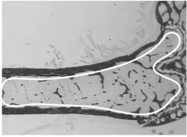
[image:3.576.77.261.56.145.2]fluorescent tetracycline compounds calcein and deme-clocycline (15 mg/kg each; Sigma Chemical Co., St. Louis, Missouri, USA) 10 days and 3 days prior to col-lection, respectively. At 15–17 weeks of age, germline and conditional Y2 knockout mice were killed by cervi-cal dislocation between 1000 and 1400 hours for col-lection of trunk blood in heparinized tubes, and plasma was immediately frozen. Both femora were excised and bisected transversely at the midpoint of the shaft. The distal halves of the right femora were fixed and embed-ded, undecalcified, in K-Plast resin (Medim-Medizinis-che Diagnostik, Giessen, Germany), and 5-µm sagittal sections were prepared for analysis using BioQuant soft-ware (R&M Biometrics Inc., Nashville, Tennessee, USA). Cortical parameters were measured at the bisection point using the proximal portion of the bone. Cortical area was calculated by subtraction of the medullary area from the total bone area, and cortical thickness was cal-culated by radius difference assuming a perfectly circu-lar shape. Sections were stained for mineralized bone (15), and trabecular bone volume, thickness, and num-ber were calculated (16) in the sample region (see Figure 2). Osteoblast parameters (osteoblast surface, osteoblast number, and osteoid surface) were estimated using sec-tions stained with von Kossa stain and toluidine blue. Only osteoblasts identified adjacent to osteoid surfaces were included in the analysis. Bone formation (miner-alizing surface, MS) was estimated using the bone surface (BS) coverage of single- and double-labeled (sLS and dLS) fluorescent bands using the equation MS = {[(0.5 ×sLS) + dLS] ×100}/BS, expressed as a per-centage of BS. Mineral apposition rate (MAR) was esti-mated by the distance between the labels divided by the time interval between injection of labels (MAR = inter-label distance divided by 7, expressed in µm/d). Bone formation rate (BFR) was calculated after fluorescence Figure 1
Effect of germline Y2 receptor deletion on cortical bone of the distal femur. Cortical area (a) and cortical thickness (b) of the femoral midshaft of male wild-type mice (WT, white bars) compared with those of Y2–/–mice (KO, black bars) at 15–17 weeks of age. Values are mean ± SD of five to eight mice per group.
Figure 2
[image:3.576.326.510.549.684.2]microscopy (Leica Microsystems, Heerbrugg, Switzer-land) as BFR = MS/BS ×MAR, expressed in µm2/µm/d) (17). For measurements of osteoclast surface and osteo-clast number, sections were stained for tartrate-resist-ant acid phosphatase (TRAP) activity as described pre-viously (18), with only multinucleated, TRAP-positive cells included in the analysis. Radioimmunoassay kits were used to determine plasma concentrations of leptin (Linco Research Inc., St. Louis, Missouri, USA), corti-costerone, free T4, testosterone (ICN Biomedicals Inc., Costa Mesa, California, USA), and IGF-1 (Bioclone Aus-tralia Pty. Ltd., Marrickville, AusAus-tralia). Plasma total cal-cium levels were determined using a colorimetric assay kit from Sigma Chemical Co.
In situ PCR. Fresh-frozen coronal brain sections were
fixed in 4% paraformaldehyde in PBS for 30 minutes, washed once (for 5 minutes) in PBS, and treated with proteinase K (5 µg/ml) for 1 minute at room tempera-ture. Sections were then rinsed for 5 minutes in PBS followed by a 5-minute wash in 100% ethanol. After drying the slides, Gene Frames (Advanced Biotech-nologies Ltd., Epsom, United Kingdom) were put around each section. One hundred microliters of PCR mix containing digoxigenin-labeled nucleotides and a combination of primers was prepared: oligo C (5′
-TTAACATCAGCTGGCCTAGC-3′) and oligo D (5′
-GGAAGTCACCAACTAGAATGG-3′) were used to verify the Y2lox/lox genotype; oligo C and oligo E (5′
-AGCATCCAGAGAAGTGCAAC-3′) were used to verify the deleted Y2–/–genotype. Each primer mixture was
pre-heated to 65°C, then added to the sections, which were then sealed off with a self-adhesive plastic cover slip. In situ PCR was carried out using an OmniSlide ther-mal cycler (Hybaid Ltd., Teddington, Middlesex, Unit-ed Kingdom), with pretreatment at 95°C for 5 min-utes followed by 40 cycles of denaturing at 95°C for 1 minute, annealing at 61°C for 1 minute, and extension at 72°C for 25 seconds. At the end of PCR, the cover slips and Gene Frames were removed and slides were washed for 5 minutes in xylene followed by 5 minutes in 100% ethanol. Staining to detect the incorporated label was performed as described earlier (8).
Statistical analyses. All data were assessed by factorial
ANOVA followed by the Fisher or contrast post-hoc tests, using StatView version 4.5 or Super-ANOVA (Abacus Concepts Inc., San Francisco, California, USA). For all statistical analyses, P < 0.05 was accepted as being statistically significant.
Results
Effects of germline Y2 receptor deletion on bone formation.
Skeletal phenotypes of Y2–/–mice were analyzed in
the femora, and cortical bone parameters were assessed at the midshaft dissection point. There was no significant difference in the cross-sectional area or the thickness of the cortical bone between wild-type and germline Y2–/– mice, although a trend
toward increased cortical bone in the knockout ani-mals can be seen (Figure 1). However, trabecular bone volume of the distal femoral metaphysis (sample region shown in Figure 2) was increased twofold in germline Y2–/– mice compared with wild-type
ani-mals, with heterozygous mice showing a tendency toward increased trabecular bone volume (Figure 3a). This increased trabecular volume of Y2–/–animals
was associated with a significant increase in the num-ber and thickness of trabecular structures (Figure 3, b and c, and Figure 4).
[image:4.576.58.368.54.155.2]Y2 receptor mRNA has been found in a variety of central and peripheral tissues (8, 19, 20). In order to determine whether bone tissue expresses Y receptors through which NPY could directly regulate bone function, we used RT-PCR to investigate the existence of mRNA for all of the five known Y receptors in total bone tissue of wild-type mice or the osteoblast cell line MC3T3 E1. Primers that are specific for each of
Figure 3
Effect of germline Y2 receptor deletion on tra-becular bone of the distal femoral metaphysis. Trabecular bone volume (a), trabecular number (b), and trabecular thickness (c) of femora from male wild-type mice (WT, white bars) compared with heterozygous Y2+/– (het, gray bars) and homozygous Y2–/– mice (KO, black bars) at 15–17 weeks of age. Values are mean ± SD of five to eight mice per group. *P < 0.05, **P < 0.01 versus wild-type mice.
Figure 4
[image:4.576.309.535.514.675.2]the receptor genes and are located on two different exons were used to ensure that only properly spliced mRNA and not genomic DNA contaminants were amplified. RT-PCR did not yield products specific for any of the Y receptors in any of these preparations, suggesting that NPY and Y2 receptor deletion influ-ences bone density by receptors in locations other than in bone itself.
Effects of hypothalamus-specific Y2 receptor deletion on
bone formation. A possible central role of Y2 receptors
in bone physiology was investigated after selective removal of hypothalamic Y2 receptors in conditional Y2 receptor knockout mice. Ten- to twelve-week old
Y2lox/loxand Y2+/+mice were bilaterally injected into the
hypothalamus with adenovirus expressing either Cre-recombinase or GFP, and femurs of these mice were collected 35 days later when mice were 15–17 weeks of age. In these experiments, the two groups of control mice (GFP-injected Y2lox/lox and Cre-injected Y2+/+
mice) were indistinguishable from each other for all parameters investigated, and data were therefore pooled into a single control group for all figures.
Appropriate positioning of the stereotaxic injection coordinates was monitored by the appearance of green fluorescence in the hypothalamus of GFP-injected animals (Figure 5a). Cre gene expression and consequent Y2 receptor gene deletion in the hypo-thalamus were confirmed at 35 days after adenovirus injection by in situ PCR analysis of coronal brain sec-tions isolated from these animals (Figure 5, b–f).
The selective deletion of hypothalamic Y2 receptors in adult mice resulted in a bone phenotype identical to that seen in germline Y2–/–animals. A twofold increase
[image:5.576.58.292.53.322.2]in trabecular bone volume was produced during the 5-week period of hypothalamic Y2 receptor deficiency (Figure 6a and Figure 7). Consistent with germline Y2 receptor knockout, hypothalamic Y2 deletion resulted in increased trabecular number and trabecular thick-ness (Figure 6, b and c). There was no indication of change in resorption as measured by osteoclast surface (Figure 8a), but osteoclast number was significantly reduced (Figure 8b). Osteoblast surface, osteoblast number, osteoid surface, and mineralizing surface were all unaffected by hypothalamic Y2 receptor deletion (Figure 8, c–f). However, the rates of bone mineral apposition and bone formation were significantly increased by the hypothalamic Y2 receptor deletion (Figure 8, g and h). This measure of the speed of bone mineralization, as indicated by the distance between the two fluorescent bands, showed that hypothalamus-specific Y2 receptor knockout animals laid down sub-stantially more bone than did control animals (Figure 9). Indeed, bone mineral apposition rate and the asso-ciated bone formation rate in the conditional knock-outs were both around twofold higher than these rates in controls (Figure 8, g and h).
Figure 5
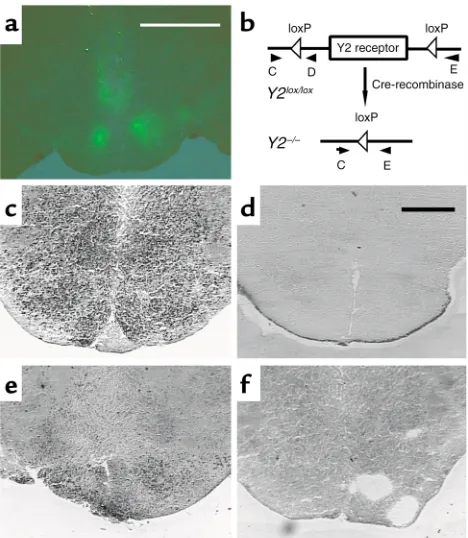
Expression of GFP and in situ PCR of the hypothalamus of Cre-ade-novirus–injected Y2lox/loxmice and germline Y2–/–mice. (a) Fluores-cence micrograph of the hypothalamus of a Y2lox/loxanimal 21 days after injection of GFP-expressing adenovirus. Bar represents 2 mm. (b) Schematic drawing of the position of the primers (oligo C, D, and E) used for in situ PCR. (c) In situ PCR on a brain section from a
[image:5.576.70.537.631.739.2]Y2lox/loxanimal using oligo C and oligo D. (d) In situ PCR on a brain section from a Y2–/–animal using oligo C and oligo D. (e) In situ PCR on a brain section from a Cre-adenovirus–injected Y2lox/loxanimal using oligo C and oligo E. (f) In situ PCR on a brain section from a Cre-adenovirus–injected Y2lox/loxanimal using oligo C and oligo E without enzyme. Bar represents 1 mm in c–f.
Figure 6
Plasma concentrations of total calcium, leptin, free T4, IGF-1, and testosterone were unaffected by germline or conditional Y2 receptor knockout (Table 1). There was no significant effect of germline Y2 recep-tor knockout on corticosteronemia, but conditional Y2 receptor knockout mice had significantly greater cor-ticosteronemia than did control mice injected with GFP- or Cre-expressing adenovirus (Table 1).
Discussion
Here we provide the first evidence that hypothalamic Y2 receptors are involved in a tonic inhibition of bone
formation. The absence of detectable levels of Y recep-tor mRNAs in bone tissue is further evidence that this effect of Y2 deficiency occurs by a central mechanism. It is noteworthy that the bone phenotype of condi-tional hypothalamic Y2 receptor knockout mice shown here is similar to that reported for mice defi-cient in leptin action (4). Trabecular bone density and the rate of bone mineralization and formation are increased in Y2 knockout mice, with no increase in osteoblast or osteoid surface, or osteoblast number. Thus deletion of hypothalamic Y2 receptors acts to release a tonic inhibition of the activity of trabecular osteoblast activity, increasing the rate of bone miner-alization and formation twofold. In contrast with the
ob/oband db/dbmice, in which osteoclast number was
increased, osteoclast number in hypothalamic Y2 receptor knockout mice was reduced, suggesting dif-ferent osteoclast regulation between the two models. However, osteoclast surface was not affected by Y2 deletion, suggesting an increase in osteoclast size in these knockouts. Such a change in osteoclast mor-phology is consistent with an increase in resorptive activity per cell (21, 22).
Both Y2 and leptin receptors are found on NPY-expressing neurons in the arcuate nucleus (10, 11). Deficiency of either the leptin receptor (1) or the Y2 receptor leads to increased NPY expression in the arcu-ate nucleus (data not shown), consistent with both receptors regulating bone formation by a common pathway. In support of this, the high-bone-density phe-notype of Y2–/–mice and ob/obmice was not increased
in Y2–/–, ob/obdouble knockout mice (unpublished
[image:6.576.62.291.53.208.2]observations from our group), indicating lack of addi-tive, independent effects of the two gene deficiencies. It is not clear whether the high hypothalamic NPY Figure 7
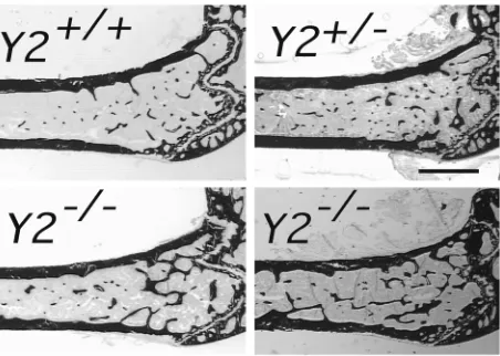
[image:6.576.92.503.472.669.2]Sagittal micrographs of the distal femoral metaphysis of control and hypothalamus-specific Y2 receptor knockout mice: Cre-adenovirus–injected wild-type control (Cre-Y2+/+), Y2lox/loxcontrol, GFP-injected control (GFP-Y2lox/lox), and Cre-injected hypothalamic Y2 receptor knockout (Cre-Y2lox/lox). All mice were males, 15–17 weeks of age. Measurements were taken 5 weeks after hypothalamic injection. Each micrograph is a representative sample. Bar represents 1 mm.
Figure 8
expression common to mice deficient in leptin action or Y2 receptors is causally related to the high bone den-sity, because 28-day intracerebroventricular NPY infu-sion actually decreased bone density (though it proba-bly also resulted in hyperleptinemia) (4). It is note-worthy that germline Y4 receptor knockout mice gen-erated in our laboratory using the same strategies used for the Y2 receptor knockout, and maintained on the same background, do not present an increased bone forming phenotype. These data collectively suggest that Y2 receptor signaling specifically is important in regulating bone mass.
NPY in the hypothalamus influences peripheral tis-sues by neuroendocrine effects. Central NPY activates the hypothalamo-pituitary-corticotropic axis (23, 24) while inhibiting activity of
the -thyrotropic (24, 25), -somatotropic (24, 26), and -gonadotropic axes (24, 26, 27). It is likely that these effects are mediated by NPY projections to neurons that secrete releasing hormones into the pituitary portal sys-tem (24). This mechanism explains why deficiency of leptin action and the subse-quently increased central NPY expression and secretion tonus not only contributes to massive obesity, but also leads to neuroendocrinologi-cal perturbations that would
be expected to decrease bone mass, namely hypercor-ticism, hypothyroid-ism (28–30), reduced soma-totropic activity, and hypogonadism (2, 4). In con-trast, germline or hypothalamus-specific Y2 receptor deletion did not induce any obvious endocrine imbal-ances that would have impacts on bone homeostasis. Knockout animals showed no significant change from controls in plasma concentrations of total calci-um, leptin, free T4, IGF-1, or testosterone, and fertil-ity was not impaired. The divergent changes in corti-costeronemia seen in germline and conditional Y2 receptor knockout mice are not likely to explain the increased bone mass phenotype observed in both knockout models. These findings suggest that Y2 receptor deficiency does not influence bone forma-tion via modulaforma-tion of humoral factors.
Another mechanism by which central NPY can influence peripheral tissues is by alterations in auto-nomic neuronal activity. This is probably mediated by NPY projections from the hypothalamus to brain stem areas where sympathetic or parasympathetic neuronal activity is modulated (31). Whereas central Y1 and Y5 receptors are the most likely mediators of the strong stimulatory effect of NPY on food intake, Y2 receptors are known to be involved in the tion of autonomic processes, such as central regula-tion of pancreatic secreregula-tion, gut motility (32, 33), and cardiovascular function (34). Notably, autonomic function is also disturbed in genetically obese rodents lacking leptin function (35, 36). Since rat bone tissue has recently been shown to contain auto-nomic fibers (37, 38), it is possible that alteration of autonomic activity in the bone presents a novel mechanism by which central regulators of bone homeostasis mediate their effects.
[image:7.576.89.246.55.258.2]These data clearly indicate a major role of hypo-thalamic Y2 receptors in the regulation of bone mass. The rapid increase in bone volume in adult mice after central deletion of Y2 receptor suggests new possi-bilities for the prevention and anabolic treatment of osteoporosis.
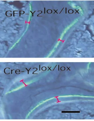
Figure 9
Effect of hypothalamus-specific Y2 receptor deletion on bone min-eralization in the distal femoral metaphysis. Fluorescence micro-graphs of male GFP-adenovirus–injected Y2lox/loxcontrol animals com-pared with Cre-adenovirus–injected Y2lox/loxknockout mice at 15–17 weeks of age, showing the greater distance between the bands in knockout mice. Bar represents 1 mm.
Table 1
Plasma total calcium, leptin, corticosterone (Cortico), free T4, IGF-1, and testosterone (Testost) concentrations
Y2+/+ Y2+/– Y2–/– GFP-Y2lox/lox Cre-Y2+/+ Cre-Y2lox/lox
Calcium (mg/dl) 10.1 ± 0.5 10.0 ± 0.4 10.0 ± 0.5 10.4 ± 1.6 8.5 ± 0.8 9.0 ± 0.4
(21) (15) (21) (5) (3) (11)
Leptin (ng/ml) 6.6 ± 2.4 7.2 ± 2.8 6.2 ± 1.6 4.6 ± 0.4 ND 4.4 ± 0.7
(6) (6) (6) (4) (5)
Cortico (ng/ml) 102 ± 32 48 ± 14 64 ± 17 54 ± 23 73 ± 38 139 ± 18A
(10) (19) (18) (5) (3) (11)
Free T4 (pmol/l) 20.7 ± 4.0 16.4 ± 2.9 20.2 ± 3.6 19.2 ± 1.1 16.3 23.3 ± 1.6
(4) (11) (8) (3) (1) (5)
IGF-1 (ng/ml) 262 ± 29 ND 235 ± 26 320 ± 58 182 ± 26 238 ± 36
(20) (17) (5) (3) (11)
Testost (nmol/l) 7.6 ± 2.4 5.1 ± 2.5 5.9 ± 2.7 ND ND ND
(5) (10) (9)
[image:7.576.209.535.574.711.2]Acknowledgments
We thank I. Saito (Institute of Medical Science, Uni-versity of Tokyo) for the Cre-adenovirus construct and Lee Carpenter for the GFP-expressing adenovirus. We thank Julie Ferguson for invaluable veterinary advice, and the staff of the Garvan Institute Biological Testing Facility. We are grateful to Sara Baker for expert tech-nical assistance. Critical review of this manuscript by John Eisman, Peter Schofield, and Trevor Lewis was greatly appreciated. This research was supported by a Garvan Project Grant donated by Ray Williams, a block grant from the National Health and Medical Research Council of Australia Centre, a Human Frontier Science Program grant (RG0045/2000-B), and a Peter Doherty Post-Doctoral Fellowship (987122), to A. Sainsbury.
1. Stephens, T.W., et al. 1995. The role of neuropeptide Y in the antiobesi-ty action of the obesegene product. Nature. 377:530–532.
2. Coleman, D.L. 1988. Classical diabetes models: past lessons and poten-tial new therapies. In Frontiers in diabetes research. Lessons from animal dia-betes II. E. Shafrir and A.E. Renold, editors. John Libbey & Co. London, United Kingdom. 253–256.
3. Erickson, J.C., Hollopeter, G., and Palmiter, R.D. 1996. Attenuation of the obesity syndrome of ob/ob mice by the loss of neuropeptide Y. Sci-ence. 274:1704–1707.
4. Ducy, P., et al. 2000. Leptin inhibits bone formation through a hypo-thalamic relay: a central control of bone mass. Cell. 100:197–207. 5. Sainsbury, A., Cusin, I., Doyle, P., Rohner-Jeanrenaud, F., and
Jeanre-naud, B. 1996. Intracerebroventricular administration of neuropeptide Y to normal rats increases obesegene expression in white adipose tissue.
Diabetologia. 39:353–356.
6. Sainsbury, A., and Herzog, H. 2001. Inhibitory effects of central neu-ropeptide Y on the somatotropic and gonadotropic axes in male rats are independent of adrenal hormones. Peptides. 22:467–471.
7. Blomqvist, A.G., and Herzog, H. 1997. Y-receptor subtypes—how many more? Trends Neurosci.20:294–298.
8. Parker, R.M., and Herzog, H. 1999. Regional distribution of Y-receptor subtype mRNAs in rat brain. Eur. J. Neurosci.11:1431–1448.
9. Naveilhan, P., Neveu, I., Arenas, E., and Ernfors, P. 1998. Complemen-tary and overlapping expression of Y1, Y2 and Y5 receptors in the devel-oping and adult mouse nervous system. Neuroscience. 87:289–302. 10. Baskin, D.G., Breininger, J.F., and Schwartz, M.W. 1999. Leptin receptor
mRNA identifies a subpopulation of neuropeptide Y neurons activated by fasting in rat hypothalamus. Diabetes. 48:828–833.
11. Broberger, C., Landry, M., Wong, H., Walsh, J.N., and Hokfelt, T. 1997. Subtypes Y1 and Y2 of the neuropeptide Y receptor are respectively expressed in pro-opiomelanocortin- and neuropeptide-Y-containing neurons of the rat hypothalamic arcuate nucleus. Neuroendocrinology.
66:393–408.
12. King, P.J., Williams, G., Doods, H., and Widdowson, P.S. 2000. Effect of a selective neuropeptide Y Y(2) receptor antagonist, BIIE0246 on neu-ropeptide Y release. Eur. J. Pharmacol.396:R1–R3.
13. Schwenk, F., Baron, U., and Rajewsky, K. 1995. A cre-transgenic mouse strain for the ubiquitous deletion of loxP- flanked gene segments includ-ing deletion in germ cells. Nucleic Acids Res.23:5080–5081.
14. Franklin, K.B., and Paxinos, G. 1997. The mouse brain in stereotaxic coordi-nates. Academic Press. San Diego, California, USA. 35–55.
15. Page, K. 1977. Bone and preparation of bone sections. In Theory and prac-tice of histological techniques.J.D. Bancroft and A. Stevens, editors. Churchill Livingstone. London, United Kingdom. 223–248.
16. Parfitt, A.M., et al. 1983. Relationships between surface, volume, and thickness of iliac trabecular bone in aging and in osteoporosis.
Implica-tions for the microanatomic and cellular mechanisms of bone loss.
J. Clin. Invest.72:1396–1409.
17. Parfitt, A.M., et al. 1987. Bone histomorphometry: standardization of nomenclature, symbols, and units. Report of the ASBMR Histomor-phometry Nomenclature Committee. J. Bone Miner. Res.2:595–610. 18. Hayman, A.R., Macary, P., Lehner, P.J., and Cox, T.M. 2001.
Tartrate-resistant acid phosphatase (Acp 5): identification in diverse human tis-sues and dendritic cells. J. Histochem. Cytochem.49:675–684.
19. Gehlert, D.R., et al. 1996. Expression cloning of a human brain neu-ropeptide Y Y2 receptor. Mol. Pharmacol. 49:224–228.
20. Goumain, M., Voisin, T., Lorinet, A.M., and Laburthe, M. 1998. Identifi-cation and distribution of mRNA encoding the Y1, Y2, Y4, and Y5 recep-tors for peptides of the PP-fold family in the rat intestine and colon.
Biochem. Biophys. Res. Commun.247:52–56.
21. Piper, K., Boyde, A., and Jones, S.J. 1995. Volumes of chick and rat osteo-clasts cultured on glass. Calcified Tissue Int.56:382–389.
22. Piper, K., Boyde, A., and Jones, S.J. 1992. The relationship between the number of nuclei of an osteoclast and its resorptive capability in vitro.
Anat. Embryol.186:291–299.
23. Sainsbury, A., et al. 1997. Chronic central neuropeptide Y infusion in normal rats: status of the hypothalamo-pituitary-adrenal axis, and vagal mediation of hyperinsulinaemia. Diabetologia. 40:1269–1277. 24. McDonald, J.K., and Koenig, J.I. 1993. Neuropeptide Y actions on
repro-ductive and endocrine functions. In The biology of neuropeptide Y and relat-ed peptides.W.F. Colmers and C. Wahlestedt, editors. Humana Press. Totowa, New Jersey, USA. 419–456.
25. Fekete, C., et al. 2001. Neuropeptide Y has a central inhibitory action on the hypothalamic-pituitary-thyroid axis. Endocrinology.142:2606–2613. 26. Pierroz, D.D., Catzeflis, C., Aebi, A.C., Rivier, J.E., and Aubert, M.L. 1996. Chronic administration of neuropeptide Y into the lateral ventricle inhibits both the pituitary-testicular axis and growth hormone and insulin-like growth factor I secretion in intact adult male rats.
Endocrinology.137:3–12.
27. Clark, J.T., Kalra, P.S., and Kalra, S.P. 1985. Neuropeptide Y stimulates feeding but inhibits sexual behavior in rats. Endocrinology.117:2435–2442. 28. Durbin-Naltchayan, S., Bouhnik, J., and Michel, R. 1983. Thyroid status
in the obese syndrome of rats. Horm. Metab. Res.15:547–549. 29. Chomard, P., Beltramo, J.L., Ben Cheikh, R., and Autissier, N. 1994.
Changes in thyroid hormone and thyrotrophin in the serum and thyroid glands of developing genetically obese male and female Zucker rats.
J. Endocrinol. 142:317–324.
30. Kaplan, M.M., and Young, J.B. 1987. Abnormal thyroid hormone deiod-ination in tissues of ob/ob and db/db obese mice. Endocrinology.
120:886–893.
31. Saper, C.B., Loewy, A.D., Swanson, L.W., and Cowan, W.M. 1976. Direct hypothalamo-autonomic connections. Brain Res.117:305–312. 32. Chen, C.H., Stephens, R.L., Jr., and Rogers, R.C. 1997. PYY and NPY:
con-trol of gastric motility via action on Y1 and Y2 receptors in the DVC. Neu-rogastroenterol. Motil.9:109–116.
33. Fujimiya, M., et al. 2000. Neuropeptide Y induces fasted pattern of duo-denal motility via Y(2) receptors in conscious fed rats. Am. J. Physiol.
278:G32–G38.
34. Mahns, D.A., Lacroix, J.S., and Potter, E.K. 1998. Inhibition of vagal vasodilatation by a selective neuropeptide Y Y2 receptor agonist in the bronchial circulation of anaesthetised dogs. J. Auton. Nerv. Syst.73:80–85. 35. Rohner-Jeanrenaud, F. 1995. A neuroendocrine reappraisal of the dual-centre hypothesis: its implications for obesity and insulin resistance. Int. J. Obes. 19:517–534.
36. Bray, G.A. 1990. The MONA LISA hypothesis. Most obesities known are low in sympathetic activity. In Progress in obesity research 1990. Y. Oomu-ra, S. Tarui, S. Inoue, and T. Shimazu, editors. John Libbey and Co. Lon-don, United Kingdom. 61–66.
37. Schwab, W., Bilgicyildirim, A., and Funk, R.H. 1997. Microtopography of the autonomic nerves in the rat knee: a fluorescence microscopic study. Anat. Rec.247:109–118.