Mutations in Ig
aa
(CD79a) result in a complete
block in B-cell development
Yoshiyuki Minegishi, … , Dario Campana, Mary Ellen Conley
J Clin Invest.
1999;
104(8)
:1115-1121.
https://doi.org/10.1172/JCI7696
.
Mutations in Btk, µ heavy chain, or the surrogate light chain account for 85–90% of patients
with early onset hypogammaglobulinemia and absent B cells. The nature of the defect in the
remaining patients is unknown. We screened 25 such patients for mutations in genes
encoding components of the pre–B-cell receptor (pre-BCR) complex. A 2-year-old girl was
found to have a homozygous splice defect in Ig
a
, a transmembrane protein that forms part of
the Ig
a
/Ig
b
signal-transduction module of the pre-BCR. Studies in mice suggest that the Ig
b
component of the pre-BCR influences V-DJ rearrangement before cell-surface expression of
µ heavy chain. To determine whether Ig
a
plays a similar role, we compared B-cell
development in an Ig
a
-deficient patient with that seen in a µ heavy chain–deficient patient.
By immunofluorescence, both patients had a complete block in B-cell development at the
pro-B to pre-B transition; both patients also had an equivalent number and diversity of
rearranged V-DJ sequences. These results indicate that mutations in Ig
a
can be a cause of
agammaglobulinemia. Furthermore, they suggest that Ig
a
does not play a critical role in
B-cell development until it is expressed, along with µ heavy chain, as part of the pre-BCR.
Article
Introduction
The pre–B-cell receptor (pre-BCR) complex is expressed transiently and at very low cell density on the surface of developing B cells, yet it plays a pivotal role in B-cell pro-duction. The successful expression of the pre-BCR marks the transition of the pro-B stage to the pre-B stage of dif-ferentiation. In addition to the membrane form of a rearranged µheavy chain, the complex includes 2 pro-teins that make up the surrogate light chain (VpreB and λ5) and 2 proteins that comprise the transmembrane signal-transduction module (Igαand Igβ). The surrogate light chain assesses the ability of the rearranged µheavy chain to bind conventional light chain before the rearrangement of the light chain genes, and the Igα/Igβ heterodimer masks the hydrophilic transmembrane domain of µheavy chain and escorts it to the cell surface. In both humans and mice, the components of the sur-rogate light chain and the Igα/Igβ heterodimer are expressed in the cytoplasm of B-cell precursors before the completion of V-DJ rearrangement (1, 2). This expe-dites cell-surface expression of the pre-BCR once a cor-rectly recombined µheavy chain has been produced.
In mice, the Igα/Igβheterodimer is also expressed on the surface of pro-B cells in the absence of µheavy chain (3). Cross-linking of this receptor in mice that are unable to rearrange µheavy chain genes induces tyrosine and serine/threonine phosphorylation of cytoplasmic pro-teins, and the differentiation of pro-B cells into pre-B cells (4). Furthermore, mice that do not make the Igβ
component of the signal-transduction module have nor-mal D-J rearrangement but impaired V-DJ rearrange-ment (5). These findings suggest that signaling through the Igα/Igβheterodimer might facilitate heavy chain rearrangement, in a manner similar to the enhanced rearrangement of light chain genes seen after successful cell-surface expression of a rearranged µheavy chain product as part of the pre-BCR (6, 7).
The Igαand Igβproteins, which are encoded by mb-1
and B29genes (8, 9), are structurally similar to CD3γ, δ,
and εchains on T cells. Each has a single extracellular immunoglobulin domain, a transmembrane domain, and an intracytoplasmic signaling domain with an immunore-ceptor tyrosine-based activation motif (ITAM) (10). Glu-tathione-S-transferase fusion proteins of the cytoplasmic domains of Igαand Igβbind to distinct sets of effector molecules, and when transfected into cell lines, chimeric proteins containing the 2 cytoplasmic tails elicit different responses (11). Igαtends to bind more readily to Src fam-ily members, and it activates tyrosine kinase more effi-ciently (11–13). However, when expressed in B cell–defi-cient mice as chimeric transgenes consisting of an IgM extracellular domain and an Igα or Igβ intracellular domain, the signaling domains of both Igαand Igβare able to induce the pro-B cell to pre-B cell transition and allelic exclusion (14, 15). This suggests that the cytoplas-mic domains of Igαand Igβare at least somewhat redun-dant in early B-cell development. This hypothesis is sup-ported by observations in mice that lack the cytoplasmic
Mutations in Ig
α
(CD79a) result in a complete block
in B-cell development
Yoshiyuki Minegishi,
1Elaine Coustan-Smith,
1Lisa Rapalus,
1Fügen Ersoy,
2Dario Campana,
1,3and Mary Ellen Conley
1,31Departments of Immunology, Hematology/Oncology, and Pathology, St. Jude Children’s Research Hospital, Memphis,
Tennessee 38105, USA
2Department of Pediatrics, Hacettepe University, 06100 Ankara, Turkey
3Department of Pediatrics, University of Tennessee, Memphis, Tennessee 38105, USA
Address correspondence to: Yoshiyuki Minegishi, Department of Immunology, St. Jude Children’s Research Hospital, Memphis, Tennessee 38105, USA. Phone: (901) 495-2497; Fax: (901) 495-3107; E-mail: [email protected].
Received for publication June 25, 1999, and accepted in revised form September 8, 1999.
Mutations in Btk, µheavy chain, or the surrogate light chain account for 85–90% of patients with early onset hypogammaglobulinemia and absent B cells. The nature of the defect in the remaining patients is unknown. We screened 25 such patients for mutations in genes encoding components of the pre–B-cell receptor (pre-BCR) complex. A 2-year-old girl was found to have a homozygous splice defect in Igα, a transmembrane protein that forms part of the Igα/Igβsignal-transduction module of the pre-BCR. Studies in mice suggest that the Igβcomponent of the pre-BCR influences V-DJ rearrangement before cell-surface expression of µheavy chain. To determine whether Igαplays a similar role, we compared B-cell development in an Igα-deficient patient with that seen in a µheavy chain–deficient patient. By immunofluorescence, both patients had a complete block in B-cell development at the pro-B to pre-B transition; both patients also had an equivalent number and diversity of rearranged V-DJ sequences. These results indicate that mutations in Igαcan be a cause of agammaglobulinemia. Furthermore, they suggest that Igαdoes not play a critical role in B-cell development until it is expressed, along with µ heavy chain, as part of the pre-BCR.
domain of Igα. These mice have only a modest reduction in the number of pre-B cells and immature B cells, but markedly decreased numbers of mature B cells (16).
In broad strokes, early stages of B-cell development in the human correspond to those seen in the mouse; how-ever, there are subtle differences that suggest some flex-ibility in the mechanisms used to regulate various stages of differentiation. In the mouse, defects in λ5 cause a block in B-cell development at the pro-B to pre-B transi-tion; however, this block is leaky, such that 4-month-old λ5-deficient mice have approximately 20% the normal number of B cells and they are able to make antibodies to T-dependent and T-independent antigens (17). By contrast, defects in λ5/14.1 in humans result in a long-lasting, profound defect in B-cell development, such that an affected patient has less than 1% the normal number of B cells at 8 years of age (18). Early stages of B-cell development appear to be more dependent on IL-7 (19–21)and less dependent on Btk in mice compared with humans (22–25). To clarify better the nature of genetic defects that result in abnormal human B-cell development, we have screened patients with early-onset hypogammaglobulinemia and absent B cells for muta-tions in genes that are expressed in early stages of B-cell differentiation. We have identified a patient with a homozygous defect in Igαproduction, and have com-pared the phenotype of this patient with that of a patient with defects in µheavy chain.
Methods
Patient. The patient with Igαdeficiency was the first-born child of healthy parents without known consanguinity. The pregnancy and delivery were uneventful, but the patient developed recurrent diarrhea associated with fail-ure to thrive within the first month of life. At 12 months of age, she was referred to Hacettepe University Hospital
for evaluation of bronchitis and neutropenia. At this time, her serum immunoglobulins were near the level of detec-tion (IgG, 10 mg/dL; IgA, 13 mg/dL; and IgM, 5 mg/dL) and isohemagglutinins were absent. Analysis of peripher-al blood lymphocytes reveperipher-aled she had less than 1% CD19+
B cells, but normal numbers of T cells and natural killer cells and normal proliferation in response to mitogens. Intravenous γ-globulin therapy was initiated, and all symptoms resolved. At 2 years, 8 months of age, her phys-ical examination was normal except for the absence of lymph nodes. Serum IgA and IgM were undetectable (< 5 mg/dL). The 13-year-old µheavy chain–deficient patient included in this study was patient no. 7 in our previous report (26). The institutional review board of St. Jude Chil-dren’s Research Hospital approved the study protocol, and informed consent was obtained from the parents.
Mutation detection. Genomic DNA was analyzed for mutations in Igαby single-strand conformation poly-morphism (SSCP) (27)using the primers indicated in Table 1. The following PCR conditions were used for all reactions: 95°C for 5 minutes followed by 30 cycles of 95°C for 45 seconds; the annealing temperature shown in Table 1 for 30 seconds; 72°C for 30 seconds with a final extension of 5 minutes at 72°C. Before SSCP analysis, PCR products from exons 1, 2, 3, and 5 were digested with
PstI, PstI + MboI, MboI, and PvuII, respectively. Fragments demonstrating altered mobility in SSCP were cloned into TA vector (Invitrogen Corp., Carlsbad, California, USA) and sequenced using M13 primers. The mutations were confirmed using a second independent PCR reaction.
Immunofluorescence staining. Peripheral blood lympho-cytes and bone marrow cells were stained using tech-niques and reagents described previously (18).
Semiquantitative RT-PCR. Total RNA was extracted from 5 × 10⁶ bone marrow mononuclear cells, using an
[image:3.612.57.536.490.720.2]RNeasy Mini kit (QIAGEN Inc., Valencia, California,
Table 1
PCR primers used to analyze early B-cell development
Annealing Size of
Forward (5′-3′) Reverse (5′-3′) Temperature Fragment (bp)
Igαgenomic DNA
Exon 1 TTGGGGTAGGGATCTGG TGTGAGACTGTGGACGC 58°C 200+160A
Exon 2 TTCTTCACTTTCCCGCATC GGTACTGGCTCTATCCCC 60°C 260+181+129A
Exon 3 AGGCTAGGGAGGGCAAGA TTGGATGGGGAACCTCAG 58°C 151+122A
Exon 4 (5′) GTAGACTCAGAGAGAACTG ATATTCATCCCCGGCATC 60°C 200
Exon 4 (3′) ACCCCTACAGAAACGATGGCAG GAGCAGTCGTCCAGGTTCAGG 66°C 229
Exon 5 AGGGTGCTGATGTTCGCT TAAGGAAGCCCCCAGAAG 58°C 135+121A
IgαcDNA
Exon 2 CGAGTCATACCAGCAGTC
Exon 3 CGGCTGTGATGATTCGGT 56°C 107
Exon 5 GTCGTCCAGGTTCAGGCC 56°C 250
Semiquantitative RT-PCR
GAPDH ATGCTGGCGCTGAGTAC TGAGTCCTTCCACGATAC 56°C 257
Igα CGAGTCATACCAGCAGTA GTCGTCCAGGTTCAGGCC 56°C 250
Igβ TCCGGCAATGTGAGCTG ACCATCCTTCAGCGTGTT 56°C 218
TdT TATGAGAGCAGCTTCTGTAAAG TCTTTAACCAGCACACTGACG 60°C 375
RAG1 CCAGCTGTTTGCTTGGCCATCCGT TTGGGATCTCATGCCTTCCAAGAT 60°C 353
VpreB CATGCTGTTTGTCTACTGCACAG TGCAGTGGGTTCCATTTCTTCC 66°C 396
λ5/14.1 ACGCATGTGTTTGGCAGC GGCGTCAGGCTCAGGTA 62°C 266
VDJµ GACACGGCCGTGTATTACTG GGAATTCTCACAGGAGACGA 56°C 150–200
VH1 GGGGTACCATGGACTGGACCTGGAG GGAATTCTCACAGGACACGAG 50°C 450–500
VH3 GGGGTACCATGGAGTTTGGGCTGAG GGAATTCTCACAGGAGACGAG 60°C 450–500
VH4 GGGGTACCATGAAACACCTGTGGTTCTTC GGAATTCTCACAGGAGACGAG 60°C 450–500
USA), and cDNA was prepared with random hexamers and Superscript RT (GIBCO BRL, Gaithersburg, Mary-land, USA). Semiquantification was performed by refer-ence to amplification of GAPDH PCR reaction. To find semiquantitative range in each PCR reaction, preparato-ry PCR amplification from 18 to 33 cycles was per-formed. Each semiquantitative RT-PCR reaction includes serial 10-fold dilutions of control cDNA to show a quantitative amplification of template cDNA. PCR primers used for semiquantitative RT-PCR analysis are shown in Table 1. Oligonucleotide sequences of RAG1, TdT, VpreB, and V-DJµwere derived from the pre-vious publication (18), and RT-PCR of RAG1 was per-formed as described elsewhere (28). PCR conditions were as indicated here except for the addition of 3 µCi of
[α-32P]dCTP. The PCR products were electrophoresed in 8%
polyacrylamide gel and were viewed by autoradiography. To evaluate the VH repertoire, the PCR products were separated in a 6% denaturing polyacrylamide gel.
Results
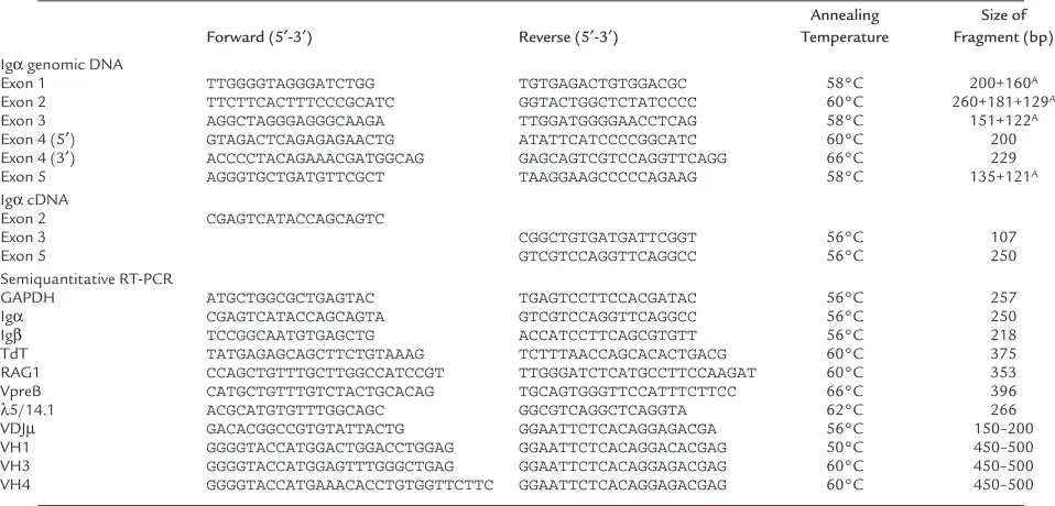
Identification of mutation in Igα.Genomic DNA samples from 25 patients with defects in B-cell development but no mutations in Btk (22, 23, 29), µheavy chain (26), VpreB, or λ5/14.1 (18)were analyzed by SSCP for muta-tions in Igαand Igβ.DNA from a 2-year-old Turkish girl with agammaglobulinemia demonstrated an altered band migration in the analysis of exon 3 of Igα (Figure 1a). Both parents of the child were heterozygous for this pattern, but analysis of DNA from 100 healthy controls did not reveal any additional samples with the same pat-tern, indicating that this was unlikely to be a polymor-phism. This region of the gene was cloned and sequenced and a single-bp substitution (A→G) at an invariant position (–2) of the splice acceptor site for exon 3 was revealed (Figure 1b).
The effects of this mutation on splicing of the Igα mes-sage were evaluated by using RT-PCR. We amplified Igα cDNA from the patient’s bone marrow and bone marrow from healthy controls and another ummunodeficient patient, using a primer from exon 2 paired with primers from either exon 3 or exon 5. When the exon 5 primer was used, the PCR product from the patient sample was approximately 120 bp smaller than the control PCR product. In addition, a faint PCR product that appeared smaller than the wild-type product was noted (Figure 2a). As expected, the sequence of the smaller PCR prod-uct from the patient demonstrated the deletion of exon 3. Because this exon encodes the transmembrane domain of Igαand is 119-bp long, the deletion of this exon would be expected to result in a truncated protein that could not be expressed as part of the pre-BCR. To determine whether any intact transcripts were produced, the exon 2 primer was used with a primer from exon 3. The PCR product from this reaction demonstrated the deletion of the first 13 bp of exon 3 and the use of a cryp-tic splice site within exon 3 (Figure 2b). These results indicate that there were no wild-type Igαtranscripts in the patient’s bone marrow.
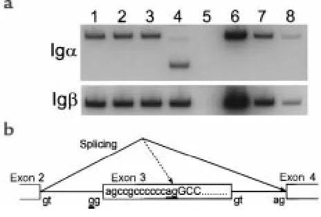
Phenotypic analysis of B cells and B-cell progenitors. The con-sequences of mutations in Igαon B-cell development were evaluated by immunofluorescence staining of
peripheral blood lymphocytes and bone marrow mononuclear cells. Like patients with mutations in µ heavy chain (26), the patient with defects in Igαhad undetectable CD19+B cells (<0.01%) in the peripheral
circulation. To determine the stage at which B-cell dif-ferentiation was blocked, bone marrow cells from the patient and an age-matched control were evaluated. For comparison, bone marrow from a patient with an amino acid substitution at an invariant cysteine in the CH4 domain of µheavy chain was analyzed in parallel. This mutation would not be expected to affect V-DJ recombi-nation, but it would prevent the normal folding and cell-surface expression of µheavy chain. Both patients had markedly reduced percentages of CD19+B-lineage cells
[image:4.612.308.536.257.611.2](9% and 2%, respectively) when compared with the healthy control (46%) (Figure 3). Triple-color immuno-fluorescence was used to examine the ratio of pro-B cells
Figure 1
(CD19+, CD34+, sIg–), pre-B cells (CD19+, CD34–, sIg–),
and B cells (CD19+, CD34–, sIg+) in the 3 individuals.
Within the CD19+population, 27% of the cells from the
healthy control expressed surface immunoglobulin, but less than 1% of the cells from either patient were sIg+,
confirming the severe block in B-cell differentiation. In both the patient with mutations in µheavy chain and the pateint with mutations in Igα, more than 75% of CD19+cells were pro-B cells, as defined by coexpression
of CD34+, whereas in the normal control patient, only
12% of the CD19+cells were CD34+. This finding
sug-gested that the B-cell differentiation was blocked at the pro-B to pre-B transition in the 2 disorders. This block was more clearly demonstrated in permeabilized cells stained for CD19, TdT, and µheavy chain (Figure 3). In both patients, more than 94% of the CD19+cell were
pos-itive for TdT and negative for cytoplasmic µheavy chain, a phenotype characteristic of pro-B cells. These findings demonstrate that B-cell development in Igαdeficiency is blocked at the pre-BCR checkpoint and that the block is identical to that seen in µheavy chain deficiency.
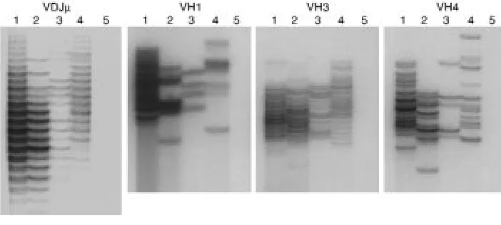
Gene expression in B-cell progenitors. Semiquantitative RT-PCR was used to evaluate the expression of genes specif-ic to various stages of B-cell differentiation. We includ-ed age-matchinclud-ed controls for each patient and analyzinclud-ed transcripts directly linked to early B-cell development and immunoglobulin gene expression. To demonstrate that PCR amplification was in the linear range, three 10-fold dilutions of control cDNA were included in each assay. GAPDH control amplification showed that equiv-alent amounts of cDNA were used in each RT-PCR amplification. The transcripts associated with the pro-B stage — TdT, RAG1, VpreB, and λ5/14.1 transcripts — were expressed in approximately equal amounts in the patient with Igαdeficiency, the one with µheavy chain deficiency, and the healthy controls (Figure 4a). Howev-er, the amount of V-DJ rearranged-µheavy chain tran-scripts were greatly decreased and equivalent in the patient with Igαdeficiency and in the patient with µ heavy chain deficiency (Figure 4b). This finding was fur-ther confirmed by using VH3-, VH4-, and VH1-specific primers to evaluate the use of each of these VH families.
To examine the diversity of the repertoire of rearranged µheavy chains, the VH-specific PCR prod-ucts were separated on a denaturing 6% polyacrylamide (sequencing) gel to allow the discrimination of tran-scripts with CDR3 regions of varying length (30). To avoid overloading, the control samples were diluted by a factor of 10 compared with the patient samples. Con-trol bone marrow samples showed a diverse repertoire, which was characterized by more than 15 bands sepa-rated from each other by 3 bp (Figure 5). The bands showed a Gaussian distribution in size with intensity of a band correlating with the total amount of µheavy chain CDR3 transcripts of that length. In both the Igα-deficient patient and the µ heavy chain–deficient patient, there were fewer bands and each band was faint and of equal intensity. The diversity of repertoire was approximately equal in the 2 patients. The degree of diversity in the 2 patients was further compared in experiments using the VH1-, VH3-, and VH4-specific primers. In every experiment, transcripts could be amplified from each patient, but the repertoire was lim-ited. The VH-specific PCR reactions were performed 4 times, and although the repertoire varied slightly in each experiment, the number of bands was always sim-ilar in the samples from the patient with mutations in Igαand the patient with mutations in µheavy chain. These results demonstrate that V-DJ rearrangement in the patient with mutations in Igαwas not decreased compared with that seen in a patient with an amino acid substitution in the constant region of µheavy chain. When coupled with the phenotypic studies, these results confirm that the block in B-cell differen-tiation in Igαdeficiency is at the pre-BCR checkpoint and is identical to that seen in patients with defects in µheavy chain.
Discussion
[image:5.612.304.536.53.210.2]The majority of patients with early-onset hypogamma-globulinemia and absent B cells are males with X-linked agammaglobulinemia (29). These patients have muta-tions in the hematopoietic-specific cytoplasmic tyrosine kinase Btk (22, 23, 29). The remaining patients appear
Figure 2
to have a heterogeneous group of disorders. We have recently shown that some of these patients have muta-tions in µheavy chain (26)or λ5/14.1 (18), both of which are components of the pre-BCR complex (31, 32). To determine whether defects in other components of the pre-BCR might result in immunodeficiency, we screened DNA from affected patients for mutations in VpreB, Igα,and Igβ. We identified a 2-year-old girl with a homozygous splice defect resulting in the truncation of Igαupstream of the transmembrane domain.
Like patients with defects in µheavy chain (26), the patient with mutations in Igαhad no detectable CD19+
B cells in the peripheral circulation. Analysis of bone marrow from this patient and a patient with an amino acid substitution in µheavy chain showed that both patients had normal numbers of pro-B cells
but an almost complete absence of pre-B cells. There is considerable variation in the number of pro-B cells in normal individu-als, and the number tends to decrease with age. This may explain the slightly higher number of pro-B cells in the patient with Igαdeficiency compared with the patient with µheavy chain defects. The amount and diversity of µ heavy chain V-DJ rearrangements in these 2 disorders were equivalent. Both patients had a small num-ber of transcripts utilizing memnum-bers of the VH1, VH3, and VH4 families.
Mice that are null for Igαhave not been described; however, mice with a truncation in Igαthat removes the COOH-terminal 40 amino acids of the cytoplasmic domain, including the ITAM motif, have a relatively mild block in the pro-B to pre-B transition (16). This suggests that the complete failure of B-cell development in the patient with a truncation in Igα before the transmem-brane domain is due to the inability of this protein to escort the µheavy chain and the Igβcomponent of the signal-transduction module to the cell surface, rather than the loss of the Igαsignaling capacity.
In contrast to our findings in the Igα-defi-cient patient, which show a block at the pro-B to pre-pro-B transition similar to that seen in µ heavy chain deficiency, studies in mice with defects in Igβare reported to show decreased V-DJ rearrangement when compared with µ-deficient mice (5). There are several possible explanations for the discordance between human Igαdeficiency and murine Igβ defi-ciency. Under some circumstances, Igβmay reach the cell surface in the absence of Igα (33). If homodimeric complexes of Igβ (34), but not Igα, are expressed, it might be possi-ble for Igβto transmit signals in the absence of Igα,whereas the reverse would not be true. Alternatively, it is possible that there are sig-nal-transduction pathways that are initiated in the cytoplasm by the ITAM-containing domain of Igβbut not Igα. Finally, despite
the overall similarity in early B-cell development in mice compared with humans, there may be differences between the 2 species in the cell-surface expression or signaling capacity of Igα/Igβ complexes.
[image:6.612.263.524.243.622.2]Complexes containing the Igα/Igβheterodimer can be detected on the surface of murine B-cell precursors before V-DJ rearrangement (3); however, attempts to document the expression of the same complex on human pro-B cells have not been successful (2, 3, 35, 36). It should be noted that it has been more difficult to demonstrate the cell-surface expression of all the components of the pre-BCR in human B-cell precursors compared with murine counterparts. In mice, surro-gate light chains form complexes with surrosurro-gate heavy chains and can be identified on the surface of pro-B
Figure 3
Flow cytometric analysis of B-cell development. Bone marrow mononuclear cells from a control (left column), µheavy chain–deficient patient (middle column), and Igα-deficient patient (right column) were stained with antibody to CD19. Both patients showed a marked reduction in the number of CD19+cells within the lymphoid gate (first row). Less
than 1% of the CD19+cells from either patient were B cells, as defined by coexpression of
CD19 and surface immunoglobulin (second row). More than 75% of the B-lineage cells in both patients were positive for CD34 (third row). CD19+permeabilized cells were stained
for cytoplasmic TdT and µheavy chain. More than 94% of the CD19+cells from both
cells in the absence of µ heavy chains (37). There is some controversy about the existence of similar com-plexes on human pro-B cells. Some groups have evi-dence suggesting that these complexes are present (38–40),but others maintain that these complexes do not occur (41). Finally, it appears that the intact pre-BCR is expressed more transiently and at lower inten-sity in human B-cell precursors compared with murine precursors (1, 42). Although these differences have sometimes been attributed to the reagents or tech-niques used to demonstrate the various proteins, true differences between the 2 species may exist.
Murine models of immunodeficiency, in which single genes have been rendered nonfunctional, have dramat-ically increased our understanding of normal lympho-cyte development. However, it is clear that that there is not always complete concordance between mice with defects in a particular gene and patients with mutations in the same gene. Detailed evaluation of patients with single gene defects of the immune system can elucidate
the pathways in lymphocyte differentiation that are invariant and highly consistent between species and those that show some flexibility and may vary from 1 species to another. In this study, we have done a side-by-side comparison of B-cell differentiation in 2 patients with defects in different components of the pre-BCR. A patient with a functionally null defect in Igαand a patient with a defect in µheavy chain that would pre-vent cell-surface expression of the antigen receptor com-plex had an identical block in B-cell differentiation. This indicates that in the human, Igαis essential for the expression of the pre-BCR, but, does not appear to play a role in B-cell development until it is expressed along with µheavy chain as part of the pre-BCR.
Acknowledgments
We appreciate the willingness of the patients and their families to participate in these research studies. These studies were supported by grants from the National Institutes of Health (AI-25129), National Cancer
Insti-Figure 4
[image:7.612.77.516.53.240.2]Semiquantitative RT-PCR analysis of B cell–specific transcripts in the bone marrow. (a) Equal amounts of cDNA from 2 healthy controls (lanes 1 and 2), a patient with µheavy chain deficiency (lane 3), the patient with Igαdeficiency (lane 4), a cDNA negative template control (lane 5), and three 10-fold dilutions of control cDNA (10×, 1×, and 0.1×) (lanes 6–8) were amplified using primers specific for TdT, RAG1, VpreB, and λ5/14.1. GAPDH was used as a control to demonstrate equal concentrations of cDNA. (b) cDNA samples were analyzed as in a, except that the 10-fold dilutions in lanes 6–8 were 1×, 0.1×, and 0.01×of the control. Primers specific for all VH family members or primers specific for VH3, VH4, or VH1 were paired with a primer form the CH1 domain of µheavy chain.
Figure 5
[image:7.612.185.536.579.737.2]tute (P30 CA21765), the Assisi Foundation, March of Dimes (FY97-0384), and American Lebanese Syrian Associated Charities, and by funds from the Federal Express Chair of Excellence.
1. Karasuyama, H., et al. 1994. The expression of Vpre-B/lambda 5 surro-gate light chain in early bone marrow precursor B cells of normal and B cell-deficient mutant mice. Cell. 77:133–143.
2. Lassoued, K., Illges, H., Benlagha, K., and Cooper, M.D. 1996. Fate of sur-rogate light chains in B lineage cells. J. Exp. Med. 183:421–429. 3. Koyama, M., et al. 1997. CD79α/CD79βheterodimers are expressed on
pro-B cell surfaces without associated µheavy chain. Int. Immunol.
9:1767–1772.
4. Nagata, K., et al. 1997. The Igα/Igβheterodimer on µ-negative proB cells is competent for transducing signals to induce early B cell differentia-tion. Immunity. 7:559–570.
5. Gong, S., and Nussenzweig, M.C. 1996. Regulation of an early develop-mental checkpoint in the B cell pathway by Igβ. Science. 272:411–414. 6. Reth, M., Petrac, E., Wiese, P., Lobel, L., and Alt, F.W. 1987. Activation of Vκgene rearrangement in pre-B cells follows the expression of mem-brane-bound immunoglobulin heavy chains. EMBO J. 6:3299–3305. 7. Tsubata, T., Tsubata, R., and Reth, M. 1992. Crosslinking of the cell
sur-face immunoglobulin (mu-surrogate light chains complex) on pre-B cells induces activation of V gene rearrangements at the immunoglobu-lin κlocus. Int. Immunol. 4:637–641.
8. Sakaguchi, N., Kashiwamura, S., Kimoto, M., Thalmann, P., and Melch-ers, F. 1988. B lymphocyte lineage-restricted expression of mb-1, a gene with CD3-like structure properties. EMBO J. 7:3457–3464.
9. Hermanson, G.G., Eisenberg, D., Kincade, P.W., and Wall, R. 1988. B29: a member of the immunoglobulin gene superfamily exclusively expressed on B-lineage cells. Proc. Natl. Acad. Sci. USA. 85:6890–6894. 10. Reth, M., et al. 1991. The B-cell antigen receptor complex. Immunol.
Today. 12:196–200.
11. Clark, M.R., et al. 1992. The B cell antigen receptor complex: association of Ig-αand Ig-βwith distinct cytoplasmic effectors. Science. 258:123–126. 12. Sanchez, M., et al. 1993. Signal transduction by immunoglobulin is
mediated through Igαand Igβ. J. Exp. Med. 178:1049–1055.
13. Kim, K.M., Alber, G., Weiser, P., and Reth, M. 1993. Differential signal-ing through the Ig-αand Ig-βcomponents of the B cell antigen recep-tor. Eur. J. Immunol. 23:911–916.
14. Papavasiliou, F., Jankovic, M., Suh, H., and Nussenzweig, M.C. 1995. The cytoplasmic domains of immunoglobulin (Ig) αand Igβindependently induce the precursor B cell transition and allelic exclusion. J. Exp. Med.
182:1389–1394.
15. Teh, Y.-M., and Neuberger, M.S. 1997. The immunoglobulin (Ig)αand Igβ cytoplasmic domains are independently sufficient to signal B cell matu-ration and activation in transgenic mice. J. Exp. Med. 185:1753–1758. 16. Torres, R.M., Flaswinkel, H., Reth, M., and Rajewsky, K. 1996. Aberrant
B cell development and immune response in mice with a compromised BCR complex. Science. 272:1804–1808.
17. Kitamura, D., et al. 1992. A critical role of λ5 protein in B cell develop-ment. Cell. 69:823–831.
18. Minegishi, Y., et al. 1998. Mutations in the human λ5/14.1 gene result in B cell deficiency and agammaglobulinemia. J. Exp. Med. 187:71–77. 19. Peschon, J.J., et al. 1994. Early lymphocyte expression is severely impaired in interleukin 7 receptor–deficient mice. J. Exp. Med.
180:1955–1960.
20. von Freeden-Jeffry, U., et al. 1995. Lymphopenia in interleukin (IL)-7 gene-deleted mice identifies IL-7 as a nonredundant cytokine. J. Exp. Med. 181:1519–1526.
21. Puel, A., Ziegler, S.F., Buckley, R.H., and Leonard, W.J. 1998. Defective
IL7R expression in T(–)B(+)NK(+) severe combined immunodeficiency.
Nat. Genet. 20:394–397.
22. Tuskada, S., et al. 1993. Deficient expression of a B cell cytoplasmic tyro-sine kinase in human X-linked agammaglobulinemia. Cell. 72:279–290. 23. Vetrie, D., et al. 1993. The gene involved in X-linked agammaglobuli-naemia is a member of the src family of protein-tyrosine kinases. Nature.
361:226–233.
24. Thomas, J.D., et al. 1993. Colocalization of X-linked agammaglobuline-mia and X-linked immunodeficiency genes. Science. 261:355–358. 25. Rawlings, D.J., et al. 1993. Mutation of unique region of Bruton’s
tyro-sine kinase in immunodeficient XID mice. Science. 261:358–361. 26. Yel, L., et al. 1996. Mutations in the mu heavy-chain gene in patients with
agammaglobulinemia. N. Engl. J. Med. 335:1486–1493.
27. Conley, M.E., Fitch, H.M., Cleveland, J.L., Parolini, O., and Rohrer, J. 1994. Screening of genomic DNA to identify mutations in the gene for Bruton’s tyrosine kinase. Hum. Mol. Genet. 3:1751–1756.
28. Meffre, E., et al. 1996. A human non-XLA immunodeficiency disease characterized by blockage of B cell development at an early proB cell stage. J. Clin. Invest. 98:1519–1526.
29. Conley, M.E., Mathias, D., Treadway, J., Minegishi, Y., and Rohrer, J. 1998. Mutations in Btk in patients with presumed X-linked agamma-globulinemia. Am. J. Hum. Genet. 62:1034–1043.
30. Gokmen, E., Raaphorst, F.M., Boldt, D.H., and Teale, J.M. 1998. Ig heavy chain third complementarity determining regions (H CDR3s) after stem cell transplantation do not resemble the developing human fetal H CDR3s in size distribution and Ig gene utilization. Blood. 92:2802–2814. 31. Melchers, F., et al. 1993. The surrogate light chain in B-cell development.
Immunol. Today. 14:60–68.
32. Rajewsky, K. 1996. Clonal selection and learning in the antibody system.
Nature. 381:751–758.
33. Costa, T.E., Franke, R.R., Sanchez, M., Misulovin, Z., and Nussenzweig, M.C. 1992. Functional reconstitution of an immunoglobulin antigen receptor in T cells. J. Exp. Med. 175:1669–1676.
34. Lassoued, K., Guglielmi, P., and Benlagha, K. 1997. Expression of the Igα and Igβmolecules in human pro-B cell lines. Bull. Acad. Natl. Med.
181:1465–1475.
35. Nakamura, T., Kubagawa, D., and Cooper, M.D. 1992. Heterogeneity of immunoglobulin-associated molecules on human B cells identified by monoclonal antibodies. Proc. Natl. Acad. Sci. USA. 89:8522–8526. 36. Okazaki, M., Luo, Y., Han, T., Yoshida, M., and Seon, B.K. 1993. Three
new monoclonal antibodies that define a unique antigen associated with prolymphocytic leukemia/non-Hodgkin’s lymphoma and are effective-ly internalized after binding to the cell surface antigen. Blood. 81:84–94. 37. Karasuyama, H., Rolink, A., and Melchers, F. 1993. A complex of glyco-proteins is associated with VpreB/lambda 5 surrogate light chain on the surface of µheavy chain–negative early precursor B cell lines. J. Exp. Med.
178:469–478.
38. Guelpa-Fonlupt, V., Tonnelle, C., Blaise, D., Fougereau, M., and Fumoux, F. 1994. Discrete early pro-B and pre-B stages in normal human bone marrow as defined by surface pseudo-light chain expression. Eur. J. Immunol. 24:257–264.
39. Meffre, E., Fougereau, M., Argenson, J.N., Aubaniac, J.M., and Schiff, C. 1996. Cell surface expression of surrogate light chain (ΨL) in the absence of mu on human pro-B cell lines and normal pro-B cells. Eur. J. Immunol.
26:2172–2180.
40. Sanz, E., and de la Hera, A. 1996. A novel anti–Vpre-B antibody identi-fies immunoglobulin-surface receptors on the surface of human pro-B cells. J. Exp. Med. 183:2693–2698.
41. Wang, Y.H., Nomura, J., Faye-Petersen, O.M., and Cooper, M.D. 1998. Sur-rogate light chain production during B cell differentiation: differential intracellular versus cell surface expression. J. Immunol. 161:1132–1139. 42. Lassoued, K., et al. 1993. Expression of surrogate light chain receptors is