http://dx.doi.org/10.4236/aar.2015.43011
How to cite this paper: Rao, K.S., Chakrabarti, S.K., Dongare, V.S., Sharath, B.S., Vikas, H.M., Chetana, K. and Deb, K.D. (2015) An Intensive Mind and Body Therapeutic Program Leads to Alteration in Gene Expression Critical to Aging Process in Peripheral Blood Stem Cells. Advances in Aging Research, 4, 89-95. http://dx.doi.org/10.4236/aar.2015.43011
An Intensive Mind and Body Therapeutic
Program Leads to Alteration in Gene
Expression Critical to Aging Process
in Peripheral Blood Stem Cells
Krishna S. Rao1, Swarup K. Chakrabarti2, Vaishali S. Dongare2, B. S. Sharath2, H. M. Vikas2,
K. Chetana1, Kaushik D. Deb2*
1Nithyananda University, Nithyananda Dhyanapeeta, Nithyananda Nagar, Bangalore, India 2DiponEd Biointelligence, Bommsandra Industrial Area, Bangalore, India
Email: *[email protected]
Received 30 2015
Copyright © 2015 by authors and Scientific Research Publishing Inc.
This work is licensed under the Creative Commons Attribution International License (CC BY). http://creativecommons.org/licenses/by/4.0/
Abstract
augments critical genes in PMBC which upregulate hematopoiesis and stem cell numbers and also controls genes that regulate age-related complications.
Keywords
MBT, Stem Cells, Aging
1. Introduction
Over the past 150 years, improvements in standards of living and advances in medical science have doubled life spans in much of the world [1]. According to 2014 world population data sheet, today, average life expectancy is increasing every day leading to gradual aging in a large population of the society.
In view of the remarkable increases in human life expectancy, there is an urgency form the biomedical com-munity to understand the connection between aging and aging-related diseases since the major burden of ill health falls on the aging population of the society. Notwithstanding of the fact that aging is a biological inevita-ble process and not a pathological condition, it is correlated with various skin and body pathologies, including degenerative disorders [2]-[4]. Furthermore aging may cause organ failure and even death over period of time.
Thus the knowledge of a panel of key genes and the underlying mechanisms that regulate their expression will be useful to understand the causes for organ failures and loss of rejuvenating organs or tissue in the body as we become older.
In the recent past integrative medicine (IM) approaches including yoga, meditation, neutraceuticals and breathing controls exercise, popularly termed as mind and body therapies [5], have been considered to have po-tential rejuvenating effects on aging human tissue/organs. It was also shown recently [6] that aging caused a de-cline in hematopoesis leading to poor blood formation and parallel decrease in stem cell numbers.
How aging affects the hematopoetic stem cells (HSC’s) and the quality of PBMNC’s remains to be deci-phered. We hypothesized that MBT might give rise to marked gene expression changes of key genes and path-ways controlling aging and rejuvenation of tissues in the body.
Therefore, the objective of this study was to evaluate the regenerative effects of our MBT on the HSC’s and PBMNC’s through expression profiling of genetic markers and key signaling pathways in people undergone meditation and in people prior to meditation.
2. Materials & Methods
2.1. Study Design & LabelingMBT program:
40 healthy adult volunteers male and female were enrolled in our intensive MBT program (Inner Awakening Yoga) for 21 days. Volunteers donated blood samples with informed consent for the differential gene expression study before and after the MBT program. Samples collected from the volunteers before program were designat-ed as BIAY group and those after the program were designatdesignat-ed as AIAY group. BIAY was considerdesignat-ed as the control group and AIAY was considered as the test group. Sample numbers 1 - 10 were pooled in as BIAY1 matched against sample numbers 1 - 10 of after samples as AIAY1 and BIAY1 Vs AIAY1. Same procedure was repeated from 10 - 20, 20 - 30 and 30 - 40 and they were named and matched respectively as BIAY2 Vs AIAY2, BIAY3 Vs AIAY3, BIAY4 Vs AIAY4. Human whole genome HG-U133_Plus_2 chips form Affymetrix (Santa Clara, CA, USA) was used for this study.
2.2. Blood Sample Collection and RNA Storing
centrifuged at 4000 rpm for 10 minutes at 4˚C. The supernatant was completely removed and discarded. After 20 minutes, visible pellets were suspended in 10 ml RNA later (Qiagen, Valencia, CA, USA). The identical procedures were carried out on the sample collected on 21 day of the program.
2.3. Microarray Data Analysis
For microarray data analysis software version GeneSpring 11.5 was used. The CEL files were imported in Af-fymetrix (Santa Clara, CA, USA) expression workflow using Technology for HG-U133_Plus_2. 5 µg of total RNA was used for GeneChip analysis.
Summarization: Like RMA, GCRMA, MAS5, PLIER 16 and LiWong were tried and finally MAS5 was opted for summarization. This also provided Flag information for the Probe sets. Baseline to median of all sam-ples was applied for this data.
Statistics: A t-test unpaired was used to identify the differentially expressing entities.
3. Results
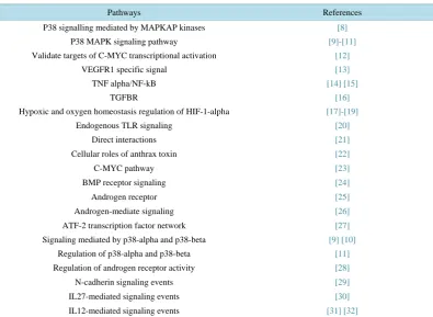
Gene ontology (GO) is a major bioinformatics tool to unify the representation of gene and gene product attributes across all species used to address the need for consistent descriptions of gene products across databas-es as opposed to gene nomenclature which maintains and develops controlled vocabulary of gene and gene products and is not unified across all species, has shown significantly enriched GO terms; an unifying vocabu-lary denoting a particular gene or gene product across species [7]. Furthermore, since the number of known pathways within cells is significantly smaller than the number of genes and GO terms that are typically profiled, the layout of data from a gene-centric view to a pathway-centered ones results a dramatic reduction in the num-ber of dimensions and can help biologist to understand and interpret the data in a manner that is not possible when viewed as a collection of individual genes. The pathway analysis of the expression data in peripheral blood stem cells in this study has revealed significant gene expression differences in 22 pathways between groups before and after the MBT program. The results are shown in Table 1.
Table 1. Microarray data reveals key pathways that show marked changes in the gene expression in PMBC due to MBT program.
Pathways References
P38 signalling mediated by MAPKAP kinases [8]
P38 MAPK signaling pathway [9]-[11]
Validate targets of C-MYC transcriptional activation [12]
VEGFR1 specific signal [13]
TNF alpha/NF-kB [14] [15]
TGFBR [16]
Hypoxic and oxygen homeostasis regulation of HIF-1-alpha [17]-[19]
Endogenous TLR signaling [20]
Direct interactions [21]
Cellular roles of anthrax toxin [22]
C-MYC pathway [23]
BMP receptor signaling [24]
Androgen receptor [25]
Androgen-mediate signaling [26]
ATF-2 transcription factor network [27]
Signaling mediated by p38-alpha and p38-beta [9] [10]
Regulation of p38-alpha and p38-beta [11]
Regulation of androgen receptor activity [28]
N-cadherin signaling events [29]
IL27-mediated signaling events [30]
[image:3.595.112.508.432.721.2]Some of the critical pathways known to regulate aging process, such as 1) pro-inflammatory TNF al-pha/NF-kB; 2) IL-12 signaling pathway; 3) hypoxic HIF-1-alpha; 4) key regulator of programmed cell death, C-MYC; and 5) P38 MAPK (mitogen-activated protein kinase) signaling pathway, found to be dysregulated in the cohorts compared to subjects prior to MBT program. In addition, TGFβ (transforming growth factor-beta) pathway, which was a positive regulator of collagen synthesis [33] and critical for enhancement of facial reju-venation, showed significant gene expression difference between control and meditated groups.
Significant genes either 2-fold up or 2-fold down were EIF4E, BMI1, TMEF2 that are known to be key play-ers in C-MYC pathway. Additional key genes of C-MYC pathway such as EP300, PKN2, HSP90AA1, TMEFF2, IREB2, CREB1 were also found to be significantly altered (more than 2-fold) in people after meditation. More-over, expression of a potent pro-inflammatory cytokine interferon-gamma went down 2.51-fold and also STAT- 2 (Signal Transducer and Activator of Transcription 2), a critical regulator of pro-inflammatory response [34]
went down 2.47-fold in the meditated group compared to pre-meditated subjects, thereby indicating a reduction of chronic inflammation in the meditated group. Chronic inflammation has been shown to induce telomere dys-function [35] and thereby can accelerate aging and age-related complications.
Besides pathway analysis, individual data for 420 upregulated and 165 downregulated entities (genes) were obtained from the microarray data. The results are presented in the supplementary Table S1.
http://www.diponed.com/publications
Activating Transcription Factor-2 (ATF2), which is implicated in DNA damage response, cell cycle and apoptosis [36], and thereby regulates cellular senescence, went up 6.86-fold after the meditation. Furthermore, ATF2 has been shown to downregulate c-Jun N-terminal kinase; a critical mediator of cellular inflammation
[36]. Additionally, RSP11 (Ribosomal protein, small subunit) was 6.5-fold down in the meditated group. It has been reported that knockdown to RSP11 increased lifespan in mice [37]. Expression of CREB1 (cAMP response element binding protein 1) was up (2.19-fold) in the meditated group which was a known regulator of a key ag-ing-related gene ATM (ataxia telangiectasia mutated) which is an early-onset disease and is characterized by signs of premature aging [38].
Very interestingly, zinc finger transcription factor GATA-2, a critical regulator of hematopoietic stem cells and progenitor cells [39] (HPCs) that eventually yield specific classes of blood cells, went up 2-fold in the me-ditated group compared to pre-meme-ditated people. Furthermore, polycomb ring finger oncogene Bmi1, which is essential for the self-renewal of adult stem cells through the repression of genes involved in cellular senescence
[40], went up 2.89-fold in the mediated group, thereby indicating enhancement of stem cells numbers in the me-ditated group which in turn can slow-down the aging process since there is an overall decline in the stem cell numbers with age.
4. Discussion
in mediated cohort. Furthermore, another critical gene under CMYC pathway such as TMEFF2 [42] (transmem-brane protein with EGF-like and two follistatin-like domains 2) was down in the meditated cohort. Genetic vari-ation of TMEFF2 has been shown to affect human longevity [43]. Fourth, in our study P38 MAPK, a key regu-lator of pro-inflammatory cytokine biosynthesis which induces low-grade chronic inflammation associated with aging was found to be affected by MBT program. Taken together, our data clearly indicated the beneficial ef-fects of MBT program by controlling the gene expression changes of well-documented gene signaling pathways associated with aging. Additionally all these data suggest that physiological changes associated with MBT pro-gram as reported previously may have integral molecular component which kicks off during a short, intensive MBT practice and may lead to long-term stable meditation effects. Thus even after discontinuation of active MBT program, the anti-aging effects could be long lasting and can result youthful appearance even at an older age.
Furthermore, in this study, there were many genes that were affected by MBT program compared with the pre-meditated regimen which was consistent with our hypothesis that MBT had specific effects on gene expres-sion in PMBCs. Importantly, in this study we clearly observed MBT had specific effects on certain aging-related genes as many of the gene expression changes were robust (more than 2-fold).
Examination of the differentially expressed individual genes upon MBT made us speculate on the cellular ef-fects of the MBT program. For example, two genes GATA-2 and BMi1 that regulate hematopoiesis were found be upregulated upon MBT program. This may augment overall stem cell numbers in the practitioner group. In a separate study (manuscript submitted for publication), our recent data clearly indicated enhancement of viable stem cells and increased telomerase activity in PMBCs due to MBT program.
Although in earlier studies, despite significant changes in gene expression that were induced by meditation, gene ontology analysis did not result in the enrichment of specific molecular pathways linked to aging. To our knowledge, ours is the first study where we have been able to identify some distinct molecular pathways and genes involved in aging. Although there were 22 molecular pathways identified in our studies, we think that a longer duration of the meditation program can reveal the most critical pathways and the genes affected by medi-tation. Thus a longer MBT program can dissect converging pathways responsible for robust gene expression changes of a very few specific molecular pathways and genes associated with aging process. Also further work is needed to figure out functional links to the gene products differentially regulated by the MBT program and the control regimens. It is critical to extend the expression changes that are observed at the mRNA levels to the pro-tein levels through proteomics analysis. It is also desirable to determine how long these MBT meditated gene expression changes effects last and thus longitudinal studies will be very useful.
5. Conclusion
In conclusion, the data presented here show that MBT have specific effects at the genetic level on some critical aging genes, hematopoietic genes, and pathways in a relatively shorter period of time. This approach can be used to systematically interrogate these molecular changes, dissecting the key molecular signals that are trig-gered by MBT program that eventually impact PMBCs.
Disclosure Statement
None of the authors has any conflict of interest to declare.
References
[1] The Academy of Medical Sciences (2009) Rejuvenating Aging Research: A Report by the Academy of Medical Scien- ces. The Academy of Medical Sciences, London.
[2] Gems, D. and Partridge, L. (2013) Genetics of Longevity in Model Organisms: Debates and Paradigm Shifts. Annual Review of Physiology, 75, 621-644. http://dx.doi.org/10.1146/annurev-physiol-030212-183712
[3] Kirkwood, T.B. (2005) Understanding the Odd Science of Aging. Cell, 120, 437-447.
http://dx.doi.org/10.1016/j.cell.2005.01.027
[4] Vijg, J. and Campisi, J.P. (2008) Puzzles, Promises and a Cure for Ageing. Nature, 454, 1065-1071.
http://dx.doi.org/10.1038/nature07216
Can-cer, 112, 2607-2616. http://dx.doi.org/10.1002/cncr.23443
[6] Hartmut, G. and Zheng, Y. (2013) Cdc42 and Aging of Hematopoietic Stem Cells. Current Opinion in Hematology, 20, 295-300. http://dx.doi.org/10.1097/MOH.0b013e3283615aba
[7] Gene Ontology Consortium. (2004) The Gene Ontology (GO) Database and Informatics Resource. Nucleic Acids Re-search, 32, D258-D261. http://dx.doi.org/10.1093/nar/gkh036
[8] Stokoe, D., Campbell, D.G., Nakielny, S., Hidaka, H., Leevers, S.J., Marshall, C. and Cohen, P. (1992) MAPKAP Ki-nase-2; A Novel Protein Kinase Activated by Mitogen-Activated Protein Kinase. TheEMBO Journal, 11, 3985-3994.
[9] Tamura, K., Sudo, T., Senftleben, U., Agnes M Dadak, A.M., Johnson, R. and Karin, M. (2000) Requirement for p38alpha in Erythropoietin Expression: A Role for Stress Kinases in Erythropoiesis. Cell, 102, 221-231.
http://dx.doi.org/10.1016/S0092-8674(00)00027-1
[10] del Barco Barrantes, I., Coya, J.M., Maina, F., Arthur, J.S. and Nebreda, A.R. (2011) Genetic Analysis of Specific and Redundant Roles for p38alpha and p38beta MAPKs during Mouse Development. Proceedings of the National Acade-my of Sciences of the United States of America, 108, 12764-12769.
http://dx.doi.org/10.1073/pnas.1015013108
[11] Ono, K. and Han, J. (2000) The p38 Signal Transduction Pathway: Activation and Function. Cellular Signalling, 12, 1- 13. http://dx.doi.org/10.1016/S0898-6568(99)00071-6
[12] Menssen, A. and Hermeking, H. (2002) Characterization of the c-MYC-Regulated Transcriptome by SAGE: Identifica- tion and Analysis of c-MYC Target Genes. Proceedings of the National Academy of Sciences of the United States of America, 99, 6274-6279. http://dx.doi.org/10.1073/pnas.082005599
[13] Koch, S. and Claesson-Welsh, L. (2012) Signal Transduction by Vascular Endothelial Growth Factor Receptors. Cold Spring Harbor Perspectives in Medicine, 2, a006502.
[14] Gilmore, T.D. and Wolenski, F.S. (2012) NF-κB: Where Did It Come from and Why? Immunological Reviews, 246, 14-35. http://dx.doi.org/10.1111/j.1600-065X.2012.01096.x
[15] Gupta, S., Bi, R., Kim, C.,Chiplunkar, S., Yel, L. and Gollapudi, S. (2005) Role of NF-κB Signaling Pathway in In-creased Tumor Necrosis Factor-α-Induced Apoptosis of Lymphocytes in Aged Humans. Cell Death & Differentiation, 12, 177-183. http://dx.doi.org/10.1038/sj.cdd.4401557
[16] Derynck, R. and Feng, X.H. (1997) TGF-β Receptor Signaling. Biochimica et Biophysica Acta, 24, F105-F150.
[17] Rezvani, H.R., Ali, N., Serrano-Sanchez, M., Dubus, P., Varon, C., Ged, C., et al. (2011) Loss of Epidermal Hypoxia- Inducible Factor-1α Accelerates Epidermal Aging and Affects Re-Epithelialization in Human and Mouse. Journal of Cell Science, 124, 4172-4183.
[18] Cho, Y.S., Bae, J.M., Chun, Y.S., Chung, J.H., Jeon, Y.K.,Kim, I.S., et al. (2008) HIF-1α Controls Keratinocyte Pro-liferation by Up-Regulating p21 (WAF1/Cip1). Biochimica et Biophysica Acta, 1783, 323-333.
[19] Rezvani, H.R., Ali, N., Nissen, L.J., Harfouche, G.,de Verneuil, H., Taïeb, A., et al. (2011) HIF-1α in Epidermis: Oxygen Sensing, Cutaneous Angiogenesis, Cancer, and Non-Cancer Disorders. Journal of Investigative Dermatology, 31, 1793-1805. http://dx.doi.org/10.1038/jid.2011.141
[20] Yu, L., Wang, L.T. and Chen, S.W. (2010) Endogenous Toll-Like Receptor Ligands and Their Biological Significance. Journal of Cellular and Molecular Medicine, 14, 2592-2603. http://dx.doi.org/10.1111/j.1582-4934.2010.01127.x
[21] Hussain, S., Wilson, J.B., Medhurst, A.L.,Hejna, J., Witt, E.,Ananth, S., et al. (2004) Direct Interaction of FANCD2 with BRCA2 in DNA Damage Response Pathways. Human Molecular Genetics, 12, 1241-1248.
http://dx.doi.org/10.1093/hmg/ddh135
[22] Lacy, D.B. and Collier, R.J. (2002) Structure and Function of Anthrax Toxin. Current Topics in Microbiology and Im- munology, 271, 61-85.
[23] Prendergast, G.C. (1999) Mechanisms of Apoptosis by c-Myc. Oncogene, 18, 2967-2987.
http://dx.doi.org/10.1038/sj.onc.1202727
[24] Mishina, Y. (2003) Function of Bone Morphogenetic Protein Signaling during Mouse Development. Frontiers in Bios-cience, 8, d855-d869. http://dx.doi.org/10.2741/1097
[25] Matsumoto, T., Sakari, M., Okada, M.,Yokoyama, A., Takahashi, S., Kouzmenko, A., et al. (2013) The Androgen Receptor in Health and Disease. Annual Review of Physiology, 75, 201-224.
http://dx.doi.org/10.1146/annurev-physiol-030212-183656
[26] Falkenstein, E., Tillmann, H.-C., Christ, M., Feuring, M. and Wehling, M. (2000) Multiple Actions of Steroid Hormo- nes—A Focus on Rapid, Nongenomic Effects. Pharmacological Reviews, 52, 513-555.
[28] Guo, Z.Y., Dai, B.J., Jiang, T.Y., Xu, K.X., Xie, Y.Q.,Kim, O., et al. (2006) Regulation of Androgen Receptor Activ-ity by Tyrosine Phosphorylation. Cancer Cell, 10, 309-319. http://dx.doi.org/10.1016/j.ccr.2006.08.021
[29] Kimita, S., Irina, S. and Mitchell, G.A. (2002) Signaling Pathway Leading to Metastasis Is Controlled by N-Cadherin and the FGF Receptor. Cancer Cell, 2, 301-314. http://dx.doi.org/10.1016/S1535-6108(02)00150-2
[30] Pflanz, S., Timans, J.C., Cheung, J., Rosales, R.,Kanzler, H.,Gilbert, J., et al. (2002) IL-27, a Heterodimeric Cytokine Composed of EBI3 and p28 Protein, Induces Proliferation of Naive CD4+ T Cells. Immunity, 16, 779-790.
http://dx.doi.org/10.1016/S1074-7613(02)00324-2
[31] Burton, J.D., Bamford, R.N., Peters, C., Grant, A.J., Kurys, G.,Goldman, C.K., et al. (1994) A Lymphokine, Provi-sionally Designated Interleukin T and Produced by a Human Adult T-Cell Leukemia Line, Stimulates T-Cell Prolifera-tion and the InducProlifera-tion of Lymphokine-Activated Killer Cells. Proceedings of the National Academy of Sciences of the United States of America, 91, 4935-4939. http://dx.doi.org/10.1073/pnas.91.11.4935
[32] Trinchieri, G. (1995) Interleukin-12: A Proinflammatory Cytokine with Immunoregulatory Functions That Bridge In-nate Resistance and Antigen-Specific Adaptive Immunity. Annual Review of Immunology, 13, 251-276.
http://dx.doi.org/10.1146/annurev.iy.13.040195.001343
[33] Duncan, M.R., Frazier, K.S., Abramson, S., Williams, S., Klapper, H., Huang, X., et al. (1999) Connective Tissue Growth Factor Mediates Transforming Growth Factor β-Induced Collagen Synthesis: Down-Regulation by cAMP. FASEB Journal, 13, 1774-1786.
[34] Reich, N.C. (2013) STATs Get Their Move on. JAK-STAT, 2, e27080. http://dx.doi.org/10.4161/jkst.27080
[35] Jurk, D., Wilson, C. and Passos, J.F. (2014) Chronic Inflammation Induces Telomere Dysfunction and Accelerates Ageing in Mice.Nature Communications, 2, 4172.
[36] Jun, H., Chantal, D., Masahide, O. and Mercola, D. (2003) The Activation of c-Jun NH2-Terminal Kinase (JNK) by DNA-Damaging Agents Serves to Promote Drug Resistance via Activating Transcription Factor 2 (ATF2)-Dependent Enhanced DNA Repair. Journal of Biological Chemistry, 278, 20582-20592.
http://dx.doi.org/10.1074/jbc.M210992200
[37] Liang, H.Y., Masaro, E.J., Nelson, J.F., Strong, R.,McMahan, C.A. andRichardson, A. (2003) Genetic Mouse Models of Extended Lifespan. Experimental Gerontology, 38, 1353-1364. http://dx.doi.org/10.1016/j.exger.2003.10.019
[38] Shackelford, R.E. (2005) Pharmacologic Manipulation of the Ataxia-Telangiectasia Mutated Gene Product as an Inter- vention in Age-Related Disease. Medical Hypotheses, 65, 363-369. http://dx.doi.org/10.1016/j.mehy.2005.02.015
[39] Rodrigues, N.P., Janzen, V., Forkert, R., Dombkowski, D.M.,Boyd, A.S.,Orkin, S.H., et al. (2005) Haploinsufficien-cy of GATA-2 Perturbs Adult Hematopoietic Stem Cell Homeostatis. Blood, 106, 477-484.
http://dx.doi.org/10.1182/blood-2004-08-2989
[40] Park, I.-K., Morrison, S.J., and Clarke, F.M. (2004) Bmi1, Stem Cells, and Senescence Regulation. Journal of Clinical Investigation, 113, 175-179. http://dx.doi.org/10.1172/JCI200420800
[41] Syntichaki, P., Troulinaki, K. and Tavernarakii, N. (2007) eIF4E Function in Somatic Cells Modulates Ageing in Cae-norhabditis elegans. Nature, 445, 922-926. http://dx.doi.org/10.1038/nature05603
[42] Chen, X.F., Overcash, R., Green, T.,Hoffman, D.,Asch, A.S. and Ruiz-Echevarría, M.J. (2011) The Tumor Suppres-sor Activity of the Transmembrane Protein with Epidermal Growth Factor and Two Follistatin Motifs 2 (TMEFF2) Correlates with Its Ability to Modulate Sarcosine Levels. Journal of Biological Chemistry, 18, 16091-16100.
http://dx.doi.org/10.1074/jbc.M110.193805