0095-1137/09/$08.00⫹0 doi:10.1128/JCM.02006-08
Copyright © 2009, American Society for Microbiology. All Rights Reserved.
One-Step, Multiplex, Real-Time PCR Assay with Molecular Beacon
Probes for Simultaneous Detection, Differentiation, and Quantification
of Human T-Cell Leukemia Virus Types 1, 2, and 3
䌤
Guillaume Besson
1and Mirdad Kazanji
1,2*
Unite´ de Re´trovirologie, Centre International de Recherches Me´dicales de Franceville (CIRMF), BP 769, Franceville, Gabon,1and
Re´seau International des Instituts Pasteur, Institut Pasteur, 28 Rue du Dr Roux, 75015 Paris, France2
Received 17 October 2008/Returned for modification 22 December 2008/Accepted 3 February 2009
A single-tube, multiplex, real-time PCR assay with molecular beacons was established in which various probes were used for the simultaneous detection, differentiation, and quantification of human T-cell leukemia virus types 1, 2, and 3 (HTLV-1, HTLV-2, and HTLV-3, respectively) and of simian T-cell leukemia virus types 1 and 3 (STLV-1 and STLV-3, respectively). The quantitative amplification of the standards with MT4 (HTLV-1) and C19 (HTLV-2) cell lines and a molecular clone of HTLV-3 was linear, with the simplex and multiplex methods having similar efficiencies. A maximum difference of 0.9 (mean, 0.4; range, 0.0 to 0.9) was found between threshold cycle values in single and multiplex reactions. The efficiency with each probe in the multiplex reaction was close to 100%, indicating strong linear amplification. The albumin gene was used to standardize the copy number. Comparable results for the detection and quantification of HTLV-1 were obtained with our new methods and with other real-time PCR methods described previously. With our new multiplex assay, however, we were able to detect and quantify HTLV-2 and -3 and STLV-1 and -3 in clinical specimens, with an excellent dynamic range of 106
to 100
copies per assay, which the other assays could not do. Thus, it will be possible to determine a wide range of HTLV types in both standard and clinical samples, with a detection of 1 to 10 HTLV copies in samples containing at least 100 cells. Furthermore, our system can provide evidence for multiple infections with the three HTLV types, with separate proviral load results. Our new method also could be used for epidemiological studies in Africa and in countries where HTLVs and STLVs are endemic.
Human T-cell lymphotropic virus types 1 (HTLV-1), 2 (HTLV-2), and 3 (HTLV-3) belong to a group of primate retroviruses with certain common epidemiological and biolog-ical properties, including tropism for T lymphocytes. HTLV-1, isolated in 1980, is the causative agent of adult T-cell leukemia/ lymphoma (37) and tropical spastic paraparesis/HTLV-1-asso-ciated myelopathy (TSP/HAM) (17). HTLV-2, isolated 2 years after HTLV-1 (24), might be responsible for rare neurological syndromes that are clinically related to TSP/HAM (21, 33). Although no tumors have been linked definitively to such in-fections (14, 22), frequent lymphocytosis was described re-cently (1). Two new retroviruses, HTLV-3 and HTLV-4, have been identified in Cameroon, but no diseases have been asso-ciated with them yet (5, 45). The finding of these different HTLV types indicates that they are more diverse and polymor-phic than previously thought (28). Previous phylogenetic anal-ysis indicated that HTLV-1 can be classified into three major lineages, Cosmopolitan, Central African, and Melanesian, with each lineage being divided into various subtypes (38). Studies of the molecular epidemiology of HTLV-2 have shown at least two major molecular subtypes, designated HTLV-2A and HTLV-2B (19).
HTLV-1 is endemic in southern Japan, some regions of sub-Saharan Africa, and the Caribbean Basin, as well as some parts of
South America and the Middle East (16), with an estimated 15 to 20 million infected persons worldwide. HTLV-2 has been shown to be endemic in various American Indian populations (2) and has been endemic for the past 15 to 25 years among intravenous drug users in Europe and North America (34). Furthermore, since 1991, sporadic cases of HTLV-2 infection have been de-tected in west and central Africa in isolated rural populations, including some Pygmies (18). No information is available yet on the epidemiology of HTLV-3 or -4 (28).
HTLV-1 and -2 are transmitted in three ways: between sex-ual partners, mainly from men to women; from mothers to infants during prolonged breastfeeding; and through transfu-sions with HTLV-infected blood cells. It has been reported that 65% of patients who received whole blood or cellular blood components from HTLV-1-seropositive donors serocon-verted against HTLV-1 (23). HTLV-1 also has been reported to be transmitted in organ transplants (40).
In Gabon, central Africa, the prevalence of HTLV-1 infec-tion is high (5% of adults in urban areas and 8.5 to 10.5% of those in rural areas) (27). As mother-to-child transmission and blood transfusion appear to be the main routes of transmission in hospitalized children, the prevention of mother-to-child trans-mission and transfusion- or transplant-associated transtrans-mission will require screening, especially of populations in which the in-fections are endemic (11).
These viruses usually are detected indirectly by enzyme-linked immunosorbent assay (ELISA), and positive samples are confirmed by Western blot analysis. Samples are consid-* Corresponding author. Mailing address: Centre International de
Re-cherche Me´dicale de Franceville, BP 769, Franceville, Gabon. Phone: 241 07 69 90 99. Fax: 241 67 72 95. E-mail: [email protected].
䌤Published ahead of print on 11 February 2009.
1129
on May 16, 2020 by guest
http://jcm.asm.org/
ered positive when a complete Western blot profile can be obtained; however, no currently available immunological assay can detect acute infection with HTLVs. Furthermore, individ-uals infected with HTLV-3 and -4 have incomplete Western blot profiles, showing an indeterminate pattern (5, 45). No direct molecular methods are available for the rapid detection of these newly discovered viruses. PCR is a good choice for detecting HTLVs but might be less sensitive for low proviral loads. A rapid, non-labor-intensive amplification technique for the accurate quantification of the viral target is real-time PCR (20), in which internal fluorescent probes provide specificity and allow the detection of amplification products that are directly related to the copy number of the target (36). Real-time PCR already has been used for detecting HTLV-1 and -2 proviral loads (10, 12), including those of subtypes 1A, 1B, 1C, 2A, and 2B; however, the method did not detect HTLV-3 or various simian T-cell leukemia viruses (STLVs).
Therefore, a rapid, specific system for evaluating the various primate T-cell leukemia viruses is needed that can be used in epidemiological and pathogenesis investigations. As the HTLV proviral load can indicate the degree of replication and usually is correlated with the onset and progression of TSP/HAM (6, 35), real-time PCR also could provide information about dis-ease progression by the quantification of the HTLV viral load. We report here a one-step (single-tube), multiplex, real-time
PCR for the simultaneous detection, differentiation, and quan-tification of proviral HTLV-1, -2, and -3; the new method also can detect STLV type 1 (STLV-1) and STLV-3. The assay was validated with samples previously described and obtained from blood donors in Franceville (southeast Gabon), where the prevalence of HTLVs, especially HTLV-1B but also 1A, 2A, and 2B, is very high (14, 28).
MATERIALS AND METHODS
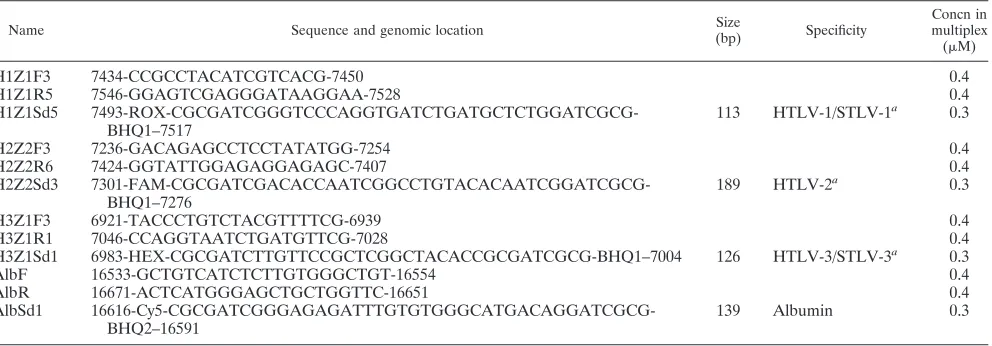
Oligonucleotide design.All of the complete genomes of HTLV-1, -2, and -3 and STLV-1 and -3 available in the GenBank database were included and aligned for the selection and design of various primers and probes (Table 1). Three pairs of primers located in theTaxgene that can detect various strains of HTLV-1, -2, and -3 and STLV-1 and -3 as well as primers for the detection of the albumin gene were designed with Beacon Designer software (Premier Biosoft, Palo Alto, CA) and are presented in Table 1. The four molecular beacon probes allow specific detection, because they are more adaptable than TaqMan probes (Table 2). The primers for the detection of albumin genes (AlbF and AlbR) were reported by Mortreux et al. (32), but we designed a new probe, AlbSd1 (5⬘-Cy5 and 3⬘-black hole quencher 2 [3⬘-BHQ2]) (Table 1).
Standards used for HTLV detection and quantification.The standard used for HTLV-1 was DNA lysate from the MT4 cell line (kindly provided by Franck Mortreux, Lyon, France). The cell line previously was characterized as having seven copies of the virus per cell (32), and the range of the MT4 standard was 105
to 100
[image:2.585.47.543.81.254.2]copies per reaction. The standard used for HTLV-2 was DNA lysate from the C19 cell line (kindly provided by Antoine Gessain, Institut Pasteur, Paris, France). It has been reported previously that this cell line carries seven copies of HTLV-2 (15, 26). The range of the C19 standard was 105to 100copies per
TABLE 1. Characteristics of primers and probes
Name Sequence and genomic location Size
(bp) Specificity
Concn in multiplex (M)
H1Z1F3 7434-CCGCCTACATCGTCACG-7450 0.4
H1Z1R5 7546-GGAGTCGAGGGATAAGGAA-7528 0.4
H1Z1Sd5 7493-ROX-CGCGATCGGGTCCCAGGTGATCTGATGCTCTGGATCGCG-BHQ1–7517
113 HTLV-1/STLV-1a 0.3
H2Z2F3 7236-GACAGAGCCTCCTATATGG-7254 0.4
H2Z2R6 7424-GGTATTGGAGAGGAGAGC-7407 0.4
H2Z2Sd3 7301-FAM-CGCGATCGACACCAATCGGCCTGTACACAATCGGATCGCG-BHQ1–7276
189 HTLV-2a 0.3
H3Z1F3 6921-TACCCTGTCTACGTTTTCG-6939 0.4
H3Z1R1 7046-CCAGGTAATCTGATGTTCG-7028 0.4
H3Z1Sd1 6983-HEX-CGCGATCTTGTTCCGCTCGGCTACACCGCGATCGCG-BHQ1–7004 126 HTLV-3/STLV-3a 0.3
AlbF 16533-GCTGTCATCTCTTGTGGGCTGT-16554 0.4
AlbR 16671-ACTCATGGGAGCTGCTGGTTC-16651 0.4
AlbSd1 16616-Cy5-CGCGATCGGGAGAGATTTGTGTGGGCATGACAGGATCGCG-BHQ2–16591
139 Albumin 0.3
a
The types and subtypes detected (only complete genomes were included) are the following: for HTLV-1, HTLV-1 WHP (AF259264), HTLV-1A ATK1 (J02029), HTLV-1A ATL-YS (U19949), HTLV-1A K30 (L03561), HTLV-1A RKI3A (AF042071), HTLV-1A TSP1 (M86840), HTLV-1B EL (M67514), HTLV-1C Mel5 (L02534), HTLV-1 (AF139170), HTLV-1 (AF033817.1), HTLV-1 Brazil (AY563953.1), STLV-1 Tan90 (AF074966), and STLV-1 TE4 (Z46900); for HTLV-2, HTLV-2 Brazil (AF139382), HTLV-2 Venezuela G2 (AF074965), HTLV-2A AF41231Hsa (AF412314), HTLV-2A K96 (AF326584), HTLV-2A MO (M10060), HTLV-2A RP329 (AF326583), HTLV-2B G12 (L11456), HTLV-2B GAB2 (Y13051), HTLV-2B GU (X89270), and HTLV-2D EFE2 (Y14365); and for HTLV-3: STLV-3 CTO604 (NC_003323), STLV-3 CTONG409 (AY222339), STLV-3 PH969 (Y07616), HTLV-3 Pyl43 (DQ462191), HTLV-3 2026ND (DQ093792), and STLV-3 TGE2117 (AY217650).
TABLE 2. Comparison ofCTvalues of the simplex and multiplex methods
Beacon probe Target(s) 5⬘reporter/3⬘quencher for a
: CTvalues for 104
copies/PCR
Single-dye assay Multiplex assay Single-dye assay Multiplex assay
H1Z1Sd5 HTLV-1/STLV-1 FAM/DABCYL ROX/BHQ1 25.1⫾0.1 25.7⫾0.1
H2Z2Sd3 HTLV-2 FAM/DABCYL FAM/BHQ1 25.2⫾0.3 25.1⫾0.3
H3Z1Sd1 HTLV-3/STLV-3 FAM/DABCYL HEX/BHQ1 25.1⫾0.1 25.7⫾0.2
AlbSd1 Albumin FAM/DABCYL Cy5/BHQ2 25.7⫾0.3 25.7⫾0.5
aFAM, 6-carboxyfluorescein: ROX, 6-carboxy-X-rhodamine; DABCYL, 4-(((4-dimethyl)-phenyl)-azo)benzoic acid.
on May 16, 2020 by guest
http://jcm.asm.org/
[image:2.585.42.546.644.716.2]reaction. The standard used for HTLV-3 was a molecular clone of HTLV-3, Pyl 43 (kindly provided by Renaud Mahieux, Institut Pasteur, Paris, France) (7) and was used in the same range as the other standards. The range of the quantitative housekeeping gene standard (albumin) was 104
to 102
cells per reaction.
DNA extraction.DNA was extracted from 200l of infected cell lines or blood samples with the QIAamp DNA mini kit (reference 51304; Qiagen) and eluted in 200l of buffer according to the manufacturer’s recommendations. For the real-time PCR assays, a 10-fold dilution series was used.
Multiplex real-time PCR.Real-time PCR was performed with a Quantitect multiplex NoROX kit (catalogue no. 204743; Qiagen) containing primer mix (Table 1). Five microliters of DNA from virus-infected cell lines or from patients, 12.5l of 2⫻master mix, primers and probes at the final concentrations (Table 1), and RNase-free water were added to a final volume of 25l. The DNA was amplified at 95°C for 15 min, followed by 50 cycles of denaturation at 94°C for 30 s, annealing at 55°C for 30 s, and extension at 72°C for 30 s. The PCR was stopped by holding the solution at 20°C for 10 min. The iQ5 cycler (Bio-Rad Laboratories, Hercules, CA) was used throughout the study for beacon assays, and the tests were systematically conducted in duplicate. We suggest that all users of this system run their samples in duplicate in order to avoid false-positive or -negative results. Two similar results are highly conclusive.
Clinical samples.To evaluate the applicability of our new multiplex, real-time PCR assay to clinical specimens, 100 blood samples were collected in Vacutainer tubes with EDTA from the blood transfusion unit at the general hospital in Franceville, Gabon. Furthermore, 19 samples of peripheral blood mononuclear cells were obtained from pregnant women who were either HTLV-1 or HTLV-2 seropositive or had HTLV-indeterminate Western blot profiles (14). We also included six samples from STLV-1-seropositive mandrills (29) and an STLV-3 molecular clone obtained by Chevalier et al. (8). DNA was extracted from 200l of collected blood. Before multiplex real-time PCR was conducted, serological tests were performed to confirm the HTLV or STLV status with an enzyme immunoassay (Vironostika HTLV1/2; Biome´rieux), and positive or borderline-positive samples were analyzed by Western blotting (HTLV blot 4; Diagnostic Biotechnology, Singapore).
RESULTS
Multiplex assay development and standardization.The best real-time PCR system for each type of virus was determined, and then each system was adjusted in the same mix to make up the multiplex. After optimization, the results of the multiplex real-time assay were compared to the results achieved in our simplex real-time PCR. Table 2 shows the reporters and quenchers on the different probes. We found a maximum dif-ference in threshold cycle (CT) values of 0.9 (mean, 0.4; range,
0.0 to 0.9) between multiplex and single reactions, which is a weak, acceptable variation.
The dynamic range of our multiplex real-time PCR was determined with 10-fold serial dilutions of standards. Standard curves were considered acceptable only when their slope was between⫺3.1 and⫺3.5 and the coefficient of correlation,R2,
was between 0.990 and 1.000, indicating a strong linear relation (Fig. 1).
The sensitivity of our multiplex assay for detecting HTLVs and albumin is shown in Fig. 1, and the linearity, sensitivity, and efficiency of PCR are indicated in Table 3 for each probe. Each standard curve was obtained in one multiplex run. For albumin, we wanted a system that could detect and quantify the cell number per sample of a minimum of 100 cells, the linearity required being between 105and 102cells. A sample with less
than 100 cells (less than 6 ng of DNA) was automatically retested (by DNA extraction and real-time PCR), as the albu-min was considered insufficient.
Multiplex detection and quantification in clinical speci-mens.The sensitivity and specificity of our multiplex real-time PCR method was tested in clinical specimens and compared to those of another real-time PCR method (Table 4). The
effi-ciency of the detection of each system was greater than 95.0%, and the standard correlations were 0.990 to 0.999.
All of the samples were albumin positive, indicating that there was sufficient DNA to perform real-time PCR and to detect any HTLVs. The slopes of all of the curves were similar, and the signals were conclusively either positive or negative despite rare high CTvalues (⬎45 was considered a negative
result). Samples from blood donors were analyzed first by ELISA. Only one sample was found to be HTLV-1 positive, but HTLV-2 and -3 were not detected (Table 4). Furthermore, in clinical samples obtained from pregnant women, subtypes A and B were detected similarly, and the results obtained with the pX system and our multiplex system were comparable (we considered that the same log scale indicated similar proviral loads). The pX method was unable to detect or quantify HTLV-2, STLV-1, or STLV-3. With our new mul-tiplex method, we were able to detect and quantify HTLV-2 in the two samples from pregnant women and also in the samples obtained from monkeys naturally infected with STLV-1 subtypes D and F and the molecular clone of STLV-3, which was obtained from an infectedPapio anubis
baboon (Table 4).
We experimentally induced coinfected samples by mixing DNA from HTLV-1 and -2, HTLV-1 and -3, HTLV-2 and -3, or the three types of HTLVs in the same tube. We were able to detect and quantify each type of HTLV independently (data not shown).
DISCUSSION
We have described the first multiplex (one step in one tube) real-time PCR method for the simultaneous detection, differ-entiation, and quantification of HTLV-1, -2, and -3 proviral loads. The one-tube method was chosen because it requires less work and sample handling, thus reducing the risk for sample cross-contamination and saving time. Like other real-time PCR systems, our method provides the linear quantifica-tion of input DNA across a broad range, low DNA consump-tion, the fast throughput of large numbers of samples, and low risks for contamination and carryover. Although a one-tube method is desirable, it is essential that the assay be highly sensitive. To reach optimal sensitivity in the one-tube assay, many combinations of primers and probes were tested, differ-ent reagdiffer-ent concdiffer-entrations were tried, and differdiffer-ent kits were used to find the best combination for all oligonucleotides. Our method was effective for the accurate detection of all of the HTLV subtypes for which complete genome sequences were available in GenBank. We found high sensitivity and specific-ity, as each dual-labeled probe used in our method could detect each type of HTLV, with no cross-reactivity to other types. Furthermore, we were able to detect and quantify STLV-1 and the STLV-3 strains previously described (8, 9, 31). Our results show that this method can accurately detect more HTLV types and subtypes than other available methods, like the pX system. In previous real-time PCR methods used for the detection and quantification of HTLVs, linear TaqMan probes were used (10, 12). In our study, we used molecular beacon probes, which are dual-labeled oligonucleotide probes that fluoresce upon hybridization with a complementary target sequence (44). The oligonucleotide is labeled at one end with a
on May 16, 2020 by guest
http://jcm.asm.org/
cent reporter dye and at the opposite end with a fluorescence quencher. Molecular beacons are designed to form a stem-loop hairpin structure in the absence of a target, forcing the fluo-rescence reporter group to approach the quencher group (dark
state). In the presence of a complementary target molecule, the molecular beacon opens, due to the formation of the more stable probe–target duplex, increasing the distance between the reporter and the quencher and restoring fluorescence FIG. 1. Sensitivity and specificity of the one-tube, multiplex, real-time PCR assay for the simultaneous detection, differentiation, and quantification of HTLV-1, -2, and -3. (A) HTLV-1, 10-fold dilutions, 105to 100copies per PCR (with standard curve); (B) HTLV-2, 10-fold dilutions, 105to 100copies per PCR (with standard curve); (C) HTLV-3, 10-fold dilutions, 105to 100copies per PCR (with standard curve); and (D) albumin, 10-fold dilutions, 104to 100cells per PCR (with standard curve).
on May 16, 2020 by guest
http://jcm.asm.org/
(bright state). The competition between hairpin formation and target hybridization makes molecular beacons more sensitive than linear probes (43). The transition between a molecular beacon’s dark and bright states allows for differentiation be-tween bound and unbound probes, with signal-to-background
ratios that can be greater than 200, making them extremely sensitive probes for nucleic acid detection (3, 42).
The thermodynamics and kinetics of molecular beacons de-pend on their structure and sequence (3, 25). The beacons are designed such that the loop sequence is complementary to the target, while the stem sequences are self-complementary but unrelated to the target sequence. Therefore, it is necessary to understand the structure-function relations of molecular bea-cons in order to optimize their performance. This may become critical in certain assays, as the addition (or deletion) of even a single nucleotide can dramatically change the behavior of the molecular beacon (43). We chose a seven-nucleotide stem for each probe (5⬘-CGCGATC…GATCGCG-3⬘), which provides a good thermodynamic range for amplification, quantification, and detection. The beacons’ efficiency can be increased or decreased by modifying the strength of the stem, with more GC content to limit stem opening and vice versa. Thus, mo-lecular beacon probes remain compliant and flexible. Further-more, these probes do not undergo hydrolysis during the elon-TABLE 3. Sensitivity and linearity of each beacon probe in the
one-step (one-tube) multiplex assay
Beacon
probe Target(s) Linearity
a Sensitivityb Efficiency (%)
Standard correlation
H1Z1Sd5 HTLV-1/ STLV-1
105–101 1 100.0 0.998
H2Z2Sd3 HTLV-2 105–101 10 94.8 0.990 H3Z1Sd1 HTLV-3/
STLV-3
105–101 1 100.0 0.998
AlbSd1 Albumin 105–102 100 100.0 0.995
a
The range of copy numbers that can be quantified in one assay. b
[image:5.585.43.283.90.173.2]The limit of detection in one assay.
TABLE 4. Comparative analysis of results obtained for clinical specimens with the pX primer method and our new multiplex assay
HTLV status
and identity Sample origin
ELISA
result Western blot pattern
Subtype (by sequencing)
Proviral load (copy no./g DNA)
pX Multiplex
HTLV-1a
BD DS26* Blood donor ⫹ Complete HTLV-1 NA 27,602 25,277
Gab112LM Pregnant woman ⫹ Complete HTLV-1 A 22 41
Gab683LB Pregnant woman ⫹ Complete HTLV-1 B 1,200 1,924
Gab958LM Pregnant woman ⫹ Complete HTLV-1 B 36 33
Gab70LM Pregnant woman ⫹ Complete HTLV-1 B 71,100 61,061
Gab109LM Pregnant woman ⫹ Complete HTLV-1 B 76 259
Gab826LM Pregnant woman ⫹ Complete HTLV-1 B 370 354
Gab197LM Pregnant woman ⫹ Complete HTLV-1 B 10 6
Gab1014FC Pregnant woman ⫹ Complete HTLV-1 B 30,800 27,454
Gab1058FC Pregnant woman ⫹ Complete HTLV-1 B 3,450 2,920
Gab1144FC Pregnant woman ⫹ Complete HTLV-1 B 21,260 46,123
Gab1123FC Pregnant woman ⫹ Complete HTLV-1 B 900 2,235
Gab1008FC Pregnant woman ⫹ Complete HTLV-1 B 1,450 1,058
Gab1089FC Pregnant woman ⫹ Complete HTLV-1 B 56,400 48,737
HTLV-2a
Gab1080FC Pregnant woman ⫹ Complete HTLV-2 B Undetectable 12,259
FE 11355* Pregnant woman ⫹ Complete HTLV-2 NA Undetectable 51,800
Indeterminatea
Gab1314PG Pregnant woman ⫹ GD21, p19, gp21, p24, p26, p28, p32, p36 85 108
Gab1527OY Pregnant woman ⫹ p19, gp21, p26, p28, p32, p36 0 0
Gab1129FC Pregnant woman ⫹ p19, gp21, p24 0 0
Gab1004LB Pregnant woman ⫹ p19, gp21, p26, p28, p36 0 0
STLV-1b
Msp 15G Wild mandrill ⫹ Complete HTLV-1 D Undetectable 16,120
Mnd 16B Wild mandrill ⫹ Complete HTLV-1 D Undetectable 6,205
Mnd 12 M Wild mandrill ⫹ Complete HTLV-1 D Undetectable 9,790
Ms 19 Wild mandrill ⫹ Incomplete (no MTA-1) F Undetectable 16,350
Ms 20 Wild mandrill ⫹ Complete HTLV-1 F Undetectable 11,556
Ms 25 Wild mandrill ⫹ Incomplete (no MTA-1) F Undetectable 3,448
STLV-3
PPA-F3c Molecular clone ⫹ GD21, p24, p36, p53, K55, MTA-1 NA Undetectable 23,504
aThe results for the samples obtained from pregnant women and the sequence data were published previously by Etenna et al. (13). An asterisk indicates that data for the sample are newly reported in this paper. The samples were first tested by ELISA and confirmed by Western blotting. NA, not available.
bThe results for the samples obtained from STLV-1-infected monkeys and the corresponding sequences were published by Makuwa et al. (29). cThe STLV-3 molecular clone obtained from an STLV-3-infected baboon (Papio anubis) was published by Chevalier et al. (8).
on May 16, 2020 by guest
http://jcm.asm.org/
[image:5.585.47.537.325.694.2]gation step, so the baseline after the assay is flat, with no residual fluorescence (41). In addition, molecular beacons al-low for the amplification of larger amplicons (100 to 200 or up to 400 bp) than TaqMan probes, which are limited to short sequences. While TaqMan probes must be used at a fixed temperature (about 60°C), the beacons are functional at an annealing temperature between 46 and 62°C. It was shown previously that molecular beacons can be used to screen blood samples for multiple viruses at once by monitoring the color of the fluorescence emission (30, 42), which TaqMan and other linear probes are unable to do.
Our assay was developed for the iQ5 cycler, but it would be easy to transfer to other equipment, because we used four filters (available on different commercial cyclers) and 96-well plates, which are common in research laboratories. Our method also could be adapted to capillaries.
Only strictly consensus regions in our sequences’ alignments were selected for detecting each HTLV type (HTLV-1, -2, and -3 and all the corresponding subtypes). After having designed primers and probes (located on theTaxgene), we confirmed the sequence homology with all of the subtypes listed in Table 1. During multiplex optimization, we determined whether the different subtypes (for example, HTLV-1A and -1B) could be detected with the same accuracy. The results were conclusive, and there is no reason that the probes would behave differently for other subtypes, as they target exactly the same sequence (no mismatches were accepted). For some other subtypes, such as HTLV-1E and -1F, only partial sequences on theEnvgene were available; however, as the sequence variation among HTLV subtypes is very low and our primers and probes have 100% homology with all subtypes, including HTLV-1A, -1B, -1C, and -1D and STLV-1 Tan90 and STLV-1 TE4, our new assay should be able to detect all of them. A similar procedure was used for the primers and probes designed for HTLV-2 (subtypes 2A and 2B) and HTLV-3/STLV-3. Furthermore, by experimentally inducing coinfection with various HTLV types, we showed that our multiplex method can identify multiple HTLV infections by quantifying the proviral load of each HTLV, which no other available system currently is able to do. Consequently, the method allows simultaneous but differential detection and quantification.
In our clinical specimens, no new HTLV-3 or STLV-3 in-fection could be detected. There are few patients positive for HTLV-3 in the world, and all have been found in Cameroon (4, 5, 39). Thus, there was very little probability that this new virus would be found in our clinical samples. Nevertheless, a large epidemiological study is ongoing in our laboratory, with more than 4,000 samples collected from rural Africa. Our method will help us to evaluate the epidemiology of this newly discovered virus in central Africa.
In conclusion, a one-tube, highly sensitive, specific, fast, mul-tiplex, real-time PCR method for measuring HTLV-1, -2, and -3 and STLV-1 and -3 proviral loads has been developed. This method could be used in studies of HTLV-related disease progression and in epidemiological studies in Africa and in countries where HTLVs and STLVs are endemic.
ACKNOWLEDGMENTS
We thank F. Mortreux (Centre Le´on Be´rard, Lyon, France) for providing MT4 cells and A. Gessain and R. Mahieux (Institut Pasteur,
Paris, France) for providing the C19 cell line and HTLV-3 and STLV-3 molecular clones.
Guillaume Besson is the recipient of a postdoctoral fellowship from the European Community. The Centre International de Recherches Me´dicales de Franceville, Gabon, is funded by the Gabonese Govern-ment, Total-Gabon, and the French Foreign Ministry.
REFERENCES
1.Bartman, M. T., Z. Kaidarova, D. Hirschkorn, R. A. Sacher, J. Fridey, G. Garratty, J. Gibble, J. W. Smith, B. Newman, A. E. Yeo, and E. L. Murphy.
2008. Long-term increases in lymphocytes and platelets in human T-lympho-tropic virus type II infection. Blood112:3995–4002.
2.Biglione, M., O. Vidan, R. Mahieux, M. de Colombo, M. de Los Angeles, A. de Basualdo, M. Bonnet, G. Pankow, M. Avila de Efron, A. Zorrilla, F. Tekaia, E. Murphy, G. de The´, and A. Gessain.1999. Seroepidemiological and molecular studies of HTLV-II, subtype b, in isolated groups of Mataco and Toba indians of northern Argentina. AIDS Res. Hum. Retrovir.15:407– 417.
3.Bonnet, G., S. Tyagi, A. Libchaber, and F. R. Kramer.1999. Thermodynamic basis of the enhanced specificity of structured DNA probes. Proc. Natl. Acad. Sci. USA96:6171–6176.
4.Calattini, S., E. Betsem, S. Bassot, S. A. Chevalier, R. Mahieux, A. Froment, and A. Gessain.19 December 2008. New strain of human T lymphotropic virus (HTLV) type 3 in a Pygmy from Cameroon with peculiar HTLV serologic results. J. Infect. Dis. [Epub ahead of print.]
5.Calattini, S., S. A. Chevalier, R. Duprez, S. Bassot, A. Froment, R. Mahieux, and A. Gessain.2005. Discovery of a new human T-cell lymphotropic virus (HTLV-3) in central Africa. Retrovirology2:30.
6.Ce´saire, R., A. Dehee, A. Lezin, N. Desire, O. Bourdonne, F. Dantin, O. Bera, D. Smadja, S. Abel, A. Cabie, G. Sobesky, and J. C. Nicolas.2001. Quanti-fication of HTLV type I and HIV type I DNA load in coinfected patients: HIV type 1 infection does not alter HTLV type I proviral amount in the peripheral blood compartment. AIDS Res. Hum. Retrovir.17:799–805. 7.Chevalier, S. A., N. L. Ko, S. Calattini, A. Mallet, M. C. Prevost, K. Kehn,
J. N. Brady, F. Kashanchi, A. Gessain, and R. Mahieux.2008. Construction and characterization of a human T-cell lymphotropic virus type 3 infectious molecular clone. J. Virol.82:6747–6752.
8.Chevalier, S. A., M. Walic, S. Calattini, A. Mallet, M. C. Prevost, A. Gessain, and R. Mahieux.2007. Construction and characterization of a full-length infectious simian T-cell lymphotropic virus type 3 molecular clone. J. Virol.
81:6276–6285.
9.Courgnaud, V., S. Van Dooren, F. Liegeois, X. Pourrut, B. Abela, S. Loul, E. Mpoudi-Ngole, A. Vandamme, E. Delaporte, and M. Peeters.2004. Simian T-cell leukemia virus (STLV) infection in wild primate populations in Cam-eroon: evidence for dual STLV type 1 and type 3 infection in agile manga-beys (Cercocebus agilis). J. Virol.78:4700–4709.
10.Dehe´e, A., R. Cesaire, N. Desire, A. Lezin, O. Bourdonne, O. Bera, Y. Plumelle, D. Smadja, and J. C. Nicolas.2002. Quantitation of HTLV-I proviral load by a TaqMan real-time PCR assay. J. Virol. Methods102:
37–51.
11.Delaporte, E., M. Peeters, J. L. Bardy, Y. Ville, L. Placca, I. Bedjabaga, B. Larouze, and P. Piot.1993. Blood transfusion as a major risk factor for HTLV-I infection among hospitalized children in Gabon (equatorial Africa). J. Acquir. Immune Defic. Syndr.6:424–428.
12.Estes, M. C., and J. S. Sevall.2003. Multiplex PCR using real time DNA amplification for the rapid detection and quantitation of HTLV I or II. Mol. Cell. Probes17:59–68.
13.Etenna, S. L., M. Caron, G. Besson, M. Makuwa, A. Gessain, A. Mahe´, and M. Kazanji.2008. New insights into the prevalence, genetic diversity and proviral load of human T-cell leukemia viruses types 1 and 2 in pregnant women in Gabon, equatorial central Africa. J. Clin. Microbiol.46:3607–3614. 14.Fouchard, N., B. Flageul, M. Bagot, M. F. Avril, O. Hermine, F. Sigaux, H. Merle-Beral, X. Troussard, J. F. Delfraissy, G. de The´, and A. Gessain.1995. Lack of evidence of HTLV-I/II infection in T CD8 malignant or reactive lymphoproliferative disorders in France: a serological and/or molecular study of 169 cases. Leukemia9:2087–2092.
15.Gallo, D., L. M. Penning, and C. V. Hanson.1991. Detection and differen-tiation of antibodies to human T-cell lymphotropic virus types I and II by the immunofluorescence method. J. Clin. Microbiol.29:2345–2347.
16.Gessain, A.1996. Epidemiology of HTLV-I and associated diseases, p. 33– 64.InP. Ho¨llsberg and D. A. Hafler (ed.), Human T-cell lymphotropic virus type 1. John Wiley & Sons Ltd., Chichester, United Kingdom.
17.Gessain, A., F. Barin, J. C. Vernant, O. Gout, L. Maurs, A. Calender, and G. de The´.1985. Antibodies to human T-lymphotropic virus type-I in patients with tropical spastic paraparesis. Lancetii:407–410.
18.Gessain, A., P. Maucle`re, A. Froment, M. Biglione, J. Y. Le Hesran, F. Tekaia, J. Millan, and G. de The´.1995. Isolation and molecular character-ization of a human T-cell lymphotropic virus type II (HTLV-II), subtype B, from a healthy Pygmy living in a remote area of Cameroon: an ancient origin for HTLV-II in Africa. Proc. Natl. Acad. Sci. USA25:4041–4045. 19.Hall, W. W., R. Ishak, S. W. Zhu, P. Novoa, N. Eiraku, H. Takahashi, M.
on May 16, 2020 by guest
http://jcm.asm.org/
d. C. Ferreira, V. Azevedo, M. O. Ishak, O. D. C. Ferreira, C. Monken, and T. Kurata.1996. Human T lymphotropic virus type II (HTLV-II): epidemiology, molecular properties, and clinical features of infection. J. Acquir. Immune Defic. Syndr. Hum. Retrovirol.13:S204–S214. 20.Heid, C. A., J. Stevens, K. J. Livak, and P. M. Williams.1996. Real time
quantitative PCR. Genome Res.6:986–994.
21.Hjelle, B., O. Appenzeller, R. Mills, S. Alexander, N. Torrezmartinez, R. Jahnke, and G. Ross.1992. Chronic neurodegenerative disease associated with HTLV-II infection. Lancet339:645–646.
22.Hjelle, B., R. Mills, S. Swenson, G. Mertz, C. Key, and S. Allen.1991. Incidence of hairy cell leukemia, mycosis fungoides, and chronic lymphocytic leukemia in first known HTLV-II-endemic population. J. Infect. Dis.163:
435–440.
23.Inaba, S., K. Okochi, H. Sato, K. Fukada, N. Kinukawa, H. Nakata, K. Kinjyo, F. Fujii, and Y. Maeda. 1999. Efficacy of donor screening for HTLV-I and the natural history of transfusion-transmitted infection. Trans-fusion39:1104–1110.
24.Kalyanaraman, V. S., M. G. Sarngadharan, M. Robert-Guroff, I. Miyoshi, D. Golde, and R. C. Gallo.1982. A new subtype of human T-cell leukemia virus (HTLV-II) associated with a T-cell variant of hairy cell leukemia. Science
218:571–573.
25.Kuhn, H., V. V. Demidov, J. M. Coull, M. J. Fiandaca, B. D. Gildea, and M. D. Frank-Kamenetskii.2002. Hybridization of DNA and PNA molecular beacons to single-stranded and double-stranded DNA targets. J. Am. Chem. Soc.124:1097–1103.
26.Lacoste, V., J. G. Judde, G. Bestett, J. Cadranel, M. Antoine, F. Valensi, E. Delabesse, E. Macintyre, and A. Gessain.2000. Virological and molecular characterisation of a new B lymphoid cell line, established from an AIDS patient with primary effusion lymphoma, harbouring both KSHV/HHV8 and EBV viruses. Leuk. Lymphoma38:401–409.
27.Le Hesran, J. Y., E. Delaporte, C. Gaudebout, A. Trebuck, D. Schrijvers, R. Josse, M. Peeters, H. Cheringou, A. Dupont, and B. Larouze.1994. Demo-graphic factors associated with HTLV-1 infection in a Gabonese community. Int. J. Epidemiol.23:812–817.
28.Mahieux, R., and A. Gessain.2 May 2008. The human HTLV-3 and HTLV-4 retroviruses: new members of the HTLV family. Pathol. Biol. (Paris) [Epub ahead of print.]
29.Makuwa, M., S. Souquie`re, S. L. Clifford, P. T. Telfer, B. Salle´, O. Bourry, R. Onanga, A. Mouinga-Ondeme, E. Wickings, K. Abernethy, P. Rouquet, F. Simon, and P. Roques.2004. Two distinct STLV-1 subtypes infecting Man-drillus sphinx follow the geographic distribution of their hosts. AIDS Res. Hum. Retrovir.20:1137–1143.
30.Marras, S. A., F. R. Kramer, and S. Tyagi.1999. Multiplex detection of single-nucleotide variations using molecular beacons. Genet. Anal.14:151– 156.
31.Meertens, L., V. Shanmugam, A. Gessain, B. E. Beer, Z. Tooze, W. Heneine, and W. M. Switzer.2003. A novel, divergent simian T-cell lymphotropic virus type 3 in a wild-caught red-capped mangabey (Cercocebus torquatus torqua-tus) from Nigeria. J. Gen. Virol.84:2723–2727.
32.Mortreux, F., M. Kazanji, A. S. Gabet, B. de Thoisy, and E. Wattel.2001. Two-step nature of human T-cell leukemia virus type 1 replication in exper-imentally infected squirrel monkeys (Saimiri sciureus). J. Virol.75:1083– 1089.
33.Murphy, E. L., J. Fridey, J. W. Smith, J. Engstrom, R. A. Sacher, K. Miller, J. Gibble, J. Stevens, R. Thomson, D. Hansma, J. Kaplan, R. Khabbaz, G. Nemo, et al.1997. HTLV-associated myelopathy in a cohort of HTLV-I and HTLV-II-infected blood donors. Neurology48:315–320.
34.Murphy, E. L., R. Mahieux, G. de The´, F. Tekaia, D. Ameti, J. Horton, and A. Gessain.1998. Molecular epidemiology of HTLV-II among United States blood donors and intravenous drug users: an age-cohort effect for HTLV-II RFLP type aO. Virology242:425–434.
35.Nagai, M., K. Usuku, W. Matsumoto, D. Kodama, N. Takenouchi, T. Mori-toyo, S. Hashiguchi, M. Ichinose, C. R. Bangham, S. Izumo, and M. Osame.
1998. Analysis of HTLV-I proviral load in 202 HAM/TSP patients and 243 asymptomatic HTLV-I carriers: high proviral load strongly predisposes to HAM/TSP. J. Neurovirol.4:586–593.
36.Niesters, H. G.2001. Quantitation of viral load using real-time amplification techniques. Methods25:419–429.
37.Poiesz, B. J., F. W. Ruscetti, A. F. Gazdar, P. A. Bunn, J. D. Minna, and R. C. Gallo.1980. Detection and isolation of type-C retrovirus particles from fresh and cultured lymphocytes of a patient with cutaneous T-cell lymphoma. Proc. Natl. Acad. Sci. USA77:7415–7419.
38.Slattery, J. P., G. Franchini, and A. Gessain.1999. Genomic evolution, patterns of global dissemination, and interspecies transmission of human and simian T-cell leukemia/lymphotropic viruses. Gen. Res.9:525–540. 39.Switzer, W. M., S. H. Qari, N. D. Wolfe, D. S. Burke, T. M. Folks, and W.
Heneine.2006. Ancient origin and molecular features of the novel human T-lymphotropic virus type 3 revealed by complete genome analysis. J. Virol.
80:7427–7438.
40.Toro, C., B. Rodes, E. Poveda, and V. Soriano.2003. Rapid development of subacute myelopathy in three organ transplant recipients after transmission of human T-cell lymphotropic virus type I from a single donor. Transplan-tation75:102–104.
41.Tse, C., and J. Capeau.2003. Real time PCR methodology for quantification of nucleic acids. Ann. Biol. Clin. (Paris)61:279–293.
42.Tsourkas, A., and G. Bao.2003. Shedding light on health and disease using molecular beacons. Brief. Funct. Genomic Proteomic1:372–384. 43.Tsourkas, A., M. A. Behlke, and G. Bao.2002. Structure-function
relation-ships of shared-stem and conventional molecular beacons. Nucleic Acids Res.30:4208–4215.
44.Tyagi, S., and F. R. Kramer.1996. Molecular beacons: probes that fluoresce upon hybridization. Nat. Biotechnol.14:303–308.
45.Wolfe, N. D., W. Heneine, J. K. Carr, A. D. Garcia, V. Shanmugam, U. Tamoufe, J. N. Torimiro, A. T. Prosser, M. Lebreton, E. Mpoudi-Ngole, F. E. McCutchan, D. L. Birx, T. M. Folks, D. S. Burke, and W. M. Switzer.2005. Emergence of unique primate T-lymphotropic viruses among central African bushmeat hunters. Proc. Natl. Acad. Sci. USA102:7994–7999.