C A S E R E P O R T
Open Access
Solid variant of aneurysmal bone cyst in the tibia
treated with simple curettage without bone graft:
a case report
Rumi Takechi
1, Takashi Yanagawa
1*, Tetsuya Shinozaki
1, Toshio Fukuda
2and Kenji Takagishi
1Abstract
The solid variant of aneurysmal bone cyst (solid ABC) is rarely encountered in long bones and appropriate treatment for this disease remains unclear. We experienced a 13-year-old boy suffering from pain in his left knee caused by solid ABC. Simple curettage of the bone lesion without any adjuvant therapy and a bone graft gave immediate pain relief. Histological examination of the surgical specimen showed typical features of solid ABC, and cycloxygenase-2 (COX-2) expression was confirmed in giant cells with a background of spindle cells by
immunohistochemistry. Magnetic resonance imaging showed that soft tissue edema surrounding the lesion was improved two months after surgery and there was no indication of recurrence two years after surgery.
If COX-2 secreted from the tumor induces soft tissue edema, simple curettage of the bone lesion seems to be a reasonable treatment for solid ABC and is able to minimize invasive treatment of the patients.
Keywords:aneurismal bone cysts, curettage, cyclooxygenase 2
Background
The term‘solid variant of aneurysmal bone cyst (solid ABC)’was first described by Sanerkin et al. as a non-cys-tic bone lesion with histological features found in the solid parts of a conventional aneurysmal bone cyst [1]. Meanwhile, Jaffe et al. first reported giant cell reparative granuloma (GCRG) as a reactive lesion to an intraosseous hemorrhage in the jaw [2], and Lorenzo et al. described GCRG in the short tubular bones of the hands and feet [3]. The histological features of solid ABC resembled those of GCRG, which indicated a close relationship between the two conditions [1].
Solid ABC commonly affects the axial skeleton and short tubular bones of the hands and feet, and is rarely encountered in the long bones. Ilaslan et al. reported 30 cases of solid ABC in the long bones, eight of which underwent magnetic resonance (MR) imaging. MR image findings showed solid bone lesions in all eight cases and surrounding edema in 50% [4]. With regard to the treat-ment, solid ABCs of short tubular bones have been
treated with resection, amputation, and curettage supple-mented with adjuvant therapies [5-7]; however, it remains unknown which treatment is most appropriate for tumors in long bones. In addition, it is obscure why solid ABC accompanies edematous lesions and how to treat the edema.
Here, we report a case of solid ABC in the left tibia treated by only curettage with radiological evaluation, and examined the cause of edema in solid ABC.
Case Presentation
A 13-year-old boy was referred to our hospital on July 2007 complaining of slight pain and swelling in his left popliteus. He initially noticed knee pain during knee motion without any major trauma in February 2007 and swelling of his left popliteal fossa two months later. On physical examination, there was swelling without tender-ness and local heat of his left posterior knee. Laboratory data were uniformly unremarkable, except for a high level of alkaline phosphatase. Plain radiographs showed an expansive and well-defined osteolytic lesion sur-rounded by a shell in the cortex of the metaphysis of the left proximal tibia. Computed tomography (CT) of the tibia showed an osteolytic expansive lesion with cortical * Correspondence: [email protected]
1
Department of Orthopaedic Surgery, Gunma University Graduate School of Medicine. 3-39-22, Showa, Maebashi, Gunma, 371-8511, Japan
Full list of author information is available at the end of the article
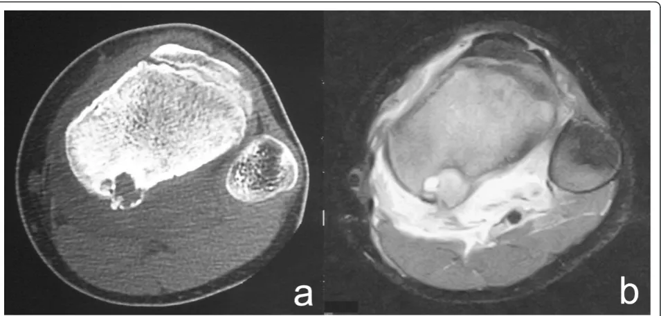
nal intensity on T2-weighted images with an edema-like soft tissue lesion adjacent to the bony lesion (Figure 1b). First, needle biopsy of the soft tissue mass was performed because the lesion was suspected to be malignant. Histol-ogy revealed that the lesion was fibromuscular with lym-phoid tissue without malignancy, and surgical treatment was performed. Intraoperative biopsy of the white soft tissue infiltrating the soleus muscle revealed that the soft tissue lesion was similar to the needle biopsy specimens pre-operation. After the soft tissue was confirmed to be reactive to inflammation and without malignancy, the bone lesion, which was occupied with solid white tissue, was curetted without adjuvant therapy, such as phenol treatment or bone grafting. Histological examination of the surgical specimens showed a solid variant of aneurys-mal bone cyst with belt-shaped giant cells against a back-ground of spindle cells and scattered osteoclasts (Figure 2a). Immunohistochemistry revealed cyclooxygenase-2 (COX-2) expressed in not only giant cells but also spindle cells (Figure 2b), which was confirmed by anti-human COX-2 antibody (IBL, Japan). The posterior knee pain immediately disappeared after surgery. On MR imaging at follow-up 2 months after surgery (Figure 3), the soft tissue edema had markedly improved. There was no recurrence of the lesion two years after the operation (Figure 4) and the operated bone exhibited good remo-deling (Figure 5a, b)
rarely affects long bones. The recurrence rate of lesions treated with simple curettage is relatively high, and therefore, amputation or curettage with adjuvant ther-apy, such as phenol and ethanol, has been applied to solid ABC in the short bones by some authors [5-7]. Oda et al. reported, however, that no patients had local recurrence after curettage or intralesional resection [8].
In the current case, simple curettage of the bony lesion without adjuvant therapy and a bone graft could cure solid ABC in a long bone. After the operation, the surrounding soft tissue edema was markedly relieved and the bony lesion completely remodeled, which was confirmed with post-operative CT and MR images. The cause of soft tissues edema surrounding the bone lesion of solid ABC remains unclear, although edema is one of the features of solid ABC on MR imaging. Some benign tumors with inflammation, such as osteoid osteoma and chondroblastoma, are known to produce prostaglandin, COX-2 and other cytokines related to inflammation [9-11]. In our solid ABC case, COX-2 was expressed in giant cells and spindle cells. If cytokines from the bone lesion of solid ABC induce soft tissue edema, simple curettage of the bony lesion seems to be a better surgery than wide resection, including edematous soft tissues. There is a possibility that the edema was provoked not only by COX-2 from the tumor but also by a small frac-ture of the shell-like cortex; however, if the cause of his
pain and edema was a fracture at the lesion, the symp-toms would have been relieved within 2 months. There-fore, we diagnosed that his pain and edema were mainly induced by the tumor.
Recently, we and Hirn et al. reported that simple cur-ettage without bone grafts for benign bone tumors, such as giant cell tumor, chondroblastoma, enchondroma, simple bone cyst, and so on, in long bones gives suffi-cient bone strength to host bones [12,13]. Our previous study showed that no patients needed orthosis 3 months after curettage for lesions of wide-ranging size (2-196 cm3). This indicates that larger solid ABCs than in the current report are expected to be cured with simple cur-ettage. Some authors reported that extensive curettage of the tumor may be more important than the use of adjuvant therapies to prevent local recurrence, which
enabled us to use no adjuvant treatment after curettage [14,15].
The typical MR imaging appearance of solid ABC is as follows: a bony lesion is heterogeneous with a predomi-nantly high signal on T2-weighted images and edema adjacent to the lesion was seen in 50% of cases [4]. These findings may lead physicians to misdiagnose this disease as malignant. Radiologists and orthopedic sur-geons should keep in mind solid ABC with lytic bone lesions that have soft tissue edema to avoid unnecessary overtreatment.
Conclusion
We experienced a solid ABC with surrounding soft tissue edema which was presumably induced by COX-2 from the tumor. Simple curettage of a bone lesion secreting
Figure 2Photomicrographs showing a) belt-shaped giant cells against a background of spindle cells and scattered osteoclasts. These features are compatible with solid ABC (hematoxylin-eosin x400). b) Immunohistochemical staining revealed COX-2 expressed in giant cells and spindle cells (x400).
Figure 3MR imaging 2 months after surgery. Soft tissue edema reduced markedly.
COX-2 achieved an excellent result with immediate reduction of the edema and good remodeling of the cur-etted lesion. To minimize surgical invasion, diagnosis of a lytic bone lesion with a sclerotic rim and surrounding soft tissue edema is important.
Consent
Written informed consent was obtained from the patient’s parents for publication of this Case report and any accom-panying images. A copy of the written consent is available for review by the Editor-in-Chief of this journal.
List of abbreviations
ABC: aneurysmal bone cyst; COX-2: cycloxygenase-2; GCRG: giant cell reparative granuloma; MR: magnetic resonance; CT: computed tomography.
Acknowledgements
We thank Ms Hiroe Miyahata from Gunma University Graduate School of Medicine for her technical assistance in IHC analyses.
Author details
1
Department of Orthopaedic Surgery, Gunma University Graduate School of Medicine. 3-39-22, Showa, Maebashi, Gunma, 371-8511, Japan.2Department
of Laboratory Sciences & Pathology, Gunma University School of Health Sciences, 3-39-22, Showa, Maebashi, Gunma, 371-8511, Japan.
Authors’contributions
RT collected data, carried out the immunohistochemistry and drafted manuscript. TY conceived of the design and helped to draft the manuscript. TF contributed to histological diagnosis. TS and KT revised manuscript critically for important intellectual content. All authors read and approved the final manuscript.
Competing interests
The authors declare that they have no competing interests.
Received: 20 December 2011 Accepted: 20 February 2012 Published: 20 February 2012
References
1. Sanerkin NG, Mott MG, Roylance J:An unusual intraosseous lesion with fibroblastic, osteoclastic, osteoblastic, aneurysmal and fibromyxoid elements.“Solid”variant of aneurysmal bone cyst.Cancer1983,
51:2278-2286.
2. Jaffe HL:Giant-cell reparative granuloma, traumatic bone cyst, and fibrous (fibro-oseous) dysplasia of the jawbones.Oral Surg Oral Med Oral Pathol1953,6:159-175.
3. Lorenzo JC, Dorfman HD:Giant-cell reparative granuloma of short tubular bones of the hands and feet.Am J Surg Pathol1980,4:551-563. 4. Ilaslan H, Sundaram M, Unni KK:Solid variant of aneurysmal bone cysts in
long tubular bones: giant cell reparative granuloma.AJR Am J Roentgenol 2003,180:1681-1687.
5. Wenner SM, Johnson K:Giant cell reparative granuloma of the hand.J Hand Surg Am1987,12:1097-1101.
6. Dahlin DC:Giant-cell-bearing lesions of bone of the hands.Hand Clin 1987,3:291-297.
7. Yoshida T, Sakamoto A, Tanaka K, Matsuda S, Oda Y, Iwamoto Y:Alternative surgical treatment for giant-cell reparative granuloma in the metacarpal, using phenol and ethanol adjuvant therapy.J Hand Surg Am2007,
32:887-892.
8. Oda Y, Tsuneyoshi M, Shinohara N:“Solid”variant of aneurysmal bone cyst (extragnathic giant cell reparative granuloma) in the axial skeleton and long bones. A study of its morphologic spectrum and distinction from allied giant cell lesions.Cancer1992,70:2642-2649.
9. Greco F, Tamburrelli F, Ciabattoni G:Prostaglandins in osteoid osteoma.
Int Orthop1991,15:35-37.
10. Kawaguchi Y, Sato C, Hasegawa T, Oka S, Kuwahara H, Norimatsu H:
Intraarticular osteoid osteoma associated with synovitis: a possible role of cyclooxygenase-2 expression by osteoblasts in the nidus.Mod Pathol 2000,13:1086-1091.
11. Uchikawa C, Shinozaki T, Nakajima T, Takagishi K:Cytokine synthesis by chondroblastoma: relation to local inflammation.J Orthop Surg (Hong Kong)2009,17:56-61.
12. Yanagawa T, Watanabe H, Shinozaki T, Takagishi K:Curettage of benign bone tumors without grafts gives sufficient bone strength.Acta Orthop 2009,80:9-13.
13. Hirn M, de Silva U, Sidharthan S, Grimer RJ, Abudu A, Tillman RM, Carter SR:
Bone defects following curettage do not necessarily need augmentation.Acta Orthop2009,80:4-8.
14. Blackley HR, Wunder JS, Davis AM, White LM, Kandel R, Bell RS:Treatment of giant-cell tumors of long bones with curettage and bone-grafting.J Bone Joint Surg Am1999,81:811-820.
15. Eckardt JJ, Grogan TJ:Giant cell tumor of bone.Clin Orthop Relat Res1986,
204:45-58.
doi:10.1186/1477-7819-10-45
Cite this article as:Takechiet al.:Solid variant of aneurysmal bone cyst in the tibia treated with simple curettage without bone graft: a case report.World Journal of Surgical Oncology201210:45.
Submit your next manuscript to BioMed Central and take full advantage of:
• Convenient online submission
• Thorough peer review
• No space constraints or color figure charges
• Immediate publication on acceptance
• Inclusion in PubMed, CAS, Scopus and Google Scholar
• Research which is freely available for redistribution