STUDIES OF EYE PIGMENTS OF DROSOPHILA. I. METHODS OF EXTRACTION AND QUANTITATIVE
ESTIMATION OF T H E PIGMENT COMPONENTS
BORIS EPHRUSSI AND JEAN LANE HEROLD The Johns Hopkins University, Baltimore, Maryland
Received October 5 , 1943
INTRODUCTION
ICROSCOPIC observation, as well as solubility tests and studies of the
M
effects of gene substitutions, have shown that the normal, wild type eye color of Drosophila melafiogaster is due to the presence of two pigments, red and brown. These two pigments are the end products of two largely inde- pendent chains of reactions. Recent work on the genetic control of eye color in Drosophila was almost entirely confined to the reaction chain leading to the formation of brown pigment. The hormone-like diffusible substances derived from tryptophane and representing intermediate links of this chain were the most intensively studied phase of this process. The study of the reaction chain resulting in the formation of red pigment was on the contrary very much neg- lected and so was the study of the end products of both reaction chains- namely, the red and brown pigments themselves.Both technical and theoretical factors were responsible for this uneven ad- vance. Technically, the diffusible nature of the hormone-like substances makes them particularly accessible for a very direct experimental attack. The inter- mediate products involved in the formation of red pigment offer no such tech- nical advantages. The origin of the red pigment, in so far as we know, is entirely intracellular and local. The study of the pigments themselves has en- countered technical obstacles, chiefly in finding suitable solvents, which have greatly retarded the understanding of their chemical nature. Only lately a most promising study by BECKER (1939,1942) somewhat clarified the chemical nature of the pigments, but this lead was broken by BECKER’S death on the Russian front.
Two theoretical considerations may be added to the above technical ones. I n the early phases of the work under discussion it was plausible to assume a rather direct relationship between the diffusible substances and the genes con- trolling their production. It was a reasonable hope that, starting from the tangible diffusible substances, it would be easier to trace back to the genes the whole chain of intermediate reactions, connecting the genes with the characters they control. This possibility had a special appeal to certain investigators, while to others the diffusible substances were of particular interest as means for the discovery of spatial and temporal correlations in development. What- ever the reason in each particular case may have been, the net result is that the chain of reactions leading to the formation of brown pigment is now rather well defined, while that leading to the formation of red is almost entirely un-
Work supported by a grant from the Penrose Fund of the AXERICAN PHILOSOPHICAL SO-
CIETY.
EYE PIGMENTS OF DROSOPHILA I49
known. The existence of distinct phases in the latter process may be inferred from the effects of some gene substitutions, but their more detailed analysis must await the introduction of new methods. It is the authors’ conviction that the study of the pigments, particularly from the quantitative angle, should permit deductions as t o the nature of the underlying reactions. It was with this expectation that the work to be reported under the above general title was begun. The purpose of this first report is to describe methods for the quantitative estimation of eye pigments of Drosophila melanogaster.
HISTORICAL
As indicated above, several different lines of investigation have provided evidence that the characteristic color of the eye of wild type Drosophila is due to the simultaneous presence of two pigments. First should be mentioned histological observations. These were inaugurated by JOHANNSEN (1924), who described in the primary and secondary pigment cells of the ommatidia the presence of two distinct types of pigment granules, some “purplish-red,” others “ochre-yellow.” JOHANNSEN also studied several mutant types, among them sepia, purple, vermilion, ruby, pink, etc., and concluded that their eyes differ from those of wild type flies in the amount and distribution of the two pigments.
Descriptions of the pigments in sections of Drosophila eyes, confirming JOHANNSEN’S findings, are given also by CASTEEL (1929), SCHULTZ (1935), and MORI (1937). The latter two workers, in addition to the histological study of the pigment in the imagoes of a variety of mutant types, have given accounts of the pigment deposition in the earlier stages of the development of the flies. It may be added that a study of the cytology of the eye of Drosophila pseudoobscura (COCHRANE 193 7) revealed in this species a pigment situation strikingly similar to that described by JOHANNSEN in D . melanogaster.
Gene substitutions have provided another and, a t first, independent line of evidence for the existence of two different pigments in the eye of Drosophila. WRIGHT (1932) was the first to suggest that the total absence of pigment in the double recessive scarlet brown may be eiplained on the assumption that the mutations a t these two loci suppress respectively one of two independent and complementary pigmentation processes. A similar conclusion was independ- ently reached by GLASS (1934), who identified the products of the comple- mentary pigmentation processes of WRIGHT with the red and yellow pigment granules of JOHANNSEN.
150 ’ BORIS EPHRUSSI AND JEAN LANE HEROLD
and has shown that (I) the eye color of any mutant, sayx, is equal to the sum of the colors of the two double recessive combinations x st and x bw, and (2) the observation of the combinations of any two eye color mutants with scarlet and with brown, say x st and x bw; and y st and y bw, permits the prediction of the color of the combination x y. This work, although not really quantitative, has lent the strongest support to the idea of two independent pigment com- ponents. The differences between the various mutant races appear to be due primarily to the independent variation in the content of the t w o pigment com- ponents.’
On the chemical side we find first an incidental remark in HERTWECK’S (1931) paper concerned with eye morphology. H ~ R T W E C K points out that while the red color of the Drosophila pigment suggests its carotinoid nature, its in- solubility in acetone, chloroform, and benzene does not support this idea. On the other hand, the solubility of the pigment in HC1 indicates that it is not a melanin.
A systematic chemical study of Drosophila eye pigments has been under- taken by SCHULTZ. Unfortunately his results were published only in the form of condensed reports (MORGAN, B+GES, and SCHULTZ 1930, 1931, 1932) and in a review (SCHULTZ 1935) devoted to general aspects of gene action. These publications contain no technical details and thus offer no lead for further in- vestigations. From SCHULTZ’S publications, however, we observe that “the red and yellow granules actually correspond to red and yellow pigments which are closely related, but can be separated by means of their solubility differences”; that these pigments have different absorption spectra, which are modified by changes of the p H and by oxidation and reduction (of smears of eyes; reversible oxidation could not be obtained in vitro); that they are water soluble, but do not dissolve in any of the usual organic solvents; and that they are dialysable -that is, of small molecular size. It will be seen below that most of these observations are corroborated by later work, but that the solubility in water solutions of the yellow pigment is negligible and that reversible oxidation and reduction of both pigments can be readily observed ilz vitro. The close relation- ship of the two pigments, according to SCHULTZ, is demonstrated by the fact that they can be converted ‘(by certain treatments” into a third, brown pig- ment. SCHULTZ’S general conclusion is that the yellow and red pigments are probably ‘‘so simply related to each other as oxidation-reduction products.”
Next should be mentioned a short note by LAKI (1935-36) who finds that a pigment extracted in water from the eye of wild type Drosophila (presumably the red component) shows the properties of a pH and redox indicator. Much more important information, however, is contained in the above mentioned work of MAINX, who, along with his histological studies, carried out some pig- ment extractions. MAINX finds in the wild type eye two pigments strikingly different in their solubilities: a red pigment, which is water soluble and a redox indicator, and a brown pigment insoluble in water; the latter is extremely sensitive to alkali, in which it can be dissolved with loss of color. Neither pig-
EYE PIGMENTS OF DROSOPHILA 1.51
ment is soluble in alcohol, ether, petroleum ether, carbon disulphide, chloro- form. These two pigments correspond to the two pigment components assumed on the basis of gene combinations (see above). The eye of the mutant st, for instance, contains nothing but the water soluble red pigment; that of the mutant bur only the water insoluble brown pigment. In vivo both pigments are thought to be bound to a protein carrier.
Finally, reference should be made to two papers by BECKER. The first (1939)
deals mainly with the eye pigments of Ephestia (also dependent on diffusible substances), but a few other pigments are also considered, among them those of Drosophila. On the whole, BECKER’S results are in agreement with those of
MAINX; however, a discrepancy appears as to the solubility of the brown com- ponent in acid water solutions. According to BECKER, the brown pigment from finely ground fly heads may be dissolved in HC1-zN. BECKER also reaches the conclusion that there are two distinct pigments in the wild type eye. The red one is very sensitive to acids and rather stable in alkali. The brown behaves in the opposite way. Both are p H and redox indicators. The red pigment (and probably the brown one) are of small molecular size. The brown pigment can be benzoylated, the red cannot. BECKER also is inclined to think that in vivo
the pigments are bound to a protein carrier; the color changes accompanying the action of denaturing agents are indicative of such a linkage. The eye of Calliphora is found by BECKER to contain one single pigment closely similar to but not identical with the brown pigment of Drosophila. For the pigments of the Drosophila and Calliphora type he proposes the generic name “Ommatins,” subdividing them into “Phaeommatins” (brown pigment of Drosophila) and “Erythrommatins” (red component of Drosophila) ; the red pigment of Drosophila, however, is included in the Ommatin group subject to the con- firmation of its close relationship with the Phaeommatins.
I n his second paper (1942) BECKER describes the presence of Ommatins in
several other groups of insects and reports a more detailed study of their properties. All Ommatins have a common physiological characteristic: their dependence on the tryptophane-kynurenine chain of reactions-that is, their chromophore group is probably derived from kynurenine. They all are different from melanins and carotinoids and show a series of common chemical charac- teristics. Prominent among these are: small molecular size, linkage with pro- teins, elementary chemical composition, color and behavior as p H and redox indicators, halochromism, stability in acids, instability in alkali, and their solubilities. They are insoluble in the usual organic solvents, poorly soluble in water, soluble in dilute alkali, very easily soluble in absolute formic acid and especially in acid alcohols. On the basis of this definition, BECKER now excludes the red component of Drosophila from the group of Ommatins.
152 ‘ BORIS EPHRUSSI AND JEAN LANE HEROLD
I n none of the studies thus far described is there any attempt to estimate quantitatively the pigments of Drosophila. The interest of developing such a technique, however, has been felt by a few investigators. Their efforts may be said to belong to two different categories, one of which is the attempt to de- termine the pigment content of individual whole eyes by reflection spectro- photometry. VAN ATTA and VAN ATTA (1931) and later FARDON and CARROLL (1937) have published the results of some measurements of this sort. The analysis of such data, however, especially in quantitative terms, seems very hazardous, particularly in view of the now well established presence of two unevenly distributed pigments. Moreover, it will be shown below that the pig- ment content is a function of many external factors and therefore subject to important individual variations. For these two reasons it will be difficult to make adequate use of techniques built on similar principles.
The other way to estimate pigment content of course is by extraction fol- lowed by some sort of measurement. Prior to the publication of BECKER’S work, the difficulties involved here were mainly in the ignorance of suitable solvents. MONOD and NEEFS in 1938 claimed to have worked out a satisfactory method. Fly heads separated from the bodies were kept for two days in absolute alcohol, then dried, ground, and extracted with water. The extracts were then rapidly boiled, centrifuged, and the pigment concentration determined photo- metrically. A similar extraction of heads of the mutant white showed that impurities may be disregarded, the absorption of light by these being of the order of one percent of the total absorption by extracts of wild type heads. The data published by MONOD and NEEFS are too few to permit a judgment on the value of the method. We will point out, however, that it disregards the presence of two pigments. From the results of MAINX and BECKER it is clear that the brown component is certainly not extracted by the MONOD and NEEFS procedure.
Early in 1940 DR. G. W. BEADLE kindly communicated to the senior author that DOCTORS E. L. TATUM and C. W. CLANCY had found it possible to extract the red pigment with 30 percent ethyl alcohol acidified with HC1 to p H 2.0 and the brown pigment with absolute methyl alcohol containing one percent by volume of dry HC1. Work started by the senior author on the basis of this communication was interrupted by the war. A paper by CLANCY (1942) dealing with the mutant claret contains a description of a technique of pigment meas- urement making use of the above solvents, as follows. Fly heads (usually 100
or more) immediately after decapitation are placed in a vial containing 2-3 cc
EYE PIGMENTS OF DROSOPHILA I53
The technique used by CLANCY will be discussed in the course of presentation of our own results. As will be seen in the following pages, an investigation of different aspects of the technique based on the use of the solvents mentioned was found to be desirable.
EXPERIMENTAL
We want t o emphasize a t this point that our aim was to work out a simple and rapid technique permitting extraction and measurement of low pigment concentrations in small volumes of solvent, these conditions being imposed by the nature of the problems to be studied with the help of this technique. We shall first report the results of extraction of the pigment from flies containing only one of the pigments-that is, from st or bw flies. Then we shall present our experiments on “double extraction” and, finally, the results of our analysis of some causes of variation in pigment content and indicate the precautions which have t o be taken to assure the maximum reproducibility of data.
Equipment
All extractions were made in glass-stoppered Pyrex glassware. Depending on the pigment concentration, either 5 and I O cc cylinders or IOO cc bottles were
used.
For the measurement of light absorption, when a sufficient amount of pig- ment extract (5 cc) was available and measurements a t a wide range of wave- lengths desired, a Coleman photoelectric spectrophotometer was used. When small volumes of extracts of low concentration were worked with and when consecutive measurements of an undisturbed sample over a period of time were desired, a Pulfrich photometer equipped with 50 mm long microcells (volume approximately I cc) and light filters S 43, S 47,
S
50, and S 53 (wave length centers of gravity a t 430,450,500 and 530 mp) was used.The results of all the absorption measurements will be given in terms of extinction, E (E=log IO& where Io is the incident, and I the transmitted light) this value being proportional to concentration. It will be remembered that E is also proportional t o the stratum thickness, L. I n the tables E has not been divided by L, since all comparisons are made between measurements obtained with the same instrument.
Unless stated otherwise, all extractions were performed in the dark a t 2 5 O C .
Red pigment component
Solvent
As indicated above, the solvent used for the extraction of the red pigment is 30 percent ethyl alcohol acidified t o pH=2.o (to be referred to below as
AEA). This p H is obtained by adding I cc of pure concentrated HC1 to 1000
I54 BORIS EPHRUSSI AND JEAN LANE HEROLD
Extraction
When heads of st flies, separated from the bodies, are placed in AEA, the solvent soon becomes colored orange yellow, which color gradually increases in intensity. After 48 hours the greater part of the pigment has diffused from the
0.4-
-
o,J-
-
0.2-
-ct UNTREATED
4 0 0 4 4 0 4 8 0 5 2 0 56C
LOSS 5 %
-:---
- _ _ _ _ - - - _ _ _
-
LOSS 15.4 %
e
z t
U
X
Y
TIME IN DAYS
I I I I I l I I
0.08
"."'I
UNTREATED -+---e- OXIDIZED 0.01
0
4 0 0 4 4 0 4 8 0 5 2 0 56C
HOURS
0 I I I I l I I
A
dA
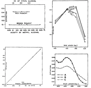
Ib 12 14 I6 10 20 22 24 26FIGURE I (above).-Absorption curves of red pigment extracted ig AEA (Coleman). FIGURE 2 (center).-Loss of absorption of an extract of red pigment on standing (Pulfrich,
FIGURE 3 (below).-Extraction of red pigment in AEA followed photometrically (Pulfrich,
470 m.4.
heads into the solution. The absorption curve of such a solution is shown in figure I. It may be seen that the light absorption is maximum a t 480 mp and decreases rapidly on both sides of this maximum.
EYE PIGMENTS OF DROSOPHILA I55
Observation of the heads after a 48 hours’ extraction shows that most eyes are completely extracted and appear nearly white, while a few still retain vari- able amounts of pigment, their color ranging from yellow to dark red. Al- though, according to CLANCY, AEA extracts the red pigment ‘readily and com- pletely,” the complete extraction under the described conditions obviously requires a much longer time. I n fact, examination of CLANCY’S data shows that in his experiments the extraction time varied from 1 2 to 38 days, and our own
experience indicates that often some unextracted heads can be found even after a week or ten days of extraction.
This very long extraction time is not only inconvenient, but also introduces an error, because, as CLANCY has already noticed, the solutions of red pigment are not quite stable. The “loss” of pigment can be followed photometrically. The results of such an experiment are shown in figure 2 where the extinction (a value directly proportional to the concentration of pigment) is plotted against time. I n this experiment an extract was filtered and kept a t 25OC. The
absorption was measured on samples of the extract a t various time intervals. It may be seen that the gradual decrease of absorption corresponds to a loss of 5 percent of pigment concentration per week. Thus a long extraction time in- troduces an appreciable error through pigment destruction.
The variability in the rate of extraction of individual heads suggested that its cause is probably mechanical and could be remedied by mechanical means. A comparison was made of the rate of extraction from (a) entire heads and (b) from heads split longitudinally (between the eyes) with the help of a chip of a safety razor blade. After 24 hours the eyes of the latter sample were nearly white: while those of the former remained strongly colored. Determination of the exact time required for the complete extraction of halved heads was made by following the light absorption of an extract. A sample of heads was placed in a large amount of AEA, and the absorption of samples of the extract was measured a t intervals. Figure 3 shows that the extraction is complete within IO hours.
PH
As pointed out by several authors (see above), the extracted red pigment shows the behavior of a p H indicator. If a few drops of dilute NaOH are added to an extract prepared in the described manner, its color turns red. Although the p H of the solvent has been found to be extremely stable and fluctuations of acidity of a stock solution may be discounted, the question was raised of how far the results of measurements would be affected by small differences in the pH of solvents prepared a t different times. Several samples of an extract of st
pigment were diluted with equal amounts of 30 percent ethyl alcohol acidified to various p H values. The final p H of each of the mixtures as well as the light absorption was measured. Figure 4 shows that the extinction, measured a t 470
mp (that is, in the vicinity of the absorption maximum) is independent of
acidity variations between pH
1.5
and 2.5.*
Longer extraction of the halved heads does not remove the remaining very pale purplish color156 BORIS EPHRUSSI AND JEAN LANE HEROLD
I n another experiment of the same type the light absorption of different samples of the same extracts adjusted to four different p H values was measured a t four different wave lengths. Figure 5 shows a marked shift of the absorption maximum towards longer wave lengths a t higher p H values. The change, cor-
001.
0 0 7 -
0 0 6 -
z
E 0 0 5 . U
f 004.
0 0 3 -
0 0 2 - 0 0 1 - rH. OF ETHYL ALCOHOL
1.5 2.0 2.5
- ~ - - - e A
- 0
WAVE LENGTH ( M p )
0 .
BROWN PIGMENT
0.1
.
I0.6s Oli 0;s 012 ais 0.~3 035 0.; o h N.
ACIDITY O f METHYL ALCOHOL
WAVE LENGTH ( u p )
430 410 500 530
FIGURE 4 (upper left).-Absorption, a t 470 mp, of red and brown pigments in solvents of diEerent acidities (Pulfrich). The curve for red pigment is referred to the upper abscissa, that for brown to the lower.
FIGURE 5 (upper right).-Effect of pH on the absorption curves of red pigment (Pulfrich).
FIGURE 6 (lower left).rRelation between concentration of red pigment and extinction (Pul-
FIGURE 7 (lower right).-Absorption curve of brown pigment extracted in AMA. A-un- frich, 470 m p ) .
treated extract, B-oxidized, C-oxidized five days later (Coleman).
responding of course to the visible color change, is particularly obvious a t 500
mp, while no significant change is seen a t 470 mp. Oxidation and reduction
EYE PIGMENTS OF DROSOPHILA I S 7 color. Therefore, the problem arose as t o whether or not the extracts prepared a t different times contain pigment in the same state of oxidation. Experiments were performed in which the light absorptions of two samples of the same ex- tract, one untreated, the other oxidized by addition of a drop of 3 percent by volume of HzOz, were measured. Figure I, representing the results of one of several concordant experiments, clearly shows that the pigment extracted in the described way is in the state of maximum oxidation. (It should be noted that this permits no conclusion as to the state of oxidation of the pigment in vivo.) No extra precaution is therefore needed to control the oxidation state of the extracts.
Applicability of the method t o determination of concentration Methods of photometric determination of concentration rest on the validity of Beer’s law, according t o which there is a linear relationship between extinc- tion and concentration. It was necessary therefore to ascertain that solutions of red pigment obey Beer’s law. A concentrated sample of red pigment was prepared. The extinctions of this solution and of several succkssive dilutions were measured with the Pulfrich photometer a t 470 mp. The resdrlt of the ex- periment is given in figure 6 , where extinction is plotted against relative con- centration. The points are experimental, the straight line theoretical. It is clear that the red pigment obeys Beer’s law within the concentration range corresponding to extinctions from 0.05 to 1.0 under the conditions of the measurements. I n terms of heads of st flies this corresponds approximately t o from 1 2 t o 250 heads per IOO cc of solvent.
I n determining the number of heads and the amount of AEA t o be used for a single measurement, two additional factors must be considered. First, in the case of the Pulfrich photometer the maximum accuracy of readings is in the neighborhood of 70 percent transmission which corresponds to an extinction of 0.155. Such a concentration is given, under the conditions of culture described below, by approximately 40 heads in IOO cc of AEA. Second, there are natu-
rally individual variations between flies, so that a greater accuracy will be reached with extracts from numerous heads. Our experience shows that samples of 30-40 heads from the same culture bottle give values rarely differing by more than z percent.
Impurities
The extracts discussed in the pre^cediing pages are quite transparent. When the heqds are split t o permit a faster extraction, small particles (mainly of brain tissue) may eventually be found suspended in the solution. But so long as st flies are dealt with-that is, so long as strongly pigmented eyes are ex- tracted-only a few heads per I cc of solvent are required t o produce a pigment
158 BORIS EPHRUSSI AND JEAN LANE HEROLD
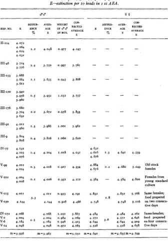
It is clear that AEA also extracts absorbing substances other than the pig- ment. To what extent these impurities affect the measurements of pigment concentration may be tested by extracting heads of flies the eyes of which contain no visible pigment. Three such experiments were performed using white (w) and white scarlet flies. The results are given in table I where the extinction (E) is calculated per ten heads in I cc of AEA. The comparison of the values obtained in these experiments with data for st (table 3, to be dis-
TABLE I
Photometric determinations (Pulfrich, 470 mp) o j absorption by impurities cztracted in AEA and
A M A . E-extinction per IO heads in I cc of solvent. N--number of heads.
EXP. NO. GENOTYPE
AND SEX
N SOLVENT E AVERAGE
B-57 VII-72
. VII-80
B-57 VII-72 VII-80
B-33 B-37
B-33 B-37
335
300
350
362 300 300
145
3 73
I79
400
W d
w;s: d
w;st d
W O
w;st O
w : s t O
W C F W C F
W O
W P
AEA 0 . 0 1 2
AEA 0.010 0.010
AEA 0 . 9
AEA 0.013
AEA 0.015 0.014
AEA 0.015
AMA 0.005
AMA 0.004 0.005
0.007
0.006 AMA
AMA 0.005
cussed later) shows that the impurities correspond to less than 0.3 percent of the total absorption of scarlet extracts. It is clear also that in working with less colored mutants, where impurities have a greater importance, appropriate corrections may easily be made.
Brown pigment component
Solvent
The acid methyl alcohol (henceforth to be referred to as AMA) used for the extraction of brown pigment is prepared by bubbling HCl gas through absolute methyl alcohol. The desired acidity of 1.0 percent by volume (ap-
proximately 0.270 N) is determined by titration with decinormal NaOH. The solvent is not stable and loses its acidity on standing.
Extraction and properties of brown pigment
Entire heads of bw flies, placed in AMA, lose their pigment rapidly; after
24 hours the eyes are white. The extract is of a brownish color. It becomes
EYE PIGMENTS OF DROSOPHILA I59
I n a fresh extract the pigment is obviously in a state of partial oxidation) and a precise study of the extracts must begin with its complete reduction or oxidation. Between these two possibilities oxidation was chosen because it was observed in preliminary experiments that reduction is frequently accompanied by appearance of turbidity.
The absorption curve of an untreated) freshly prepared brown pigment ex- tract in AMA is represented in figure 7 (curve A). Curve B shows the absorp- tion of the same solution after it has been oxidized under the conditions to be described in the next section. It may be seen that both curves have a peak at
450 mp and probably a second peak in the near ultra-violet and that the ab- sorption in the major part of the range and especially a t the peak is increased by oxidation.
Curve C represents the absorption of a fraction of the same extract kept five days a t room temperature and oxidized a t the end of this period. Its compari- son with curve B shows that the extract is perfectly stable a t room temperature. The time required for complete extraction of bw flies may be determined by the procedure already used for the red pigment. Figure 8 shows that the ex- traction is complete in less than 24 hours.
The extracts of brown pigment contain no gross turbidity. They may be used for photometric measurements directly. The amount of extracted im- purities has been determined by extracting heads of white flies. The extracts were oxidized and their absorption measured a t 470 mp. The data recorded in table I compared to those in table 4 (to be discussed later) show that the im- purities are responsible for approximately one percent of the total absorption of the extract of bw heads. The reservations made in connection with the im- purities contained in red pigment solutions are equally valid here.
Oxidation
The precautions required for the oxidation of brown pigment may be il- lustrated by the following experiment. A fresh solution of brown pigment is divided into eight samples of 5 cc each. To each of the samples one drop of hydrogen peroxide of a known and different concentration is added. The con- trol receives one drop of distilled water. After three hours the absorptions of the different samples are measured a t 470 mp. Figure g shows that the samples oxidized with H202 of 1.0, 2.0, 5.0) 10.0 percent by volume show identical ex- tinctions) all higher than that of the control, and that the samples oxidized with higher concentrations of HZOZ show a decrease of absorption. I n other words, drastic oxidation destroys the pigment. The destruction is faster in stronger H202 solutions. The measurements repeated after 24 and 48 hours (see figure) show a similar destruction in samples to which lower concentrations of Hz02 were added. The samples oxidized with 1.0 and 2 . 0 vol. percent HzOz are stable for 48 hours. This experiment indicates the concentration of H2OZ
I 60 BORIS EPHRUSSI AND JEAN LANE HEROLD
HOURS
Ib Ib 20 25 2 i 2's z i 3b
0 24
0 16-
0 0 6 -
0 0 4 -
0 02,
0 I 2 3 4 5 10 2 0 50 I
H ~ O , CONCENTRATION ( V O L U M E S ~ )
I
eta 0
T I M E IN MINUTES
0 2 0 4 0 6 0 80 100 120 140 160
FIGURE 8 (above).-Extraction of brown pigment in AMA followed photometrically (Pulfrich,
FIGURE 9 (center).-Absorption of brown pigment extracts oxidized with various concentra-
FIGURE IO (below).--Oxidation of brown pigment followed photometrically (Pulfrich, 470 470 mP).
tions of H202 (Pulfrich, 470 mp).
4.
EYE PIGMENTS OF DROSOPHILA 161
9 0 bw
17' 200 25' 30'
TEMPERATURE
4004
I
I 2 3 4
AGE IN DAYS
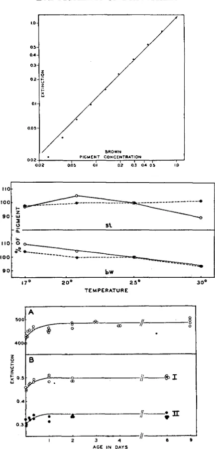
FIGURE I I (above).-Relation between concentration of brown pigment and extinction (Pul- frich, 470 mp).
FIGURE 1 2 (center).-Effect of temperature on the pigment content in bzt and st d d (Pul-
frich, 470 mp). E calculated per I O heads in one cc of solvent. Details in text.
FIGURE 13 (below).-Change in content of brown pigment with the age of bw d d. Figures
A and B represent the results of two different experiments. Curves A and B-I give the extinctions of oxidized extracts, curve B-I1 those of untreated extracts. E calculated per IO heads in I cc of A M A (F'ulfrich, 470 mp).
162 BORIS EPHRUSSI AND JEAN LANE HEROLD
Determination of relative concentration
The applicability of Beer’s law was tested for the brown pigment by a pro- cedure similar to that employed with the red pigment. All tested samples were oxidized as described in the preceding paragraph. Figure 1 1 shows that the brown pigment follows Beer’s law within the concentration range correspond- ing to extinctions from 0.06 to 1.0 as measured by the Pulfrich photometer.
This corresponds to from 1 2 to zoo heads of bw flies grown under our standard
culture conditions (see below) in IO cc of solvent. The number of heads for
optimum photometric determinations was found to be of the order of 40 heads
per IO cc of AMA.
Double extractiofi
Now that the behavior of the brown and red pigments in the solvents used for their extraction is known, the question may be raised whether these same solvents may be used for a “double extraction”-that is, for separation of the two pigments from eyes containing both (wild type eyes for example). First to be ascertained in this connection is the behavior of each of the pigments with respect to the solvent so far used for the extraction of the other.
If st heads are placed in AMA, it is observed that this solvent extracts the red pigment very easily and completely. A double extraction, beginning with the extraction of the brown component in AMA is therefore impossible. On the other hand, if bw heads are placed in AEA for a period of time sufficient for the extraction of red pigment, the solvent remains colorless to the naked eye. It seems possible therefore to extract, from a wild type head for example, first the red pigment with AEA and to follow this extraction by an extraction with AMA to remove the remaining brown pigment.
On closer examination, however, difficulties appear which have already been noticed by CLANCY (1942)) who, as stated above, used double extraction with the reservation that the method is open to criticism. I n the first place, the sol- vent used for the extraction of red pigment (AEA) seems to render insoluble a part of the brown,pigment. Thus bw eyes, placed first in AEA for 24 hours, then extracted 24 hours in AMA, retain a slight but distinct purplish tinge, while control eyes, extracted in AMA directly, become white. Second, AEA
does extract some brown pigment. How serious these two circumstances are may be tested by experiments in which equal samples of bw heads are extracted either directly in AMA or after pretreatment with AEA. Five such experiments were performed, and the photometric determinations of pigment concentration consistently indicated loss of pigment which, in the different experiments, amounted to 25.6, 26.2, 27.0, 29.6, and 30.6 percent (for details compare columns B, C, and D, table 2 ) . These losses are much greater than assumed by
CLANCY. He measured the absorption of the extract of bw heads in AEA after
EYE PIGMENTS OF DROSOPHILA 163
TABLE 2
Comparison of the amounts of brown pigment obtained by double extradwn of wild type and by double and direct extraction of brown jeies. Wild type and brown jeies were grown togefher in tke same bottles. In each experiment the three extractions were practiced on identical numbers of heads o f j e k s
of the same sex in the same volume of solvent. E-extinction* of a sample of 470 mp; M-average of samples of the same experiment.
A B C D E F
EXP. NO.
AND LOSS OF
BROWN BROWNIN B I N % C IN %
WILD TYPE BROWN
INsTRu- DOUBLE DOUBLE DIRECT DOUBLE OF OF
EXTRACTION EXTRACTION EXTRACTION EXTRACTION A A
hlENT USED
%
E M E Y E Y
I . 298
1.328 26.2 70.5 95.5
N - 8 5 1.369
Pulfrich 1.410 0.973
-
-
- -
0.283 0.410Coleman 0.283 0.283 0.405 0.408 30.6
VII-88
0.926 - 0.610 - 0.867
-
29.6 66.0 93.7Coleman
E - 6 3
Pulfrich 1.180
-
0.808-
1.108-
2 7 . 0 68.5 92.8- - 0.821
-
1.105-
25.6-
-
B-29
Pulfrich
m=27.8 m=68.3 m e 9 4 . o
*
Not corrected for number of heads and volume of solvent.What parts are played by the solubility in AEA and by the fixation of a certain fraction to AMA in the loss of brown pigment during double extraction is not known, but we believe that the greater part of the loss is due to the first factor.
These experiments, performed on bw eyes, do not necessarily mean that the same situation obtains in the extraction of wild type eyes. Certain differences in the behavior of pigments in these two types do exist. For instance, wild type eyes retain much less color after double extraction than bw eyes; however, there is no evidence that the brown pigment from wild type eyes is less soluble in
AEA than that from bw eyes, and so long as this is not proved, the method of double extraction as attempted here and used by CLANCY must be considered inadequate for the estimation of brown pigment.
There still remains the possibility (which could be of real advantage in certain cases) that the measurement of the red pigment extracted with AEA
164 BORIS EPHRUSSI AND JEAN LANE HEROLD
certain number of st heads and (b) an equal number of st heads plus the same number of bw heads. The extinctions of two such samples were found t o differ by less than one percent. It is clear therefore that the red pigment may be estimated in extracts ‘contaminated” by some brown pigment, provided the proportion of red is far in excess of brown pigment.
Numerous other attempts a t double extraction will not be described. It will only be pointed out that theoretically determination of the respective concen- trations of two pigments in a mixture is possible when the absorption spectrum of each is known, when one differs sufficiently from the other, and when there is no interaction between them. I n such cases the absorption values of the mixture a t two adequately selected wave lengths permit the calculation of the concentration of each of the components. However, such determinations are always delicate, and our tests of this procedure, by measuring the absorp- tion of mixtures of brown and red pigment solutions of known concentrations in AMA, have yielded unsatisfactory results.
I n view of these considerations, use of the double extraction was restricted t o a few special cases and we relied chiefly on separate extractions of the two pigment components, from flies having only one of them a t a time. The validity of this method will be discussed after some factors of variation of the pigment content in flies of a given genotype have been considered.
Factors of variation of pigment content
Preliminary determinations of the content of brown and red pigments in the eyes of the mutants bw and st of our stock raised without special precautions under the usual conditions of Drosophila work have shown great variations in the values obtained. Within the same stocks, determinations made a t different times may give values differing by more than 50 percent. ,Variations of such magnitude would overshadow diff ere.nces due t o genotype, in a comparative study of different mutants for example. Since it is known that external factors influence many characteristics of the flies, including their size, standardization of the culture conditions was attempted. Moreover, the eye color in Drosophila changes with the age of the flies; the causes of the age-change are unknown, and it was thought that eventually it may be responsible for a part of the variations encountered. Pigment content, as a function of age, was therefore investigated in some detail.
Culture conditions
The procedure adopted for this investigation is as follows.
EYE PIGMENTS OF DROSOPHILA 165
cent of Fleischmann's dry powdered brewer's yeast. No Moldex is used. The food (approximately 80 cc per bottle) is slanted and "painted" with a fresh yeast suspension immediately prior to the transfer of larvae. Preliminary ex- periments have shown that the weight of flies is n o t affected by the number of larvae grown per bottle, as long as that number is below 175. I n our experi- ments IOO to 1 2 0 larvae were raised in one bottle.
These operations are performed a t 25OC in accurately controlled incubators.
The importance of temperature as a factor of variation in pigment content was shown by experiments of which figure 1 2 (solid lines) gives an example. I n
this experiment st and bw larvae were raised a t 25°C up to the moment of
pupation and thereafter transferred to four different temperatures (17", 21', 25", and 30°C). The hatched flies were aged (see below) before decapitation.
Figure 1 2 shows clearly that the amount of both red and brown pigments
undergo considerable and independent variations with temperature. Age change
I n several experiments bw and st flies, raised in large numbers under the above described conditions, were isolated within the hour after emergence. At definite times thereafter they were decapitated, the heads extracted, and the absorption of the extracts determined. The results of some of these experi- ments are represented in figures 13 t o 17. Figure 1 3 A shows for the mutant bw
t h a t the absorption increases during the first 14 hours, a t which time the curve reaches a plateau. Figure 1 3 B , curve I, shows a similar result of another experi-
ment of the same type. It is a control to curve I1 (same figure) which represents the absorptions of the untreated extracts. This curve is exactly parallel to the curve for oxidized extracts (control). Curves A and
B
of figure 14 give again the absorptions of extracts of bw eyes from flies of different ages: flies of seriesA (curve A) are controls grown under standard conditions, those of series
B
(curve
B)
have been raised under conditions of crowding and undernutrition(150 larvae per vial containing IO g. of food of the same formula without dry yeast). Here the size of the flies was much smaller, the absorption per head much lower, but the general shape of the curve is again similar to that of the control.
Figure 15 shows the absorption curves of extracts from heads of freshly hatched and ten day old bw flies. Here the ordinates are given as log E in order t o make the shape of the curves independent of concentration differences. The shapes of the two curves are strictly similar; the pigments extracted from young and old flies are identical, and the increase in absorption must therefore be attributed to change in pigment content.
166 BORIS EPHRUSSI AND
I 2 3 4 5
AGE I N DAYS
JEAN LANE HEROLD
5.0
c
r 4.01
I
I1 2 3 4 5 6 7
ACE I N DAYS
WAVE LENGTH (Up) ,
400 440 480 5 2 0
-
1 0 .-
1.5-
CI
'a
U
3
V
-
o-.O- 0-4 HOURSCc 24 HOURS 2.0 -
I .
6WAVE LENGTH (Mp.) 400 4 4 0 480 5 2 0
FIGURE 14 (upper left).-Change in content of brown pigment with the age of bw 88. A-
flies grown under standard conditions, B-flies grown under conditions of crowding (Pulfrich,
47OW).
FIGUFS 15 (upper right).-Absorption curves of brown pigment extracted from bw Q 0 of dif-
ferent ages. E calculated per IO heads in I cc of AMA (Coleman).
FIGURE 16 (lower left).-Change in content of red pigment with the age of d and U 3 3. E
calculated per IO heads in I cc of AEA (Pulfrich, 470 mp).
FIGURE 17 (lower right).-Absorption curves of red pigment extracted from st Q Q of different
ages. E calculated per IO heads in I cc of AEA (Coleman).
The practical conclusion from these experiments is that quantitative pig- ment measurements must be made on flies which have reached the age of stable pigment concentration. In the experiments reported here, unless other- wise stated, hw flies used for extraction were aged for a t least two days, st and v
EYE PIGMENTS OF DROSOPHILA 167
Analysis of data
The methods of culture, extraction, and measurement described in the pre- ceding paragraphs, applied to the mutants bw and st, have furnished the data contained in tables 3 and 4. Inspection of these shows that, in spite of the standardized conditio*ns, there remains a considerable variability in the values obtained. I n column “E” (extinction of extracts of individual samples of flies) the greatest differences between the highest and lowest values, in percentage of the lowest, are as great as 15.8 for st 3 , 7.8 for st 0
,
15.1 for bw 8 , and 17.9for bw 9 . I n the data contained in the column “Average E” (average of the
values given by several samples in a single experiment) the variations are slightly smaller. Here the greatest differences (in the above order) are 14.5, 7.8, 13.8, and 15.3 percent. It will be observed that these differences are con- siderably greater than the differences observed between the different samples within any particular experiment, recorded in the third and eighth columns of tables 3 and 4. This fact shows that the greater part of the variation is due not to the methods of measurement, but to variations in the conditions of develop- ment of the flies from experiment to experiment. Several possible reasons have been considered,
I. It was thought that the condition (age, nutrition, size) of the females used as progenitors could have an effect on the pigment content of the progeny. An experiment was performed in which the content of red pigment was com- pared in the offspring of (a) young st females, raised under standard conditions and of (b) old, undersized st females from crowded stock bottles. The results, recorded in table 3 (experiments V-gg and V-102) clearly show that the age and condition of the females do not detectably influence the pigment content of the progeny. The differences between the two kinds of offspring are 0.3 per- cent for the males and 2.1 percent for the females; these are not greater than the usual differences between different samples of a single experiment. These data are in accord with the finding of WARREN (1924) that the size of eggs in Drosophila is independent of external conditions and the size and age of the females.
2. Although the food used in our experiments was always prepared according to the same formula, it was thought that media prepared on different days might be different, due to slight variations in cooking time or to differences in the yeast or in the contaminating bacterial flora. The sections a t the bottom of
tables 3 and 4 contain experiments in which females were allowed to lay for several days; larvae hatched on successive days were raised on media freshly prepared on each of these days. It may be seen that the differences between the values obtained here are greater than the usual differences between different samples of single experiments, recorded in the upper sections of the same tables. From these results it may be concluded that to test the effects of gene substitu- tions ceteris paribus, different types of flies must be raised simultaneously on the same media. I n the experiments described in the next section the st, bw,
and wild type flies were actually raised in the same culture bottles.
I 68 BORIS EPHRUSSI AND JEAN LANE HEROLD
TABLE 3
Photometric determinations (Pulfrich, 470 mp) of red pigment i n the mutant scarlet. E-extinction per I O heads in I cc A E A .
d d 9 9
COR-
m m n
DIFFER- AVER-
COR- BECTED
AVERAGE
E ENCE AGE
DIFFER- AVER- WEIGHT
E W . N O . E ENCE AGE OF d d AVERAGE
% E I N Y G S . % E
11-124 111-46 111-153 ID-I20 111-176 111-91 111.9 Iv-77 v-99 v-102 V-I15 V-130 IV-172 V-2 V-6 v-14 4.072 4.064 4.024 4.032 3.724 3.776 3.664 3.688 3.612 3.940 3.956 3.960 3.680 3,704 3.632 4.012 3.960 3.824 3.808 4.132 4.103 4.076 4.012 4.024 4.004 4.008 4.012 4. I44 4.068 4.184 4.048 4,004
1 . 2
1.4
2 . 1
0 . 5
2 . 0
1.3
0 . 4
1.4
0.3
0 . I
3.3 4.5 4.048 3.750 3.655 3.952 3.672 3.986 3.816 4.104 4.018 4.006 4.012 4. I44 4.068 4.004 4.048 4.184 0,977 0.997 0.945 1.052 0.958 1.001 1.060 1.018 0.927 0.951 0.935 0.928 1.052 0.984 0.946 0.972 4.143 3.761 3.868 3.757 3.833 3.982 3.600 4.031 4.334 4.212 4.291 4.466 3.867
4 . 0 6 9
4.423 4.165
-
-
-
-
-
-
-
4.632 4.676 4.684 4.676 4.584 4.616 4.832 4.748 4.484 4.572 4.644 4.528-
-
-
-
-
-
-
4.641 4.680 4.584 4.832 4.748 4.484 4.644 4.528 4.572 4.559Old stock 5.049 females
Females from 4’820 young standard
culture
5.168 Same female; food prepared
tive days 5 . I 16 on two consecu-
4.262 Same females; 4.646 food prepared
4.658 tive days 4. go9 on four consecu-
”3.936 m ‘3.967 m=4.050 “4.640 m-4.635m94.799
the question was raised as to whether or not the pigment content should be corrected for these weight differences. I n tables 3 and 4 the columns ‘Corrected average E” contain the data resulting from adjusting the average extinction to I mg of eight.^ The differences between the lowest and highest values in
EYE PIGMENTS OF DROSOPHILA 169 TABLE 4
Photometric determination (Pulfrich, 470 mp) of brown pigment in the mutant brown. E-extinction per I O heads i n I cc A M A .
d d 9 9
COR- RECTED AVERAGE
E
DIFFER- AVEB-
COR- RECTED
E ENCE AGE
DIFFER- AVER- WEIGHT
%
OF d d AVERAGE
EXP.NO. E ENCE AGE
% E I N M G S .
-
1-3 0.523
0 . 5 0 9 2 , 8 0.513 0,514
111-3 0.532 0.547 2 . 8 0.547
1-106 ' 0.498 0 . 5 0 0
0 . 5 0 1 0 . 5 0 1
0,497
0 . 5 0 1
0 . 5 0 0 0 . 5 0 0
111-76 0.565 0.572 0 , 5 7 0
0 . 8
. 0.567
IV-I15 0 . 5 2 9
Iv-I27 0 . 5 3 1 6 . 2 IV-139 0.562
0 . 5 1 5 0.968
0 . 5 0 0 0.969
0 . 5 4 4 1.035
0.637
0.532
z:?:
6 . 9 0.619 0.639 0.6200 . 5 2 9 0.981 0.531 1.000
0.562 0.987
0.539 0.576 0.576 0 . 5 8 7 Same females; 0.531 0.626 1 1 . 3 0.626 0.626 foodprepared 0.569 0.641 0.641 0.649 on three con- secutive days
m = o . 5 4 9 m=o.627 m = o . 6 2 5 m=o.629 m=o.528 m = o . 5 3 7
170 BORIS EPHRUSSI AND JEAN LANE HEROLD
data of figure 14 show that the pigment content of bw flies raised under condi- tions of crowding is approximately 5 5 per cent of that of control flies. The difference in weight, however, is much less (0.651 mg and 0.969 mg, respec- tively) and, after correction by weight, the value for pigment content in the flies from the crowded cultures still is only about 80 per cent of the control. Further data pointing to similar conclusions will be found in table 5 , to be discussed in the next section.
Validity o j the method of separate extractions
As stated above, the unsatisfactory results of “double” extractions have led us to prefer in our work the separate extraction of the two pigment components from flies in which the formation of one of the components is suppressed by a mutant gene. This second method is obviously based on MAINX’S (1938) state- ment that the total pigmentation of a given mutant, x , is equal to the sum of pigments contained in the two double-recessives,
x
st and x bw. However,MAINX’S statement is a deduction from purely visual observation. Whether or not it holds when translated into strict quantitative terms is evidently an open question which has to be solved in each individual case because there may or may not be interaction between the main gene studied and the genes introduced to eliminate one of the pigments. Here we will limit the discussion to the case
of the wild type eye.
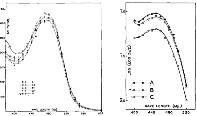
First, are the two pigments contained in the wild type eye qualitatively identical with those separately extracted from the mutants st and bw? Since it has been shown that the determination of red pigment extrazted from st with
AEA is not measurably affected by contamination with small amounts of brown pigment, we have compared AEA extracts of wild type eyes with similar extracts of st eyes. Figure 18 shows that the absorption curves of the two ex- tracts are identical. The same figure shows absorption curves of extracts of three other mutants: vermilion (v), cinnabar (cn) and cardinal (cd), all three be- longing, together with st, to the so-called vermilion group of mutants. Their phenotypes resemble very closely the phenotype of st: their eyes contain very little or no brown pigment (SCHULTZ 1935; MAINX 1938). The figure shows that the red pigment extracted from these mutants is similar to that contained in wild type and st eyes. Observation of heads after extraction with AEA shows completely white eyes in v and cn, a slight purplish tinge in st, and a consider- ably darker purplish color in cd. This color is exactly similar to that of bw eyes after double extraction and is undoubtedly due to the presence of a small amount of brown pigment. From the qualitative point of view, then, the genes
v, m, and st may all be used for the suppression of brown pigment. The greater amount of brown pigment in cd calls for caution.
EYE PIGMENTS OF DROSOPHILA 171
of differences in concentration the ordinates in this figure are given as log E. The three curves have definitely similar shapes.
For reasons which will not be discussed here we chose to use for the suppres- sion of brown pigment the gene st. It therefore became important to ascertain whether or not the eyes of the mutant st contain quantitatively the full amount of red pigment-that is, whether the gene st suppresses the formation of the brown pigment only. Several experiments were performed in which wild type and st flies were raised under similar standard conditions (see above). They were grown in the same bottles. Table
5
contains the data obtained. It may be seen that the pigment content in st, as compared with wild type is 99.4 percent> 400 4 4 0 480 5 2 0
FIGURE 18 (left).-Absorption curves of red pigment extracted from identical numbers of eyes
of wild type, cd, st, cn, and v 9 9 (Coleman).
FIGURE 19 (right).-Absorption curves of brawn pigment extracted A-from wild type 3 3 by
“double” extraction, B-from h 8 8 by direct extraction in AMA, C-from bw 8 3 by ‘double”
extraction. E calculated per IO heads in I cc of AMA (Coleman).
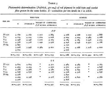
in 3 3 and 97.4 percent in 0 0 when calculated from the E values and re- spectively 97.7 percent and 101.2 percent when calculated from the “Average E” values. I n these experiments a systematic difference in weight appears to exist between the flies of the two genotypes. The correction by weight slightly exaggerates the difference in pigment content of females. After correction, the pigment content in st 3 3 is 101.8 percent and in st Q Q 105.7 percent of that in the wild type. The value of the correction by weight has already been dis- cussed. It is clear that the contents of red pigment in st and wild type are practically equal. It must be emphasized, however, that the two stocks were not isogenic.
The question as to the relative amounts of brown pigment in the eyes of
BORIS EPHRUSSI AND JEAN LANE HEROLD
TABLE 5
Photometric determination (Pulfrich, 470 mfi) of red pigment i n wild type and scarlet
p e s grown in the same bottles. E-extinction per ten heads in I cc A E A .
WILD TYPE SCARLET
EXP. NO.
WEIGHT OF CORRECTED 3 3 I N PGS. AVERAGE E
E AVERAGEE
WEIGHT O* CORRECTED
67 67 I N YGS. AVERAGE E
E A V E R A G E E
IV-171 4.160 4.160 1 . 0 7 2 3.884 V-I 4.124 4.124 1.024 4.028 V-5 4.348 4.348 1.020 4.264 V-13 4.328 4.328 1.090 3.976 IV-69 3.892
3.928 3.948 0.984 4.012 4.024
~
_
_
_
~
-m=4.115 m=4.182 m=1.038 m'4.033
4.068 4.068 1.052 3.868 4.004 4.004 0.984 4.068 4.184 4.184 0.946 4.424 4.048 4.048 0,972 4.168 4.132
4.104 4.068 1.018 4.000 4.076
-___--
m=4.088 "4.074 m=o.g94 m=4.106
IV-171 4.624 4.624 v-I 4.404 4.404 V-5 4.568 4.568 V-13 4.656 4.656 IV-69 4.328
4.404 4.348 4.316
--
m=4.471 m=4.520 m=4.357
*
4.484 4.484 4.264
4.572 4.572 4.648
4.644 4.644 4.908
4.528 4.528 4.656
4.616 4.640 4.560
4.632
4 . 6 7 6
--
m=4.593 "4.574 m=4.607
See corresponding experiment in upper part of the table.
It will be recalled that during double extraction a part of the brown pigment is lost, but whether or not the same amount of brown pigment is lost in the extraction of bw and wild type eyes is not known. Table 2 contains the data on brown pigment obtained by double extraction of wild type and bw eyes and by direct AMA extraction of bw eyes. If the amounts of pigment extracted from wild type and bw eyes by double extraction are compared, it appears that bw eyes contain approximately 30 percent less pigment than eyes of wild type. I n this connection it will be noted that wild type and bw eye< after the extrac- tion with AEA is terminated, both appear dark purple, but that the color is definitely darker in wild type. If, qn the other hand, the comparison is made between the amount of brown pigment extracted from wild type eyes by double extraction and the amount of the same pigment extracted from bw eyes directly with AMA, the difference found is much smaller (6 percent). The important point is that even in this case direct extraction of bw eyes with
EYE PIGMENTS OF DROSOPHILA I73
RECAPITULATION
The experiments reported above provide the basis of a technique for pigment extraction and measurement which is currently used in this laboratory. This technique is briefly as follows:
Flies grown under “standard conditions” are aged after emergence for a t least two days in the case of bw and a t least five days in the case of st.
The flies are decapitated, and the heads are placed in the solvents. Entire
bw heads are used, while st heads are split into halves with a safety razor blade. Acid ethyl alcohol is used for the extraction of red pigment; acid methyl alcohol for the‘extraction of brown. The first of these solvents is perfectly stable. The acidity of the second must be frequently checked.
The extractions of pigment in both these solvents are complete in less than
24 hours a t 25OC.
The red pigment extract, eventually filtered, may be used without further treatment for photometric determination of the absorption. The brown pig- ment must be oxidized with hydrogen peroxide. One and one-half to two hours should be allowed for the oxidation a t 25°C.
The optimum number of heads per sample depends of course on the instru- ment used for the determination of absorption. Using a Pulfrich photometer with 50 mm micro-cells the highest accuracy was found a t 470 mp with ex- tracts from 40 st heads in IOO cc AEA and from 40 bw heads in I O cc AMA.
These numbers of heads are sufficiently great for individual differences to be cancelled.
This technique fulfills the requirements defined a t the beginning of this paper. It is a rapid procedure, permitting work with small amounts of material and involving a minimum of manipulation.
DISCUSSION
The various observations made in the course of this technical study of pig- ment extraction are in agreement with the general conclusions of MAINX and
BECKER. The relative genetic and physiological independence of the two pig-
mentation processes, controlled by the genes st and bw, is paralleled by the clear differences in the properties and behavior of their end products. This conclusion, in which we claim no originality, is emphasized here because a different picture is frequently assumed on the basis of SCHULTZ’S 1935 paper. T o quote only one example, we read in GOLDSCHMIDT’S book (1938, p. 32):
“Both the red and yellow pigments are water soluble and may be separated chemically, and the red pigment can be oxidized into the yellow one. Re- ciprocally, the tan pigment of the early pupa may be reduced to red by action of HIS. This means that only one pigment, in different states of reduction, is present.’’ Similar statements are found in WADDINGTON’S book (1939) and elsewhere.