A Novel Murine Gene,
Sickle tail
, Linked to the
Danforth’s short tail
Locus,
Is Required for Normal Development of the Intervertebral Disc
Kei Semba,*
,†Kimi Araki,* Zhengzhe Li,* Ken-ichirou Matsumoto,*
,†Misao Suzuki,
‡Naoki Nakagata,
§Katsumasa Takagi,
†Motohiro Takeya,** Kumiko Yoshinobu,
††Masatake Araki,
††Kenji Imai,
‡‡Kuniya Abe
§§and Ken-ichi Yamamura*
,1*Division of Developmental Genetics, Institute of Molecular Embryology and Genetics, Kumamoto University, Kumamoto 862-0976, Japan, †Department of Orthopaedic Surgery, Kumamoto University School of Medicine, Kumamoto 860-8556, Japan,‡Division of Transgenic
Technology, Institute of Resource Development and Analysis, Kumamoto University, Kumamoto 860-0811, Japan,§Division of Reproductive Engineering, Institute of Resource Development and Analysis, Kumamoto University, Kumamoto 860-0811,
Japan,**Department of Cell Pathology, Kumamoto University School of Medicine, Kumamoto 860-8556, Japan, ††Division of Bioinformatics, Institute of Resource Development and Analysis, Kumamoto University,
Kumamoto 860-0811, Japan,‡‡Institute of Developmental Genetics, GSF-National Research Center for Environment and Health, 85764 Neuherberg, Germany and§§Technology
Development Team for Mammalian Cellular Dynamics, BioResource Center, RIKEN, Tsukuba, Ibaraki 305-0074 Japan
Manuscript received July 29, 2005 Accepted for publication September 26, 2005
ABSTRACT
We established the mutant mouse line, B6;CB-SktGtAyu8021IMEG(SktGt), through gene-trap mutagenesis in
embryonic stem cells. The novel gene identified, calledSickle tail(Skt), is composed of 19 exons and en-codes a protein of 1352 amino acids. Expression of a reporter gene was detected in the notochord during embryogenesis and in the nucleus pulposus of mice. Compression of some of the nuclei pulposi in the intervertebral discs (IVDs) appeared at embryonic day (E) 17.5, resulting in a kinky-tail phenotype showing defects in the nucleus pulposus and annulus fibrosus of IVDs inSktGt/Gtmice. These phenotypes were
dif-ferent from those inDanforth’s short tail(Sd) mice in which the nucleus pulposus was totally absent and replaced by peripheral fibers similar to those seen in the annulus fibrosus in all IVDs. TheSktgene maps to the proximal part of mouse chromosome 2, near theSdlocus. The genetic distance between them was 0.95 cM. The number of vertebrae in both [Sd1/1SktGt] and [Sd SktGt/1 1] compound heterozygotes was
less than that of Sd heterozygotes. Furthermore, the enhancer trap locusEtl4lacZ, which was previously
reported to be an allele ofSd, was located in the third intron of theSktgene.
T
HE notochord is an integral component of theaxial structure of vertebrates, functions as a signal-ing center dursignal-ing embryogenesis, and plays essential roles in patterning of both somites and the neural tube (Ang and Rossant 1994; Wilson et al.1995; Chiang et al.1996). In addition, the notochord has major roles in vertebral column formation. In the mouse, the noto-chord is a continuous rod of constant diameter ex-tending from the hypophysis to almost the tip of the tail at embryonic day (E) 9.5. At E10.5–E11.5, signals from the notochord induce the migration, proliferation, and fusion of the sclerotome to form a continuous and unsegmented perichordal tube around the notochord and neural tube. At 12.5, mesenchyme acquires a
char-acteristic metameric pattern of densely packed areas caudally and loosely packed areas cranially. Some densely packed cells move cranially and give rise to the annulus fibrosus of the future intervertebral disc (IVD). The re-maining densely packed cells fuse with the immediately caudal loosely packed cells to form the cartilaginous primordia of the vertebral bodies. Notochord cells located in the vertebral body of cranial regions start to relocate into intervertebral regions (Paavolaet al.1980; Rufai et al.1995; Aszodi et al.1998). At E13.5, the vertebral regions are enlarged and chondrified. The notochord proliferates and undergoes hypertrophy to form the ge-latinous center of the intervertebral disc, called the nucleus pulposus. This nucleus is surrounded by the circularly arranged fibers of the annulus fibrosus. These two structures together constitute the IVD (Langman 1969; Theiler1988). At E14.5, nearly all chondrocytes are hypertrophied. Starting from E14.5, the annulus fibrosus can be subdivided into a fibrous outer annulus and a cartilaginous inner annulus. At E16.0, notochord cells complete relocation from vertebral regions into
Sequence data from this article have been deposited with the EMBL/ GenBank Data Libraries under accession nos. AB125594, AB125595, and AB033043.
1Corresponding author:Division of Developmental Genetics, Institute of
Molecular Embryology and Genetics, Kumamoto University, 4-24-1 Kuhonji, Kumamoto 862-0976, Japan.
E-mail: [email protected]
intervertebral regions. Failures in somite, neural tube, and notochord formation are closely correlated with vertebral malformations. However, the mechanisms that underlie the formation of IVDs are largely unknown.
In the mouse, several mutations are known to affect the formation of the vertebral column due to functional defects in the notochord, includingDanforth’s short tail (Sd) and the enhancer trap line (Etl4lacZ
).Sd, located on chromosome 2 (Laneand Birkenmeiser1993; Alfred et al. 1997), is a semidominant mutation affecting the development of the vertebral column and the urogen-ital system (Dunnet al. 1940; Gruneberg1953, 1958). At E9.5, the notochord shows discontinuities. At E11.5, mesenchymal organization around the notochord is ab-normal. At E13.5, the notochord is fragmented and does not show proliferation and dilatation. Chondrifi-cation is much reduced in vertebral regions. Thus, an early reduction of the notochord results in cellular degeneration in the sclerotome, leading to reduced vertebral bodies and a characteristic short tail due to a reduced number of caudal vertebrae. HomozygousSd animals show a similar but much more severe tailless phenotype. In the enhancer trap line,Etl4lacZ
, a reporter (lacZ) gene was inserted near theSdlocus and was ex-pressed in the notochord, mesonephric mesenchyme, and apical ectoderm ridge (Gossleret al. 1989; Korn et al. 1992; Maatmanet al. 1997; Zachgoet al. 1998). Etl4lacZ
homozygotes exhibited kinks in the caudal re-gion of their tails and a synergistic genetic interaction betweenEtl4lacZand
Sdwas observed. Genetically,Etl4lacZ
and Sd are separated by 0.75 cM. Interestingly, at-tenuation or enhancement of the Sd phenotype was observed when theEtl4lacZinsertion was in acis- or
trans-conformation, respectively. This suggests thatEtl4lacZ is
an allele ofSd, presumably by trapping acis-regulatory element of theSdgene, and thatSdis a gain-of-function mutation (Zachgoet al.1998). Nevertheless, neither the cis-element nor the trapped gene has been identified.
In this study, we report a new mutant mouse line,SktGt
, obtained by gene-trap mutagenesis in embryonic stem (ES) cells.SktGt/Gt
mice exhibit a kinky tail in the caudal vertebral columns due to malformation of the IVDs. The gene identified,Sickle tail(Skt), was expressed in the notochord, its derivative nucleus pulposus, and in the mesonephros. Interestingly, theSktgene maps to the prox-imal part of mouse chromosome 2, near the locus forSd, and thelacZinsertion site inEtl4lacZwas found to be
lo-cated in the third intron of theSktgene. Furthermore, a cumulative effect of theSktGtmutation on theSdmutant
was observed.
MATERIALS AND METHODS
Generation and genotyping of mutant mice:The gene-trap method using the pU-8 trap vector was previously described (Arakiet al. 1999). Chimeric mice were produced by the
ag-gregation method using ES gene-trap clones and morulas of ICR (Charles River, Wilmington, MA) mice, and the chimeric mice were mated with C57BL/6 (CLEA) females to obtain F1
heterozygotes. Sd mice were purchased from the Jackson
Laboratory (Bar Harbor, ME) and propagated byin vitro fer-tilization.Sd1/1SktGtmice were generated by matingSd1/1
1mice (C57BL/6 genetic background) toSktGt/Gtmice with a
C57BL/6 genetic background. One heterozygote carrying the
Sdmutation and theSktGtinsertion on the same chromosome
(Sd SktGt/1 1;cis-configuration) was obtained through mating
betweentrans-heterozygotes and wild-type C57BL/6 mice.Sd
mice were distinguished by external inspection. Genotyping forSktGtalleles was done with PCR using tail genomic DNA
as a template. For the wild-type allele, the 59 primer, GTS (59-CCACCCCTACATGTGTCTTT-39), and the 39primer, GTA
(59-CGAGTAAGTAACATCCCTCC-39) located in the 14th
in-tron, were used to generate a 339-bp wild-type fragment. To detect the trapped allele, the 59primer, called Z1 (59-GCGTT
ACCCAACTTAATCG-39), and the Z2 (59-TGTGAGCGAGTAA
CAACCCG-39) located inlacZgene, were used to generate a 320-bp fragment.
Skeletal preparations: After tail skins were peeled off, tails were fixed in 95% ethanol for 3 days. Tails were cleared by placing in 1% KOH for 1 day and were stained in alizarin red for 1 day until the bone was red. Excess stain was removed with 2% KOH. After removing excessive alizarin red stain, tails were transferred to glycerol (Hoganet al. 1994).
To count the number of vertebral bodies, we examined all mice by X-ray photography. We counted the number of nor-mal vertebral bodies, that is, those without obvious nor- malforma-tions, but did not consider the size reduction observed in the vertebral bodies ofSdmutants.
Cloning of genomic DNA and cDNA: Plasmid rescue to obtain flanking genomic DNA was performed as described (Arakiet al. 1999). The 59-end of the cDNA of the trapped gene was isolated by 59-rapid amplification of cDNA ends (59 -RACE) using the 59-RACE system (Invitrogen, Carlsbad, CA). Total RNA from a B6;CB-SktGtAyu8021IMEGgene-trapped ES clone
was extracted by using Sepasol-RNA I (NACALAI TESQUE, Kyoto, Japan), and then poly(A)1RNA was isolated with an oligo(dT) column (Takara Biomedicals, Shiga, Japan). First-strand cDNA synthesis from 1mg of poly(A)1RNA was per-formed with reverse transcriptase from ReverScript (Wako,
Osaka, Japan) and with the primer SA13 (59-TCTGAAACT
CAGCCTTGAGC-39) in the splice acceptor (SA) sequence.
After dCTP tailing with terminal deoxynucleotidyl transferase (Invitrogen), cDNA was purified using a QIAquick nucleotide removal kit (QIAGEN, Chatsworth, CA). The initial PCR was
performed using the primer SA10 (59-AGCAGTGAAGGCTGT
GCGA-39) in the SA sequence and the anchor primer (59
-GGCCACGCGTCGACTAGTACGGGiiGGGiiGGGiiG-39)
(In-vitrogen). Then, nested PCR was performed using primer 63
(59-GCTTGTCCTCTTTGTTAGGG-39) in the SA sequence and
the amplification primer (59-GGCCACGCGTCGACTAGTAC-39)
in the anchor primer sequence. Amplified fragments were then sequenced directly by the dideoxy-chain termination method using Big Dye terminator cycle sequencing (Perkin-Elmer, Foster City, CA).
RT–PCR analysis:RT–PCR was performed using the Ther-moscript RT–PCR system (Invitrogen) according to the man-ufacturer’s instructions. The PCR was performed using the primers a–f in the sense and antisense sequences in theSktgene. The sequences of the primers used are as follows: primer-a,
59-TCACCATGAAGATGCTGGAG-39; primer-b, 59-CTACAG
TAAGCACTCGCTGAC-39; primer-c, 59-ACTCCTCAGCCTTG
ATGAAC-39; primer-d, 59-GTGGTGGTAAGTCCTGATCC-39;
primer-e, 59-GCCACCTTAAAGACACTAGG-39; and primer-f,
94°for 1 min, 55°for 2 min, and 72°for 2 min using 0.5 units of Taq polymerase for 30 cycles.
Northern blot analysis: Total RNA and poly(A)1 mRNA isolated from ES cells and embryo and adult tissues were electrophoresed on a 0.7% denaturing formaldehyde-MOPS-containing agarose gel and transferred to a positively charged nylon membrane (Roche). After baking at 80°for 1 hr, the membrane was prehybridized and then hybridized using the
Skt gene-specific RNA probes and the lacZ RNA probes
prepared using DIG RNA labeling and detection kit (Roche).
Detection ofb-galactosidase (lacZ) activities:Whole-mount X-gal staining was performed according to the method of Allenet al. (1988). Samples were fixed for 30 min at room temperature in fix solution [1% formaldehyde, 0.2% glutar-aldehyde, and 0.02% NP-40 in phosphate-buffered saline (PBS)]. Fixed samples were washed two times in PBS and incubated overnight at 30° in staining solution (5 mm
po-tassium ferricyanide, 5 mm potassium ferrocyanide, 2 mm
MgCl2, 0.5% X-gal in PBS). Samples were rinsed twice in PBS,
postfixed in 4% paraformaldehyde, and made transparent using benzylalcohol/benzylbenzoate (1:2) after dehydration with a series of ethanol steps (25, 50, 70, 100, and 100%, 1 hr each). Adult tissues were fixed in 4% paraformaldehyde in PBS. Tissue sections of 10mm were prepared and stained over-night at 30°with X-gal in staining solution. After staining, sec-tions were counterstained with Fast Red. For the section of the intervertebral discs, after X-gal stained tails were refixed in 3.7% formaldehyde/PBS, the caudal vertebral bones were demineralized in Plank–Rychlo solution and embedded in paraffin according to standard procedures. Sections of 8mm were prepared and counterstained with Nuclear Fast red.
Construction and transfection of theSktexpression vector:
To clone the full open reading frame (ORF) of theSktgene, RT–PCR was performed using the Thermoscript RT–PCR sys-tem. The initial PCR was performed using the primer ORFS1 in the sense strand upstream from the start codon in theSkt
gene (59- ACCGGAGTGGAGACTAGTTG-39) and primer ORFA1
in the antisense strand downstream from the stop codon in the
Sktgene (59-TGCATGAGGCCTTGAACGATACAG-39). Then,
nested PCR was performed using the sense-strand primer ORFS2 (59-TTTCTGCGAGCTTTCCGAAC-39) and the
antisense-strand primer ORFA2 (59-ACCTTGGTCCTAATAGGATCTG
GC-39). The PCR conditions used were 94°for 1 min, 58°for 1 min, and 72°for 3 min using 1.0 unit of LA Taq polymerase (Takara Biomedicals) for 25 cycles. The 4.1-kb PCR products were cloned into the pGEM-T vector (Promega, Madison, WI). This cDNA ORF was confirmed by sequencing and cloned into the pCAGGS expression vector (Niwa et al. 1991).
Trans-fection into BMT10 cells (Gerardand Gluzman1985) was
carried out by the lipofection method using LipofectAMINE reagent (Invitrogen).
Western blot analysis: BMT10 cells and 40 pieces of the nucleus pulposus of caudal IVDs of adult mice were homog-enized in 23sample buffer (100 mmTris HCl pH 6.8, 4% SDS,
12%b-mercaptoethanol, 20% glycerol). Extracts were electro-phoresed on a 6.0% polyacrylamide gel, transferred to a nitro-cellulose filter (Immobilon, Millipore, Bedford, MA), and detected using anti-Skt antibodies with the ECL detection system (Amersham, Arlington Heights, IL).
Immunohistochemistry:Tails were fixed in 4% paraformal-dehyde in PBS. The caudal vertebral bones were demineral-ized in 0.24mEDTA2Na, 0.22mEDTA4Na solution for 48 hr and embedded in paraffin blocks. Sections of the IVDs were immunostained with anti-Skt antibodies by the avidin–biotin complex method (Vector Laboratories, Burlingame, CA). Sec-tions were counterstained with hematoxylin.
RESULTS
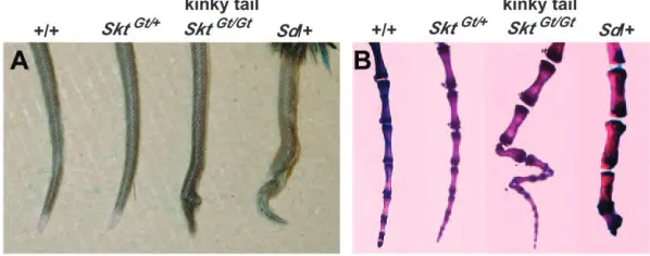
Generation ofSickle tailmutant mice:A gene-trap ES clone was isolated by the exchangeable gene-trap method using the trap vector pU-8 (Arakiet al. 1999). We ob-tained eight chimeric mice of which three were germ-line chimeras. Heterozygous animals appeared normal and were fertile. About half (35/66) of the homozy-gotes, however, showed a peculiar kinky-tail phenotype (Figure 1A and Table 1). This mutant mouse line was designated as B6;CB-SktGtAyu8021IMEG, in whichSkt means
Sickle tailbecause of the characteristic shape of the tail. Shortened and curved caudal vertebrae were apparent by the age of 2 weeks and were restricted to the 20–25th caudal vertebrae (Figure 1B). In contrast, heterozygous Sdmice showed short tails with truncation of vertebral columns at the 6th caudal vertebral body on average (Figures 1B and 7A) as reported previously. In SktGt/Gt
mice, no other skeletal abnormality was observed by bone X-ray examination (data not shown).
Figure 1.—(A) Tail phenotype of 8-week-old
mice.SktGt/Gtmice had kinked tails compared to
wild-type,SktGt/1, and heterozygous
Sdmice. (B) Alizarin red whole-mount preparations of the tails of 8-week-old mice. SktGt/Gt mice confirmed
this kinky-tail phenotype. Sd mice showed
de-creased numbers of vertebrae with truncation at the caudal vertebrae.
TABLE 1
Summary of genotyping of 4-week-old mice fromSktGt
heterozygote matings
Gt/Gt
1/1 Gt/1 Kinked Normala
60 151 35 (53%) 31
a
Characterization of the integration site of the trap vector: To characterize the gene-trap locus, we cloned and sequenced genomic DNA fragments flanking the gene-trap vector. A single copy of the vector was inte-grated into the genome as determined by Southern blot analysis using genomic DNA samples extracted fromSktGt
mice. Three base pairs of genomic DNA were deleted at the integration site of the trap vector. Using PCR am-plification on genomic DNA samples, the genotype of offspring from the heterozygous intercross was easily determined (data not shown).
Identification of theSickle tail gene: To identify the gene trapped, we performed 59-and 39-RACE. The se-quence of the ORF of the trapped gene was determined by compiling sequences of 59- and 39-RACE products and of EST that showed 100% homology to the RACE prod-ucts. We thus obtained a cDNA sequence comprising 5930 nucleotides (accession no. AB125594) that encodes a putative protein of 1352 amino acids with a predicted molecular weight of 147 kDa (Figure 2A). This gene was termedSkt. The protein contains a proline-rich region (amino acid residues 298–364) and a coiled-coil region
(amino acid residues 626–656) as determined by anal-ysis using Lupas’s algorithm (Lupaset al.1991). An ATG codon is located at nucleotide positions 559–561. This codon is most likely the initiation codon, because there is a Kozak sequence surrounding this ATG codon (Kozak 1996):i.e., there is a G residue following the ATG codon and an A residue three nucleotides upstream. A BLAST search of the amino acid sequence deduced from theSkt cDNA sequence revealed 80.6% homology with an un-characterized human protein, KIAA 1217 (accession no. AB033043), and no other evolutionarily conserved pro-tein was identified.
Sequence comparison ofSktcDNA with the murine genome sequence in the public Mouse Genome Re-sources (http://www.ncbi.nlm.nih.gov/genome/guide/ mouse/) revealed that theSktgene consists of 19 exons spanning.300 kb and that the ATG codon is located in the second exon (Figure 2B). The trap vector was in-tegrated in the 14th intron, resulting in the disruption of the Skt protein at position 998 (arrowhead in Figure 2, A and B). Sequence analyses of fusion transcripts revealed the presence of two fusion transcripts with theb-geo se-quence: Skt-a, containing the 1st–14th exons, and Skt-b,
lacking 33 bp of the 13th exon fromSkt-a(Figure 2B). In any case, a truncated protein lacking 355 amino acids encoded by exons 15–19 in the C-terminal region is ex-pected to be produced from theSktGtallele.
RT–PCR and Northern blot analyses:We performed RT–PCR using wild-type and SktGt/Gt
embryos at E10.5. RT–PCR with primers a and b located within the 59 -region of the integration site and detected the expected band in both embryos (Figure 3A). Using RT–PCR with two primer pairs—c in the 59-region and d in the 39 -region of the integration site and primers e and f within the 39-region of the integration site—we could not de-tect any product inSktGt/Gtembryos (Figure 3, B and C),
indicating the absence ofSktmRNA containing exons 15–19 inSktGt/Gtembryos.
To analyze the size and expression pattern of Skt mRNA, we performed Northern blot analysis using the Skt-specific probe in the 59-region (see Figure 2B). As shown in Figure 3D, a major band of 8 kb and a minor band of 6.5 kb were detected in wild-type ES cells. In wild-type whole embryos at E10.5, the 8- and 6.5-kb bands were also detected, although the expression levels were low. Surprisingly, Northern blot analysis using mRNA
Figure 3.—Analyses ofSkt transcripts. (A–C) RT–PCR analyses using E10.5 embryos to detect
Skttranscripts in wild-type (1/1) andSktGt/Gt
em-bryos. The transcripts containing nucleotide sequences upstream of the insertion site of the trap vector were detected in both wild-type and
SktGt/Gt embryos (A). The transcripts containing
nucleotide sequences downstream of theSkt se-quence were not detected inSktGt/Gtembryos (B
and C). M, molecular marker. (D) Northern blot analyses to detectSktmRNA in the wild-type ES cells, E10.5 embryo, and 8-week-old mice, using theSkt-specific probe in the 59-region (see Figure 2B). Total RNA (10mg) from TT2 ES cells, wild-type E10.5 embryos, mRNA (5mg) from wild-type organs, and aSktRNA probe were used for North-ern blotting. (E) NorthNorth-ern blot analyses to detect
Sktandb-geofusion transcripts. Total RNA (20mg) from TT2 and heterozygous (Gt/1) ES cells, wild-type (1/1), heterozygous (Gt/1), and
ho-mozygous (Gt/Gt) adult brains was used for
Northern blotting. The Skt RNA probe or lacZ
from wild-type adult organs revealed the presence of four different mRNA transcripts, 5.5, 6.5, 7.0, and 8.0 kb. The faint 7.0-kb and strong 6.5-kb bands in the brain, the 7.0- and 5.5-kb bands in the heart, the 7.0-kb band in the testis, the 8.0- and the 5.5-kb bands in the lung, and the 8.0-kb band in the intestine were detected (Figure 3D). We then examined the presence of theSkt andb-geofusion mRNA (Figure 3E). InSktGt/1
ES cells, an expected 10.5-kb band representing the fusion transcript containingb-geowas detected with both the SktandlacZprobes, although the intensity was weaker than that of the 8-kb band of the endogenous transcript. In theSktGt/1and
SktGt/Gtadult brains, two bands of 10.5
and 9.5 kb were detected with theSktprobe (Figure 3E, left), and these bands were also hybridized with thelacZ probe (Figure 3E, right), confirming that both transcripts corresponded to the fusion mRNA containing b-geo. This result indicates the existence of alternative splicing in the upstream region of the insertion site. Northern blot analysis using mRNA from wild-type adult heart revealed the presence of two different transcripts, the 7- and 5.5-kb bands when theSktprobe was used. How-ever, in theSktGt
adult heart, one 9.5-kb band was detected with thelacZprobe (data not shown). This result indi-cates that alternative splicing in the heart may occur in the downstream region of the insertion site, leading to the production of the 7- and 5.5-kb bands. The largest size of mRNA is 8 kb, which is larger than that of the predicted cDNA sequence comprising 5930 nucleotides (accession no. AB125594). As described below, the anti-Skt antibodies recognized only an150-kDa protein cor-responding to the predicted molecular weight of 147 kDa in extracts from the nucleus pulposus of the caudal IVDs (see lane 4 in Figure 6A). Thus, the 8-kb mRNA may contain untranslated regions at both the 59- and 39-ends and splicing may occur in each untranslated region. Fur-ther study will be required to determine the untrans-lated regions.
Formation of vertebral column andb-geoexpression inSktGt
mice: We examined formation of the vertebral column inSktGt
mice during the embryonic, fetal, and postnatal periods (E8.0, E8.5, E9.0, E9.5, E10.5, E11.5, E12.5, E13.5, E14.5, 15.5, E16.5, E17.5, E18.5, E19.5, newborn, and 2 weeks of age) by histological analysis with and without X-gal staining. Before E8.0, theb-geo gene was expressed in the chorion, but not in the embryo (Figure 4A). At E8.5 (Figure 4B) and E9.0 (Figure 4C), intense staining was detected in the notochord. At E11.5, the notochord and the mesonephros expressedb-geo strongly (Figure 4, D–F). In addition, sections of E11.5 embryos showed b-geo expression in the epithalamus sulcus, roof of the neopallial cortex, lens vesicle, inner layer of retina, heart (atrium and ventricle), surface of hepatic primordium, infundiblum, surface ectoderm, hind gut, and mesenchyme of the limb bud (data not shown). No abnormality was found in both vertebral and intervertebral regions up to E16.5. In the SktGt/Gt
embryo at E17.5, compression was observed in some IVDs (Figure 4, I and J) of theSktGt/Gtembryo, but not in
the wild-type embryo (Figure 4, G and H). In theSktGt/Gt
neonate, the X-gal-positive cells in the nucleus pulposus were shifted to the periphery and the alignment of the vertebral bodies was undulated (Figure 4, M and N). At this stage, sizes of IVDs are the same as those of wild-type mice. In 2-week-oldSktGt/Gt
mice, the X-gal-positive no-tochord cells were dislocated to the left or right side and the nuclei pulposi were smaller and eccentric, resulting in the disappearance of normal IVDs (Figure 4, Q and R). In theSktGt/1
embryo and mice, the X-gal-positive cells were positioned in the center of the vertebrae at any stage (Figure 4, G, K, and O). Interestingly, the expres-sion of the reporter gene was much lower in the upper IVDs than in the fifth to seventh caudal IVD, coinciding with a relationship between the phenotype of theSktGt/Gt
and strong expression in the caudal region of the tail.
Histological analyses of IVDs ofSktGt/GtandSdmutant
adult mice:InSktGt/Gtadults, theb-geogene was also
ex-pressed in many tissues such as the corpus callosum in the brain, uriniferous tubules in kidney, cardiac muscle, Sertoli’s cells in testes, and basal cells and outer root sheaths of hair follicles in skin (data not shown).
In normal adult mice, the nucleus pulposus was lo-cated in the center of IVDs at all levels of the vertebral column (Figure 5, A, D, G, and J). However, the nucleus pulposus in the IVDs of the tail region ofSktGt/Gt
mice contained an aggregation of notochord-like cells with fewer vacuoles than normal and were dislocated to the periphery (Figure 5, H and K). The nucleus pulposus in the upper regions appeared normal (Figure 1, B and E). Although histochemical analyses of embryos and new-borns in theSdmutant were reported by Paavolaet al. (1980) and Theiler(1988), the histochemical analyses of the IVDs inSdmutant adult mice have not previously been performed in detail. Thus, we analyzed the IVDs of the whole spine inSd1/1 1mutant mice. Surprisingly, the IVDs were totally occupied by peripheral fibers sim-ilar to those seen in the annulus fibrosus and no nucleus pulposus was found within the IVDs (Figure 5, C and F). The degeneration of the nucleus pulposus in the center of caudal IVDs was occasionally observed (Figure 5, I and L). Although tail kinks ofSktGt/Gtmice were restricted
to the 20–25th caudal vertebrae (Figure 1B), an irregular boundary with direct contact between the nucleus pulpo-sus and annulus fibropulpo-sus (Figure 5, O and P) was observed in the 5–25th caudal IVDs ofSktGt/Gtmice, compared to
the sharp boundary and fixed space observed between the nucleus pulposus and the fibrous layers of the an-nulus fibrosus in normal mice (Figure 5, M and N). In addition to the abnormalities of the nucleus pulposus in IVDs, the annulus fibrosus development was also im-paired inSktGt/Gt
observed in the regions that did not have kinks inSktGt/Gt
mice (Table 1). Surprisingly, histological analysis of exter-nally normal tails ofSktGt/Gt
mice revealed the presence of similar IVD abnormalities such as dislocation of the nucleus pulposus and thin fibrous layers of the annulus fibrosus (Figure 5S) as found in kinked tails (Figure 5, Q and R). This IVD phenotype was observed over 10 gen-erations in the progeny backcrossed to C57BL/6. Thus, the primary phenotype ofSktGt/Gtmice is the deformity of
IVDs in the tail region. These histological pictures were clearly distinct from those ofSdmice.
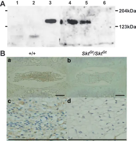
Analysis of Skt protein using anti-Skt antibody:The anti-Skt antibodies recognized an150-kDa protein cor-responding to the predicted molecular weight of 147 kDa in the lysates of BMT10 cells transiently expressing the SktcDNA by the CAG promoter as well as in extracts from the nucleus pulposus of the caudal IVDs. No band was detected in extracts from untreated BMT10 cells and BMT10 cells transfected with mock expression vector
(Figure 6A). This is consistent with the notion that ATG at positions 559–561 and TAA at positions 4615–4617 in theSktcDNA are the start codon and termination codon, respectively. As expected, the amount of Skt protein was decreased inSktGt/1
mice and was below the detectable level inSktGt/Gt
mice (Figure 6A). After immunohistochem-ical staining, the nucleus pulposus cells from upper cau-dal vertebral discs were positively stained in wild-type mice (Figure 6B, a and c), but not inSktGt/Gtmice (Figure 6B,
b and d). At higher magnification, the staining was ob-served mainly in the cytoplasm of the nucleus pulposus cells, indicating cytoplasmic localization of the Skt pro-tein. Since we could not produce antibodies against the N terminus, the expression of the truncated protein was not confirmed inSktGt/Gt
mice.
Relationship ofSktwithEtl4lacZ
orSdloci:Both the ex-pression pattern of theSktgene and the phenotype of SktGt
mutant mice were quite similar to those ofEtl4lacZ
mu-tant mice (Zachgoet al. 1998). Furthermore, by examining
Figure 4.—b-gal expression and histological analyses in SktGtmice. At E7.5 (A), the chorion
was stained with X-gal, but the midline region of the embryo was not stained. At E8.5 (B) and E9.0 (C) intense staining was detected in the no-tochord. At E11.5 (D–F), the notochord and the mesonephros expressed b-geostrongly in whole-mount X-gal staining (D), the frontal section (E), and the sagittal section (F). Sagittal sections of the tail bud ofSktGt/1
(G and H) and SktGt/Gt
(I and J) embryos at E17.5. At E17.5, some IVDs were compressed in the tail bud ofSktGt/Gt( J).
Sag-ittal sections of the tail tips of newbornSktGt/1
(K and L) andSktGt/Gt(M and N) mice. In theSktGt/Gt
neonate, the vertebral body alignment was un-dulated (M and N). Sagittal sections of the tail tips of 2-week-old SktGt/1 (O and P) and
SktGt/Gt
(Q and R) mice. In the 2-week-oldSktGt/Gtmice,
the reported primer sequences of the Etl4lacZ
locus (Maatmanet al. 1997), we found that theEtl4lacZlocus is located in the third intron of theSktgene (Figure 2B) and that the distance between the integration sites ofEtl4lacZ
andSktGt
was 237 kb.
To examine the genetic distance and interaction be-tween theSktGt
andSdlocus, we crossedSktGt
mice withSd mice to produce the compound heterozygote (Sd1/1
SktGt
;trans-configuration). Then, these compound het-erozygotes were used to analyze the recombination rate
Figure 5.—Histological analyses of IVDs of
SktGt/GtandSdmutant adult mice. Sagittal sections
of the thoracic and caudal IVD from 8-week-old adult wild-type (A, D, G, and J), SktGt/Gt (B, E,
H, and K), and heterozygous Sd mice (C, F, I,
and L). (D, E, F, J, K, and L) Higher magnifica-tion of the area indicated by the boxes in A, B, C, G, H, and I, respectively. Arrowheads indicate the dorsal side. Axial sections of the upper caudal IVD in wild-type (M and N) andSktGt/Gt(O and P)
8-week-old mice. (N and P) Higher magnification of the area indicated by the boxes in M and O, respectively. The arrowheads in P indicate an ir-regular boundary with close contact between the nucleus pulposus and annulus fibrosus. (Q– S) The 20–25th caudal IVDs of SktGt/Gt
8-week-old mice. Impaired development of the annulus fibrosus inSktGt/Gtmice was demonstrated by the
between the two loci. As shown in Table 2, one com-pound heterozygote carrying theSdmutation and the SktGt
insertion on the same chromosome (Sd SktGt
/1 1; cis-configuration) and two wild-type mice were obtained
among 249 mice obtained by mating the trans
com-pound heterozygote with the wild-type C57BL/6 mice, demonstrating that the Sd and SktGt
mutations were genetically separated. The genetic distance was calcu-lated to be0.95 cM by combining the data from mating the [Sd SktGt/1 1] (cis) compound heterozygote with the
wild-type mice (Table 2).
phenotype in heterozygousSdmice in either the trans-or thecis-configuration, the number of vertebrae was de-termined by X-ray analysis. HeterozygousSd[Sd1/1 1] mice (n ¼ 22) had a variable number of vertebrates and the vertebral columns were truncated at the sixth caudal vertebral body on average. Bothtrans[Sd1/1
SktGt
] (n ¼23) and cis[Sd SktGt
/1 1] (n ¼10) com-pound heterozygous mice had shorter tails, in which the vertebral columns were truncated at the second and third caudal vertebral body on average (Figure 7A). We examined whether the phenotype of [Sd SktGt
/1SktGt
] neonatal mice is more severe than those oftrans[Sd1/1
SktGt] and
cis[Sd SktGt/1 1] compound heterozygous
mice. Approximately 80% of [Sd SktGt/1
SktGt] neonatal
mice died within 48 hr of birth and all mutant mice died within 2 weeks. In addition, as shown by making alcian blue/alizarin red whole-mount preparations of [Sd SktGt/1SktGt] neonatal mice, [Sd SktGt/1SktGt] neonatal
mice had shorter tails than those oftrans[Sd1/1SktGt]
andcis[Sd SktGt/1 1] compound heterozygous mice, in
which the vertebral columns were truncated at the fourth sacral vertebral body on average, suggesting a cumulative effect of theSktGt
mutation on theSdmutant. To examine the pathologic effect of theSktmutation on the Sdphenotype, we carried out histological ana-lyses on the IVDs ofSd1/1SktGt
andSd SktGt
/1 1 mu-tant mice. Both compound heterozygous mice showed IVD histology similar to that seen in Sd 1/1 1 mu-tant mice. The nucleus pulposus was totally absent and replaced by peripheral fibers similar to those seen in the annulus fibrosus in all IVDs (Figure 7B, a–h). These results suggest that theSktmutation did not affect the histological picture of IVD in theSdmutation. In addi-tion, the expression pattern of theb-geogene in the tail notochord was the same in both trans [Sd1/1 SktGt]
andcis[Sd SktGt
/1 1] compound heterozygous embryos at E9.5 and E13.5 (Figure 7C). The notochord of both transandciscompound heterozygous embryos was thin and clearly stained at E9.5 (Figure 7C, a and d), and the X-gal-positive notochord was similarly fragmented in bothtransandciscompound heterozygous embryos at E13.5 (Figure 7C, b, c, e, and f). This suggested that the Sdmutation did not affectSktexpression. In addition, this trans–cis test demonstrated that the double het-erozygotes exhibited indistinguishable phenotypes in
Figure6.—Detection of the Skt protein. (A) Western blot analysis to detect Skt protein using extracts from untreated BMT10 cells (lane 1), BMT10 cells transfected with vector (lane 2), BMT10 cells transfected with theSktexpression vec-tor (lane 3), and extracts of the nucleus pulposus of caudal IVDs from 8-week-old wild-type mice (lane 4), SktGt/1 mice
(lane 5), andSktGt/Gtmice (lane 6). An150-kDa protein
cor-responding to the predicted molecular weight of 147 kDa was detected in lanes 3, 4, and 5, but not in lane 6. The amount of Skt protein was reduced in theSktGt/1
mutant (lane 5) and was below the detectable level inSktGt/Gt(lane 6). (B)
Immunohis-tochemistry of frontal sections of the nucleus pulposus in up-per caudal IVDs from adult 8-week-old mice using purified anti-Skt antibodies. Skt protein was detected in the cytoplasm of nucleus pulposus cells in wild-type (a and c), but notSktGt/Gt,
mice (b and d). (c and d) Higher magnification of the area indicated by the boxes in a and b, respectively. Bars, 200mm.
TABLE 2
Distribution of the haplotypes among the 315 offspring of the backcrossSd1/1SktGtorSd SktGt/1 1 3C57BL/6
Sd1/1SktGt3C57 BL/6 Sd SktGt/1 1 3C57 BL/6
1 1/1SktGt Sd1/1 1 Sd SktGt/1 1 1 1/1 1 1SktGt/1 1 Sd1/1 1 Sd SktGt/1 1 1 1/1 1
144 102 1 2 0 0 24 42
regard to notochord degradation whetherSdandSktGt
were in atrans-or in acis-configuration.
DISCUSSION
We established a new recessive trap line, B6;CB-SktGtAyu8021IMEG
, which had a deformity in caudal IVDs. The insertion site of the trap vector is in the 14th intron of the novel geneSkt, located on chromosome 2, near the locus forDanforth’s short tail.In addition, we found that the enhancer trap locus Etl4lacZ, which was
pre-viously reported to be an allele ofSd, was located in the third intron of theSktgene.
Structure, expression, and function of the Sktgene:
The sequence of the trapped gene,Skt, was obtained using the gene-trap ES clone. TheSktgene contains 19 exons encoding a novel protein of 1352 amino acids with a proline-rich region in the N terminus (amino acid positions 298–364) and a coiled-coil domain in the middle (amino acid positions 626–656). Although the role of the proline-rich region in many proteins is not
clear yet, a proline-rich region located in the amino-terminal region has been shown to be important for proper folding in cytochrome P450s (mitochondrial, microbial, and microsomal P450s) (Kusanoet al.2001a,b). Thus, the proline-rich region in the Skt protein may have a similar function in protein folding. The coiled-coil motif was first described by Crick (1952) and by Paulingand Corey(1953) as the main structural ele-ment of a large class of fibrous proteins that included keratin, myosin, and fibrinogen, mediating dimeriza-tion, heterodimer formadimeriza-tion, or trimerization (for a re-view see Lupas1996). These proteins provide a scaffold for regulatory complexes such as tropomyosin and a protective surface for pathogens. As the Skt protein is localized in the cytoplasm, it is conceivable that the Skt protein may provide structural elements or act as a scaffold for regulatory complexes.
Insertion of the gene-trap vector into the 14th intron of theSktgene, downstream of the coiled-coil domain and the proline-rich region, resulted in production of two fusion transcripts with theb-geosequence: one,Skt-a,
Figure7.—(A) Schematic of
axial levels and severity of
verte-bral malformations in
com-pound mutant 8-week-old mice. A single solid circle indicates the level of the terminal verte-bral body for a single mouse. In bothtransandciscompound mutant mice, the degree of ver-tebral malformation is more se-vere than that in heterozygous
Sd mice (trans, P , 0.005; cis,
P,0.003, Mann–WhitneyU-test). There was no significant
differ-ence between trans [Sd 1/1
SktGt] (n ¼ 23) and cis [Sd SktGt/1 1] (n¼10) mice (P¼
0.956, Mann–Whitney U-test).
(B) Histological analyses of ver-tebral columns in the Sd 1/1
SktGt and Sd SktGt/1 1 mutant
mice. HE staining of midsagittal sections in thoracic spines of
Sd 1/1 SktGt(a and c) and Sd SktGt/1 1(b and d) mice and
in sacral spines ofSd1/1SktGt
(e and g) and Sd SktGt/1 1
(f and h) mice. Arrowheads indi-cate dorsal sides. (c, d, g, and h) Higher magnification of the areas indicated by the boxes in a, b, e, and f, respectively. (C) Whole-mount X-gal staining in the
Sd1/1SktGtandSd SktGt/1 1
mu-tant embryos. The b-gal expres-sion in the tail notochord oftrans -heterozygous embryos [Sd 1/1
SktGt] at E9.5 (a) and E13.5 (b
and c) and of cis-heterozygous embryos [Sd SktGt/1 1] at E9.5
33 bp of the 13th exon from Skt-a (see Figure 2B). Northern blot analysis showed that the amount of fusion transcripts from the trapped allele was lower than that of the wild-type allele. Therefore, it is also possible that the insertion of the gene-trap vector resulted in decreased mRNA stability. As tail phenotypes are recessive, in-sertion of the trap vector may cause a hypomorphic or null mutation, but not a dominant-negative mutation. Although we detected four types of mRNA transcribed from the wild-type Skt allele, further analysis will be required to elucidate the function of these mRNA.
The expression patterns of theSktgene can be mon-itored by X-gal staining, because the expression of the reporter gene,b-geo, is under control of the regulatory region of theSktgene. At E11.5,b-geowas expressed mainly in the notochord and mesonephros at high levels, and in other tissues as described in theresults. In theEtl4lacZ line, thelacZ reporter gene was also expressed in two main tissues, the notochord and the mesonephros (Zachgo et al. 1998). In both lines, lacZ expression was first detected at E8.5 in the presumptive notochord cells. In the Etl4lacZ
line,lacZ expression was detected in the future IVDs at E13.5 and persisted up to E14.5. However, nolacZexpression was detected in the IVDs of newborn and adult mice. On the other hand,lacZ ex-pression in the IVDs persisted to adult stage in theSktGt
line. These results suggest that the enhancer located near theEtl4lacZlocus is not sufficient to express the
lacZ gene in the IVDs of adult mice.
Histochemical analyses of the vertebral column re-vealed the differences betweenSdmice andSktGtmice.
As histological data onEtl4lacZmice were not described
by Zachgoet al.(1998), it is not clear whether the IVD histology is similar to that seen in Sd or SktGt mice.
As reported previously, the development of both the vertebral column and the urogenital system is affected inSdmutation, suggesting that theSdgene is required for formation of derivatives from both the paraxial and the intermediate mesoderm. In addition, both vertebral bodies and IVDs are affected inSdmice. The notochord shows discontinuities as early as E9.5, resulting in the total absence of the nucleus pulposus at all levels and is replaced by peripheral fibers similar to those of the annulus fibrosus inSdadult mice. All vertebral bodies are reduced in a dorso-ventral direction and the number of tail vertebrae is reduced, leading to shortening or absence of the tail. However, inSktGtmice, the
compres-sion and dislocation of the nucleus pulposus were first observed at E17.5 and were restricted to the tail region. Interestingly, the size of the nucleus pulposus was similar to that in wild-type mice until birth. After birth, the nucleus pulposus did not expand and was dislocated to the periphery, resulting in a kinky-tail phenotype in adults. These observations suggest thatSdacts at an early stage of mesoderm development involving both sclero-tome and notochord development and thatSktacts during
and hypertrophy of the nucleus pulposus. Although massive apoptosis in notochord cells was observed in embryos with targeted disruption ofJun(Behrenset al. 2003) orSox5//Sox6/(Smitsand Lefebvre2003), no apoptotic notochord cells were observed in the SktGt/Gt
embryos at E16.5 and in neonatal mice. Thus, apoptosis is not the cause of compression or dislocation of the nucleus pulposus inSktGt
mice. As the Skt protein con-tains the coiled-coil motif that is involved in the forma-tion of mechanically rigid structures, it is possible that the nucleus pulposus lacking Skt protein may not be capable of sustaining mechanical loads, leading to com-pression or dislocation of the nucleus pulposus.
Although the IVD is formed from two components of developmentally different origins, the nucleus pulposus and the annulus fibrosus, interaction of the nucleus pulposus and annulus fibrosus is not clear yet. InSdmice, disappearance of the notochord cells occurs at early stages of development. Theiler(1988) reported that the fibers of the annulus fibrosus are reduced inSdheterozygous embryos and newborns. However, our results suggest that the annuli fibrosi are not reduced in Sdheterozygous adult mice. Thus, the annulus fibrosus may be able to completely compensate for the loss of the nucleus pulpo-sus during growth after birth. InSktGt
mice, the thin an-nulus fibrosus was observed together with abnormalities in the nucleus pulposus. At present, it is not known whether the thin annulus fibrosus is caused by the direct effect ofSktdeficiency or indirectly caused by defects in the nucleus pulposus.
The relationship among theSktGt,Etl4lacZ, andSd
mu-tations:Our breeding studies revealed that the genetic distance between the Skt andSdloci was 0.95 cM. We believe that theSktgene is distinct from theSdgene for the following reasons. First, anti-Skt antibody detected the predicted size of protein deduced from theSktgene. If the Sktwas part of the Sdgene, we should have de-tected a larger protein by Western blot analysis. But, we could not detect any such protein. Second, ourtrans– cis test demonstrated that both double heterozygotes exhibited indistinguishable phenotypes, regardless of whether SdandSktGt
were in atrans-or a cis-configura-tion. On the basis of the results by Zachgoet al.(1998), Sdis a gain-of-function mutation. As theSktis a recessive mutation, producing a hypomorphic or null allele, the Sd phenotype is expected to be attenuated in the cis-configuration. Therefore, the phenotype in double heterozygotes in thetrans-configuration might be more severe than that in thecis-configuration if theSktgene is part of theSdgene.
We have shown that theEtl4lacZ
Etl4lacZ. As the expression patterns of
Etl4lacZ are quite
similar to those inSktGtmice and as
Etl4lacZ mice have
similar kinks in the tail region (Zachgoet al.1998), the lacZgene in the enhancer trap vector may be expressed under the control of this possible node/notochord en-hancer. Zachgoet al.(1998) reported that theEtl4lacZlocus is localized0.75 cM distal toSdand that theSd phe-notype is attenuated whenEtl4lacZ
is present incis. These results suggest that the possible enhancer sequence in the fourth intron of theSktgene functions also as the enhancer for theSdgene and that the insertion of an enhancer trap vector in the third intron, as found in the Etl4lacZline, may eliminate the enhancer function for the
Sdgene, resulting in the attenuation of theSdphenotype. Future study on the functions of the Skt and Sd pro-teins may provide important clues for understanding the mechanisms for the development of notochordal cells and their differentiation into the nucleus pulposus cells.
We thank T. Iwamura, K. Miike, K. Haruna, and H. Hino for helpful and critical discussions and comments on the manuscript, and Michiyo Nakata and Ikuyo Kawasaki for technical assistance. This work was supported in part by a Grant-in-Aid on Priority Areas from the Ministry of Education, Science, Culture, and Sports of Japan and a grant from the Osaka Foundation for Promotion of Clinical Immunology.
LITERATURE CITED
Alfred, J. B., K. Rance, B. A. Taylor, S. J. Phillips, C. M. Abbott
et al., 1997 Mapping in the region of Danforth’s short tail and the localization of tail length modifiers. Genome Res.7:108–117. Allen, N. D., D. G. Cran, S. C. Barton, S. Hettel, W. Reiket al.,
1988 Transgenes as probes for active chromosomal domains in mouse development. Nature333:852–855.
Ang, S., and J. Rossant, 1994 HNF-3bis essential for node and
no-tochord formation in mouse development. Cell78:561–574. Araki, K., T. Imaizumi, T. Sekimoto, K. Yoshinobu, J. Yoshimuta
et al., 1999 Exchangeable gene trap using the Cre/mutated lox system. Cell. Mol. Biol.45:737–750.
Aszodi, A., D. Chan, E. Hunziker, J. F. Batemanand R. Fassler,
1998 Collagen II is essential for the removal of the notochord and the formation of intervertebral discs. J. Cell Biol.143:1399– 1412.
Behrens, A., J. Haigh, F. Mechta-Grigoriou, A. Nagy, M. Yaniv
et al., 2003 Impaired intervertebral disc formation in the ab-sence ofJun.Development130:103–109.
Chiang, C., Y. Litingtung, E. Lee, K. E. Young, J. Cordenet al.,
1996 Cyclopia and defective axial patterning in mice lacking Sonic hedgehog gene function. Nature383:407–413.
Crick, F. H., 1952 Is alpha-keratin a coiled coil? Nature170:882–883.
Dunn, L. C., S. Gluecksohn-Schoenheimerland V. Bryson, 1940 A
new mutation in the mouse affecting spinal column and urogenital system. J. Hered.31:343–348.
Gerard, R. D., and Y. Gluzman, 1985 New host cell system for
reg-ulated simian virus 40 DNA replication. Mol. Cell. Biol.5:3231– 3240.
Gossler, A., A. L. Joyner, J. Rossantand W. C. Skarnes, 1989 Mouse
embryonic stem cells and reporter constructs to detect develop-mentally regulated genes. Science244:463–465.
Gruneberg, H., 1953 Genetical studies on the skeleton of the
mouse. VI. Danforth’s short-tail. J. Genet.51:317–326. Gruneberg, H., 1958 Genetical studies on the skeleton of the
mouse. XXII. The development of Danforth’s short-tail. J. Embryol. Exp. Morphol.6:124–148.
Hogan, B., R. Beddington, F. Costantiniand E. Lacy, 1994
Manip-ulating the Mouse Embryo: A Laboratory Manual, pp. 379–381. Cold Spring Harbor Laboratory Press, Cold Spring Harbor, NY. Korn, R., M. Schoor, H. Neuhaus, U. Henseling, R. Sonininen
et al., 1992 Enhancer trap integrations in mouse embryonic stem cells give rise to staining patterns in chimaeric embryos with a high frequency and detect endogenous genes. Mech. Dev.39:
95–109.
Kozak, M., 1996 Interpreting cDNA sequences: some insights from
studies on translation. Mamm. Genome7:563–574.
Kusano, K, N. Kagawa, M. Sakaguchi, T. Omuraand M. R. Waterman,
2001a Importance of a proline-rich sequence in the amino-terminal region for correct folding of mitochondrial and soluble micro-bial p450s. J. Biochem.129:271–277.
Kusano, K., M. Sakaguchi, N. Kagawa, M. R. Waterman and
T. Omura, 2001b Microsomal p450s use specific proline-rich
se-quences for efficient folding, but not for maintenance of the folded structure. J. Biochem.129:259–269.
Lane, P. W., and C. S. Birkenmeiser, 1993 Urogenital syndrome
(us): a developmental mutation on chromosome 2 of the mouse. Mamm. Genome4:481–484.
Langman, J., 1969 Medical Embryology, pp. 134–135. Williams & Wilkins,
Baltimore.
Lupas, A., 1996 Coiled coils: new structures and new functions.
Trends Biochem. Sci.21:375–382.
Lupas, A., M. VanDykeand J. Stock, 1991 Predicting coiled coils
from protein sequences. Science252:1162–1164.
Maatman, R., J. Zachgo and A. Gossler, 1997 The Danforth’s
short tail mutation acts cell autonomously in notochord cells and ventral hindgut endoderm. Development124:4019–4028. Nishizaki, Y., K. Shimazu, H. Kondofand H. Sasaki, 2001
Iden-tification of sequence motifs on the node/notochord enhancer of Foxa2 (Hnf3b) gene that are conserved across vertebrate spe-cies. Mech. Dev.102:57–66.
Niwa, H., K. Yamamuraand J. Miyazaki, 1991 Efficient selection
for high-expression transfectants with a novel eukaryotic vector. Gene108:193–200.
Paavola, L. G., D. B. Wilson and E. M. Center, 1980
Histo-chemistry of the developing notochord, perichordal sheath and vertebrae in Danforth’s short-tail (Sd) and normal C57BL/6 mice. J. Emboryol. Exp. Morphol.55:227–245.
Pauling, L., and R. B. Corey, 1953 Compound helical
configura-tions of polypeptide chains: structure of proteins of the alpha-keratin type. Nature171:59–61.
Rufai, A., M. Benjaminand J. R. Ralphs, 1995 The development of
fibrocartilage in the rat intervertebral disc. Anat. Embryol.192:
53–62.
Smits, P., and V. Lefebvre, 2003 Sox5 and Sox6 are required for
notochord extracellular matrix sheath formation, notochord cell survival and development of the nucleus pulposus of interverte-bral discs. Development130:1135–1148.
Theiler, K., 1988 Vertebral malformations. Adv. Anat. Embryol.
Cell Biol.112:1–99.
Wilson, V., L. Manson, W. C. Skarnes and R. S. Beddington,
1995 The T gene is necessary for normal mesodermal morpho-genetic cell movements during gastrulation. Development121:
877–886.
Zachgo, J., R. Kornand A. Gossler, 1998 Genetic interactions
sug-gested that Danforth’s short tail (Sd) is a gain-of-function muta-tion. Dev. Genet.23:86–96.