T cell development and lineage commitment :
studies based on differential gene expression in thymocyte subsets
Bruno M. C. Silva-Santos
A thesis su b m itte d for th e d eg ree of D octor of P h ilo so p h y
a t th e U n iv ersity of L o n d o n
L ab o rato ry of L y m p h o cy te M o lecu lar B iology C an cer R esearch UK
44 L in co ln 's In n F ields, L o n d o n W C 2A 3PX
ProQuest Number: U642532
All rights reserved
INFORMATION TO ALL USERS
The quality of this reproduction is dependent upon the quality of the copy submitted.
In the unlikely event that the author did not send a complete manuscript and there are missing pages, these will be noted. Also, if material had to be removed,
a note will indicate the deletion.
uest.
ProQuest U642532
Published by ProQuest LLC(2015). Copyright of the Dissertation is held by the Author.
All rights reserved.
This work is protected against unauthorized copying under Title 17, United States Code. Microform Edition © ProQuest LLC.
ProQuest LLC
789 East Eisenhower Parkway P.O. Box 1346
To m y p a ren ts, Eduardo and L tdia
Bruno Miguel de Carvalho e Silva Santos
Laboratory of Lymphocyte Molecular Biology Cancer Research UK, London
Supervisor: Dr. Michael J. Owen
D epartm ent of Im m unology University College London Supervisor: Dr. Benjamin Chain
Gulbenkian PhD Program me in Biology and Medicine Instituto Gulbenkian de Ciência, Oeiras (Portugal)
Supervisors: Dr. Ana Ponces Freire, Dr. Antonio Coutinho
Program a Praxis XXI, BD/18594/98
Abstract
T lym phocytes develop prim arily in the thym us, w here lym phoid progenitors im ported from foetal liver or adult bone m arrow go through a series of differentiation events that produce m ature T cells bearing antigen-specific T cell receptors (TCR).
Early thymocyte developm ent (before TCR expression) is m arked by two crucial events: a p versus T cell lineage commitment; and "p-selection". The two T cell lineages derive from a common thymic progenitor and are defined by the TCR isotype expressed on the cell surface, «P or yô. "p-selection" consists of a checkpoint in the differentiation of the aP lineage, in which only precursors that receive signals from the pre-TCR - a complex m ade of a new ly synthesised TCRP chain and the invariant protein p T a - are selected for further m aturation.
Since very little w as know n about the genetic program m e that accompanies these tw o processes, we used cDNA-RDA (Representation Difference Analysis) to identify genes differentially expressed in thym ocyte subsets representative of distinct lineages.or developmental stages.
The ICER isoform of the CREM gene was identified as differentially expressed betw een lineage-committed thymic aP and yÔ T cell populations. Although thymic developm ent was unperturbed in CREM/ICER'^' mice, w e dem onstrate that subsequent to the DN4 stage of thymocyte differentiation, ICER is a robust m arker of the yÔ T cell lineage. ICER expression is not observed in aP-com m itted DP or SP thymocytes, or in aP T cells from the lym ph node and spleen. Furtherm ore, w e show that ICER expression is a characteristic of developm ental lineage rather than the type of TCR that is expressed, which supports a non-instructive mechanism for the lineage divergence. In addition, the analysis of ICER expression in subsets of less well characterised intestinal intraepithélial lymphocytes (lELs) allowed us to propose a refinem ent to the conventional aP /y ô classification of T cells that incorporates TCRap(+)CD8aa(+) lELs as having a "yS-like" profile.
and ICER expression. M oreover, ICER expression can be induced in pre-TCR- deficient pre-T cells by CD3 signalling, and this induction is dependent on an intact MAPK pathw ay. These data suggest that ICER is a dow nstream target of pre-TCR signalling in pre-T cells. Consistent w ith this, ICER(+) pre-T cells have the phenotype and developm ental behaviour of ^-selected thymocytes.
Ackowledgements
Professional
I would like to thank Dr. Mike Owen for his supervision and support during our common time at ICRF (now CR UK); Dr. Doreen Cantrell, for her suggestions and advice throughout my PhD; Dr. Adrian Hayday, for his enthusiastic input to my work via our discussions and precious help regarding the related publications.
To Dan Pennington, I am extremely grateful for his guidance, both technical and intellectual. Our collaboration over these four years was the milestone of my PhD. Thank you so much, "Boss"!
I am also thankful to the other members of the Lymphocyte Molecular Biology Lab 1998-2001: Ludovica Bruno, for her important suggestions concerning my PhD project, and for introducing me to the "FACS world"; César Trigueros, for our collaborations, particularly in the IL-7R project; Diane Maurice, Juli Miller, Jenny Buckland, Anette Them and Angela Denzel, for our discussions and - above all - our friendship, which made the lab such a pleasant place to be in; Jenny Dunne, Theresa Higgins, Eddy Wang, Katsuto Hozumi, Stéphane Mancini and Anthony Boureux, for their assistance with techniques and general help (in particular Anthony's computer skills!).
To Dan Pennington and Jenny Buckland, I am also very grateful for their thorough and critical reading of this thesis.
Elsewhere at ICRF, I would like to thank: Roman Spoerri, Ulrica Marklund and Patrick Costello for their friendly collaborations; Dr. Caetano Reis-e-Sousa for all the initiatives of the "Immunology Supergroup"; Dr. Facundo Batista for his support during the writing of this thesis; and those responsible for the central services and facilities that kept my experiments running - Iain Goldsmith, Gary Martin, Tracy Grafton and Cheryl Young at Clare Hall; Gill Hutchinson and Julie Bee in the animal house; Graham Clark in the sequencing lab; and, last but not least, a very special thanks to all the FACS lab experts - Derek Davies, Cathy Simpson, Ayad Eddaoudi, Gary Wames and Aaron Rae: without your cell sorting, this thesis would have been much thinner!
Being part of the Gulbenkian PhD Programme for Biology and Medicine, I am grateful to its former director. Dr. Antonio Coutinho, and former co-ordinator. Dr. Paulo Vieira, and my national supervisor. Dr. Ana Ponces Freire, for their support and advice. And to my colleagues of the 5^ Programme - namely, Rita Nunes, Lufs Graca, Mario Gomes-Pereira, Filipe Madeira, Susana Nery and Tiago Magalhâes - for the great time we had together!
My work was funded by the Portuguese Government (Fundaçâo para a Ciência e Tecnologia, Programa Praxis XXI) and by Cancer Research UK (previously ICRF).
Personal
Science apart, these four years also meant that I left my Portuguese Motherland for England. Absolutely vital for the maintenance of my mental health and happiness were:
- In my new town London, my partners at home, who had to put up with me everyday after work: Neü Thorington (during the last year, which included this thesis!); and Joige Vasconcelos (during the first three years). Thank you so much for your companionship !
- Also in London, my dear friends Rita Nunes (you, again?), Monica Dias, Anita Gomes, Zé Leal, Paulo Pereira, Andrea Gaspar, Christian Dillon and Piero Bassu: your friendship made me 'Teel at home".
- Back in Lisbon, my faithful friends Maria de Lurdes (Mima!) Elias, Ana Pamplona, Pedro Lamosa, Rita Lemos, Patricia Medeiros, Pedro Gomes, Isabel Abreu, Sofia Nunes, Agostinho Leite, David BenazuHm and Paula Fareleira: thank you for making me feel as if I had never "left home"!
- Elsewhere in the World, I couldn't forget my precious friends Manuel Ostheider ('brotha"), Pedro Mattos and Cristina Santos.
Finally, this is my thesis - and I am who I am because of my family:
- my Parents (to whom this Thesis is dedicated), Eduardo and Lidia, who have always believed in me and given me so much strength and love. There are no words to express the extent of my gratitude, so I'll just say that I love you dearly.
- my brother Joâo, with his constant presence in my life and his great personality. - my aunt Helena (Lele!) and my cousins Nuno and Luis, and young Francisco.
Table o f contents
Abstract 2
Acknow ledgem ents 4
List of figures and tables 11
List of abbreviations 14
Chapter I : Introduction --- 16
1. T cells and the thymus in the immune system 171.1. Pioneering w ork on thymectomy 17
1.2. The T cell lineage within haematopoiesis 19
1.3. T cell receptor, the hallmark of T cells 23
2. T cell developm ent 26
2.1. Thym us organogenesis 26
2.2. Thymic m icroenvironm ent 28
2.3. Developm ental stages of murine thymocytes 30 2.4. The role of pre-TCR in thymocyte development 36
2.4.1. pTaas part of pre-TCR 36
2.4.2. "P-selection" 37
2.4.3. Components of pre-TCR signalling pathway(s) 38 2.4.4. Transcription factors involved in "P-selection" 43 2.5. The role of TCRaP in thymocyte development 48
2.5.1. Positive and negative selection 48
2.5.2. CD4/CD8 lineage commitment 51
2.5.3. Signalling pathways and TFs downstream ofTCRaP 53 2.6. TCR-independent signalling pathways in T cell developm ent 58 2.6.1. Pro-and anti-apoptotic pathways 58
2.6.2. înterleukin-7/ÎL-7R signalling 61
2.6.3. Wnt signalling 63
3. T cell lineage commitment: aP versus yô 69
3.1. yô T cell biology 69
3.2. Models for the a P / yô lineage split 76
3.3. Analysis of TCR rearrangem ents in T cell subsets 81 3.4. Analysis of TCR transgenic and gene-deficient mice 85 3.5. TCR-independent mechanisms in a P vs. yÔ cell differentiation 91
3.5.1. IL-7 / IL-7R signalling 91
3.5.2. Notch signalling 93
4. Objectives of the studies presented in this thesis 95
Chapter II : Methods --- 96
1. Cellular biology - general methods 971.1. Preparation of m urine cells 97
1.2. Depletion of CD4(+)/CD8(+) T cells 98
1.3. Cell staining w ith antibodies and chemicals 98
1.4. Flow cytometry analysis and cell sorting 101
1.5. Foetal thymic organ cultures 102
2. Molecular biology - general methods 103
2.1. Protein 103
2.1.1. Protein extraction 103
2.1.2. SDS-polyacrylamide gel electrophoresis 103
2.1.3. Western blotting 104
2.2. DMA 105
2.2.1. Extraction of genomic DNA 105
2.2.2. Preparation of plasmid DNA 105
2.2.3. Polymerase chain reaction 106
2.2.4. Agarose gel electrophoresis 107
2.2.5. Purification and radio-labelling of D NA probes 108 2.2.6. Restriction fragment length polymorphism -PCR 108
2.2.7. Cloning - general procedures 111
2.3. RNA 115 2.3.1. R N A extraction and DNase treatment 115
2.3.2. Reverse transcription and RT-PCR 116
2.3.3. Real time (quantitative) PCR 117
2.3.4. Northern and virtual northern blotting 119 2.3.5.Probing of Atlas cDNA array 121 2.3.6.Probing ofcDNA library filter array 122 3. Representation difference analysis 123
3.1. Synthesis of double-stranded cDNA 125
3.2. G eneration of tester and driver representations 126
3.3. First subtractive hybridisation 127
3.4. Second subtractive hybridisation 129
3.5. Isolation, sequencing and identification of differentially
expressed genes 130
Chapter III : Results --- 132
1. Genes differentially expressed in aP versus yô thymocytes 1331.1. RDA analysis of DP vs. yô thymocytes 133
1.2. Differential expression of candidate genes 139
1.3. Pattern of expression of candidate genes 141
1.4. Prelim inary studies on candidate genes 145
1.4.1. Ly-49A 148
1.4.2. Sugano EST 151
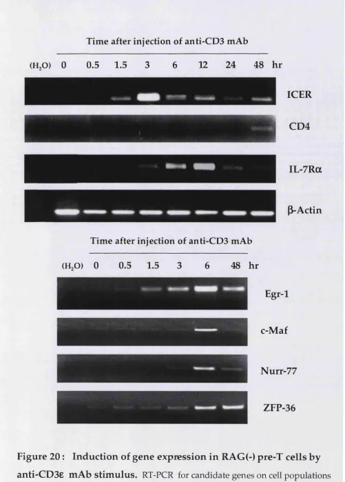
2. Identification of pre-TCR responsive genes 155
2.1. RDA analysis of TCRp(+) vs. TCRp(-) pre-T cells 155 2.2. RDA analysis of RAG(-) pre-T cells unstim ulated vs. stimulated
w ith anti-CD3e antibody 158
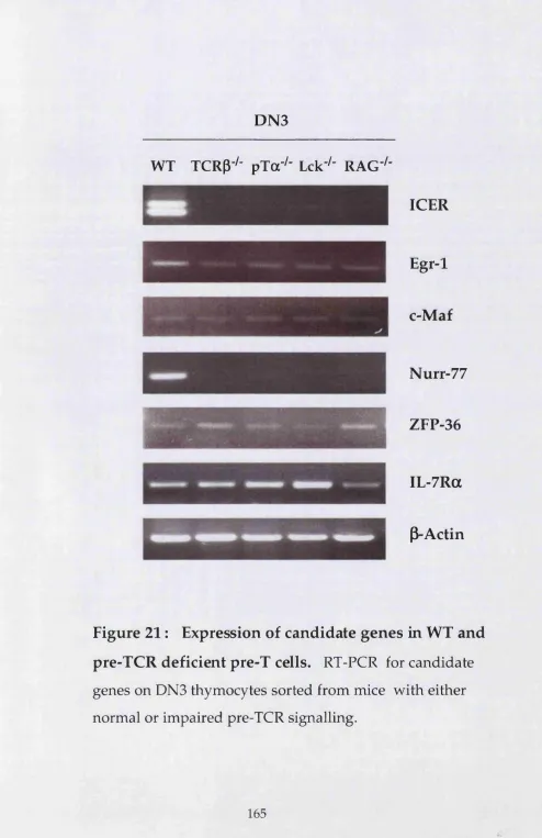
2.3. Expression of candidate genes: induction and dependence
on pre-TCR signalling 163
3. ICER in aP versus y5 T cell lineage com m itm ent 174 3.1. Identification of CREM isoforms expressed in yô thymocytes 174 3.2. Pattern of expression of ICER in the thym us 177 3.3. ICER expression and the status of TCR gene rearrangem ents
in pre-T cells 177
3.4. Analysis of the thym us of CREM/ICER deficient mice 182 3.5. Role of cyclic-AMP signalling in ICER expression during
thym ocyte developm ent 182
3.6. Generation and analysis of CD2-ICER transgenic mice 184 3.7. Pattern of expression of ICER in peripheral lym phoid tissues 186 3.8. ICER expression in mouse m utants for TCR 192 3.9. Lineage potential of pre-T cells expressing different levels
of ICER 194
4. ICER, P-selection and yS thymocyte developm ent 198 4.1. ICER expression in pre-T cells undergoing P-selection 198 4.2. ICER expression in pre-T cells w ith im paired pre-TCR
signalling 200
4.3. Induction of ICER expression by signalling through the
CD3 complex 200
4.3.1. ICER expression in response to anti-CD3e antibody 200
4.3.2. Involvement of the MAPK pathway 204
4.4. Analysis of the DN com partm ent of ICER deficient mice 206 4.5. Analysis of pre-T cells expressing different levels of ICER 207
4.5.1. Developmental potential 207
4.5.2. Phenotypic analysis 210
4.6. Gene expression in yô thymocytes developing in the
absence of P-selected cells 213
4.7. Requirem ent of a norm al (P-selected) composition of the
thym us for ICER expression. 215
4.8. Cross-talk betw een a p and yô lineages during yô
Chapter IV : D isc u ssio n _______________________________ 220
1. G enes d ifferentially expressed betw een aP and yÔ T cells 222
2. Pre-TCR responsive genes 228
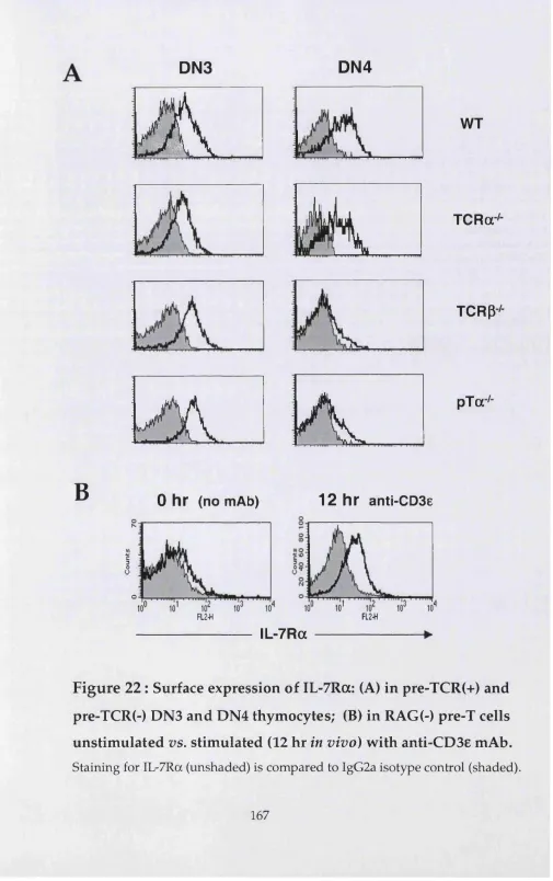
2.1. Role of IL-7 receptor in the DN to DP transition 229
3. ICER as a m arker for the yô T cell lineage 233
4. Pre-TCR d ep en d en t expression of ICER in pre-T cells 239 5. Cross-talk betw een a p and yô T cell differentiation 243
6. C onclusion 249
List of figures and tables
Figures
1 Model for murine haematopoiesis. 22
2 Early thymocyte development. 32
3 Proximal molecules associated with pre-TCR signalling. 39 4 T cell developmental blocks caused by gene deletion. 47
5 Models for the aP/yÔ T cell lineage split. 80
6 Structure of the human CD2 expression cassette. 114
7 Outline of representation difference analysis. 124
8 FACSorting of TCRa'^* thymocytes for RDAnalysis. 134 9 Summary of RDA analysis of yô vs. DP thymocytes. 136 10 RT-PCR for candidate genes in wild type yô and DP thymocytes. 140 11 FACS profiles of WT thymocytes in cell sorting experiments. 142 12 RT-PCR for candidate genes in WT haematopoietic lineages. 143 13 Ly49A protein surface expression in NK and T cells. 149 14 Sugano EST: sequence and linkage to IL-2Rp transcript. 152 15 IL-2Rp protein surface expression in thymic subsets. 154 16 FACS plots (CD25 vs. CD44) for WT and TCRP^ DN thymocytes. 155 17 FACS plots (CD25 vs. CD44) used in the purification of cells for RDA
analysis of anti-CD3e mAh stimulated vs. unstimulated RAG(-) cells. 159 18 FACS profiles of sorted populations used for RDA analysis:
CD25, CD69 and CD2 surface expression; forward scatter. 160 19 Cell cycle status of sorted populations used for RDA analysis. 161 20 Induction of gene expression in RAG(-) pre-T cells by anti-CD3e mAb. 164 21 Expression of candidate genes in WT and pre-TCR deficient T cells. 165 22 Surface expression of IL-7Ra in pre-TCR(+) and pre-TCR(-) DN3 and
DN4 thymocytes. 167
23 Effect of blocking antibodies to IL-7R complex on DN4 to DP transition. 169 24 Comparison of WT and IL-7Ra-deficient DN4 thymocytes:
ceU cycle status, cell death and intracellular TCRP expression. 171 25 Effect of blocking antibodies to IL-7R complex on proliferation and
26 Identification of CREM isoforms expressed in yô thymocytes. 175 2 7 Real-time PCR expression profile for ICER in the thymus. 178 28 Pre-TCR levels and ICER expression in WT pre-T cells. 180 29 Pre-TCR levels and TCR gene rearrangement status of WT pre-T cells. 181 30 FACS analysis of thymocyte development in CREM'^'mice. 183 31 Effect of cyclic-AMP on thymocyte development {in vitro) and ICER
expression. 185
3 2 Generation and screening of CD2-ICER transgenic mice. 187 33 FACS analysis of thymocyte development in CD2-ICER transgenic mice. 188 34 Real-time PCR expression profile for ICER in lymph nodes and spleen. 190 35 Real-time PCR expression profile for ICER in intra-epithelial lymphocytes. 191 3 6 ICER expression in TCRa-deficient and TCRÔ-deficient mice. 193 3 7 ICER expression in TCRaP transgenic and TCR^-deficient mice. 195 3 8 Lineage potential of pre-T cells expressing different levels of ICER. 197 39 ICER-LacZ protein expression in pre-T cells of CREM/ICER-LacZ mice. 199 40 ICER mRNA expression in pre-T cells of mice with deficient pre-TCR
signalling. 201
41 Induction of ICER expression by anti-CD3e mAh stimuli. 203 42 Effect of MAPK pathway inhibitors on the induction of ICER-LacZ
protein expression by anti-CD3e mAb stimuli. 205
43 FACS plots (CD25 vs. CD44) of WT and CREM"^" DN thymocytes. 206 44 Developmental potential of pre-T cells expressing different levels of
ICER-LacZ protein. 208
45 Phenotypic analysis of DN3 cells expressing different levels of
ICER-LacZ protein. 211
46 Gene expression in pre-T cells expressing different levels of
ICER-LacZ protein. 212
4 7 Gene expression in yÔ cells of TCRP"^" and pTa'^' mice. 214 48 ICER expression in foetal and adult pro-T and yô thymocytes. 215 4 9 ICER expression in the thymus of WT —> RAG^ bone marrow chimera. 217 5 0 Induction of ICER expression in yô thymocytes mediated by DP cells. 219 51 Comparison of proliferation and effector functions of WT and
TCRp-/- y5 cells. 245
Tables
1 Genes isolated from the RDA subtractive hybridisation of yd and DP
thymocytes. 137
2 Candidate gene expression (mRNA) in haematopoietic lineages. 141 3 cDNA sequences with no matches in GenEMBL databases,
obtained from the RDA subtraction DP - yd cells. 145 4 Genes isolated from the RDA subtraction of TCRP (+) and TCRP(-)
DN3 thymocytes. 156
5 Genes isolated from the RDA analysis of RAG(-) thymocytes stimulated
List of abbreviations
APC antigen presenting ceU
BCR B cell receptor
CD cluster of differentiation
cDNA complementary DNA
CLP common lymphoid progenitor
CMP common myeloid progenitor
CRE cyclic-AMP response element
CREM cyclic-AMP response element modulator
dATP deoxyadenosine 5'-triphosphate
dCTP deoxycytosine 5'-triphosphate
dGTP deoxyguanosine 5'-triphosphate
dTTP deoxythymidine 5'-triphosphate
DC dendritic cell
DETC dendritic epidermal T cell
DN double negative (CD4' CDS') thymocyte DN l DN thymocyte subset 1 (CD44^ CD25')
DN2 DN thymocyte subset 2 (CD44^ CD25^)
DN3 DN thymocyte subset 3 (CD44" CD25^)
DN4 DN thymocyte subset 4 (CD44' CD25 )
DNA deoxyribonucleic acid
DP double positive (CD4^ CD8^) thymocyte
E embryonic day
ECL enhanced chemiluminescence
FACS fluorescence-activated cell sorting FTOC foetal thymic organ culture
g gravitational constant (-9.8 m/s^)
GAPDH glyceraldehyde 3-phosphate
GFP green fluorescent protein
HSC haematopoietic stem cell
ic intracellular
ICER inducible cyclic-AMP early repressor
lEL intra-epithelial lymphocyte
ISP immature single positive (CD8^) thymocyte
ITAM immunoglobulin family tyrosine-based activation motif
kb kilobase
kD kilodalton
KO "knock-out"
-L ligand
LCR locus control region
loxP locus of cross-over in PI bacteriophage
fe immunoglobulin
M(j) macrophage
MAPK mitogen-activated protein kinase MHC major histocompatibility complex
mRNA messenger RNA
n number of replicate experiments
NK natural killer cell
NT no treatment
PCR polymerase chain reaction
PI propidium iodide
pTa pre-TCRa chain
PAGE polyacrilamide gel electrophoresis
PTK protein tyrosine kinase
-R receptor
RAG recombination activating gene
RDA representation difference analysis
RSS recombination signal sequence
RNA ribonucleic acid
rpm revolutions per minute
RT reverse transcriptase
SCID severe combined immunodeficiency
SH Src-homology domain
SP single positive (CD4^ or CD8^) thymocyte
Tg transgenic
tRNA transfer RNA
TCR T cell receptor
V-D-J variable-diversity-junctional (rearrangement of TCR genes)
Chapter I :
1 T cells and the Thymus in the Immune System
"The outstanding feature of the developm ent of im m unobiology in the last 10 years has been the recognition of the function of the lym phocyte and of the im portance of the thym us in the im m une process''.
M. Burnet (Nobel Prize of Medicine and Physiology), 1966
N ow adays, T cells are one of the m ost well know n cell types w ith the general public. This is the result of the extensive coverage in the lay press of AIDS (acquired immunodeficiency syndrom e), one of the m ost devastating diseases of m odern times. In stark contrast, before 1960, neither T cells nor their preferential producing organ, the thym us, were considered to play any role in im m unity. In fact, only 45 years ago MacLean and collaborators concluded from their research that "the thym us gland does not participate in the control of the im m une response" (MacLean et al., 1957). Even though the thym us was already know n to be a lym phocyte-producing organ, its relevance w as not recognised by the com m unity of imm unologists. Peter M edaw ar (Nobel Prize laureate himself) suggested in 1963 that "w e shall come to regard the presence of lym phocytes in the thym us as an evolutionary accident of no very great significance" (M edawar, 1963).
W hat then was the turning point?
1.1 Pioneering work on thymectomy
not later than a few days after birth were very susceptible to infections, had a m arked deficiency of lymphocytes in the blood and in lym phoid organs, and w ere unable to reject foreign skin grafts. In contrast, it had been know n for a long time th at thym ectom y of adult mice did not have such dram atic consequences - which was w hy the thym us had, until then, been disregarded as a vital lym phoid organ. Miller then show ed that if adult thym ectom ized mice w ere exposed to total body irradiation, the recovery of the lym phoid system w as thym us-dependent (Miller, 1962). Indeed, thym us implants allowed the developm ent of a norm al im m une (lymphoid) system in both neonatally thym ectom ized and irradiated adult thym ectom ized mice. Im portantly, if the thym us tissue used for the im plant was obtained from a foreign strain of mice, the neonatally thym ectom ized recipients became specifically immunologically tolerant to the histocompatibility antigens of the donor, implying that the thym us was the site w here self tolerance is established.
A nother striking phenotype of neonatally thym ectom ized mice w as that lym phocyte deficiency was seen in areas associated w ith cellular im m une responses, and not in areas w here antibody-producing cells resided (Parrott et al., 1966). Subsequently, the introduction of genetically m arked cells into neonatally thym ectom ized mice established beyond doubt and for the first time that antibody- producing cells (B cells), derived from the bone m arrow , were different from thym us-derived cells (T cells). It also dem onstrated that T cell communication with B cells was essential for antibody production (Mitchell, 1968).
1.2 The T cell lineage within haematopoiesis
All cellular components of the blood, from leukocytes of the im m une system to erythrocytes of the respiratory system, derive from haem atopoietic stem cells (HSC). These special cells are pluripotent (generate m any different cell types) and are also capable of self-renewal, unlike the cells they generate.
The production of all blood cell types from HSC is term ed haematopoiesis. This process is characterised by a continuous loss of potency, as im m ature pluripotent cells give rise to m ore m ature cells w ith more lim ited potential.
In adult mammals, HSC are present in the bone m arrow . H ow ever, in foetal life they initially localise to the yolk sac blood islands, and only produce cells of prim itive erythroid lineages (required for oxygen transport). This is called ''prim itive haematopoiesis". Later, HSC appear in the liver and spleen of the em bryo, and finally in the bone m arrow . D uring this second phase of haem atopoiesis, term ed "definitive", cells of erythroid, myeloid and lym phoid lineages are formed.
reconstitution (of limphopaenic recipients), unlike the HSC of the yolk sac. Definitive haematopoiesis therefore seems to derive from the em bryo, and not from the yolk sac.
D uring their m aturation, HSC first lose the ability to self-renew while still m aintaining their full developmental potential. These cells isolated from m urine bone m arrow are term ed short-term (ST)-HSC, because they self-renew for only 6 weeks in transplantation experiments (reviewed in Kondo, 2001). This stage precedes the generation of distinct blood cell types.
There have been several descriptions of the developm ental relationships betw een haemapoietic cell lineages (reviewed in Keller, 1999) - Figure 1.
On receiving differentiation signals, HSC commit to either the lym phoid or the myeloid lineage, thereby losing their pluripotency. W hereas lym phoid cells (T, B and NK cells) play a role in both adaptive and innate im m unity, m yeloid cells (i.e., m acrophages and granulocytes) essentially belong to the innate im m une system.
Recently, clonogenic lymphoid- and myeloid-lineage com m itted progenitors have been identified in adult m ouse bone m arrow . Com m on lym phoid progenitors (CLP) are the m ost im m ature lym phoid-com m itted precursors identified to date. These differentiate into T, B and NK lineages b u t not into m yeloid lineages. Their phenotype is IL-7Ra(+) Lin(-) Sca-l(low) c-kit(low), w here Lin represents m ature lineage m arkers, IL-7Ra is the a chain of the IL-7 receptor, Sca-1 is stem cell antigen 1 and c-kit is stem cell factor receptor (reviewed in Akashi,
2000).
The cytokine IL-7 is indispensable for both T and B cell developm ent. The IL-7 receptor is composed of a specific a chain and the com m on cytokine receptor y chain (yj (Kondo et al., 1994). Mice that are genetically deficient for IL-7 or IL-7R have a severe reduction in their T and B cell com partm ents (Feschon et al., 1994). Mice deficient for y^ also lack NK cells (Cao et al., 1995) due to an im pairm ent in the form ation of IL-15 receptor, for which y^, is also necessary.
lym phoid-restricted zinc-finger transcription factors (TFs). In their functional absence, mice are devoid of all lym phoid lineages (Georgopoulos et al., 1994).
The next step in T cell differentiation is the comm itm ent of CLPs to the T lineage and involves transcription factor GATA-3 (reviewed in Kuo, 1999). GATA-3 belongs to the GATA family of zinc-finger TFs, of which m em bers - 1 /-2 /-3 are highly expressed in haematopoietic cells, w hereas m em bers - 4 /-5 /-6 predom inate in heart, gut and muscle. GATA-3 is expressed in both T and NK lineages, and was first identified as a TF that binds to the T cell receptor a chain gene enhancer. Targeted disruption of the GATA-3 gene in mice resulted in embryonic lethality at E ll (Pandolfi et al., 1995), which precluded an analysis of its role in T cell developm ent. To overcome this difficulty. Ting et al (Ting et al., 1996) com plem ented RAG-2'^ blastocysts (Chen et al., 1993), from which no T or B cells can be generated, with GATA-3-deficient ES cells. The resultant RAG-2^ GATA-3^ chimeric mice had norm al B cell populations, b u t completely lacked T cells. Detailed analysis of the thym us of these animals dem onstrated that GATA-3 is necessary for the survival and developm ent of the earliest T cell com m itted CD4(-) CD8(-) thym ocytes or their precursors, defining it as the earliest know n TF required specifically for T cell lineage commitment.
T cell developm ent occurs preferentially in the thym us, from CLP precursors im ported from the bone m arrow . It is still unknow n w hether CLPs hom e directly to the thym us in vivo. The earliest identified thymic progenitors are CD4(low) CD8(-) CD44(+) CD25(-) c-kit(+). A lthough the m ajority of these cells seem to be T cell-committed, there are also some that can differentiate into B, NK and lym phoid dendritic cells (DC) at a low frequency (Ardavin, 1993; Wu, 1991). Consequently, it is possible that the earliest thymic progenitor population contains a small num ber of CLP that have hom ed to the thymus. Because the thymic m icroenvironm ent (see 2.2) is best suited for T cell differentiation, CLP m ay preferentially develop into T cells in the thym us (Akashi et al., 2000). This contrasts w ith w hat happens to CLP in the bone m arrow , w here the environm ent favours B cell developm ent.
pro-T
c-myb A iolos PU.l GATA-3
IL-7R+
GATA-1 GATA-2
0
c-myb PU.1++ Aiolos^
GATA-3 c-myb Aiolos
P U .r " GATA-3
IL-7R
Epo-R+ GMP
CMP
MEP GATA-l
GATA-2++ c-myb^ PU.1 + Aiolos'
GATA-1+ GATA-2+ c-myb"*” PU.l"^ Aiolos' GATA-3'
T cell
NK cell
Bcell
Monocyte
Granulocyte
Megakaryocyte
Erythrocyte
1.3
T cell receptor, the hallmark of T cells
The T cell receptor genes were initially identified as cDNAs differentially expressed betw een T and B cells, that respected tw o criteria: w ere encoded by gene segm ents that had undergone rearrangem ent only in T cells; and had sequence similarity to im m unoglobulin (Ig), which had been previously identified as the B cell receptor (Kronenberg et al., 1986).
The TCR and Ig genes are indeed structurally related: each is composed of multiple variable (V), diversity (in some cases) (D) and joining (J) gene segm ents th at are subjected to rearrangem ent during lym phocyte developm ent. This process is m ediated by the same enzym es (RAG - recom bination activation gene - recombinases) in both cell types, b u t is tightly regulated in a lineage-spedfic m anner (the TCR only rearranging in T cells, and Ig only in B cells) (Yancopoulos, 1986).
M urine TCR genes are spread betw een three lod: p, which comprises 800 kb on chrom osom e 6; y, 205 kb on chrom osome 13; and a /5 , a com m on locus to TCRoc and TCRÔ, 1000 kb on chrom osome 14. W ithin the a / 5 locus, J and C (and D) segm ents are spedfic for either TCRa or TCRÔ, w hereas the majority of V segm ents can be incorporated in both chains (reviewed in G raw under, 1998).
Somatic recom bination involves V (to D) to J rearrangem ents that proceed betw een gene segments m arked by ""recombination signal sequences"' (RSS). These are DNA patterns consisting of a palyndrom ic heptam er (CACAGTG) and a nonam ere (ACAAAAACC) spaced by either 12 or 23 bp (term ed RSS-12 and RSS- 23, respectively). Rearrangem ents follow the ""12/23"" rule, which imposes the involvem ent of one RSS-12 and one RSS-23 gene segments. The process is catalysed by RAG (recombination activation gene) enzym es (-1 and -2), and is fadlitated in term s of DNA locus accessibility by HMG (high m obility group) proteins, w hich are capable of m odifying the conformation of DNA (""DNA bending proteins") (reviewed in Graw under, 1998).
each T cell to assemble its TCR, depending on the lineage it commits to: aP T cells use a and P; yô T cells use y and 5 chains (Kronenberg, 1986; Marrack, 1987; Raulet, 1989; Stronünger, 1989; Davis, 1988).
Both chains of the TCR have, similarly to Ig, an arnino-tenninal variable (V) region, a constant (C) region, and a short hinge region w ith a cysteine residue that forms the inter-chain disulfide bond. Each chain spans the cell m em brane lipid bilayer by a hydrophobic transm em brane dom ain, the notable feature of which is the presence of positively charged amino a d d residues, im portant for the association w ith the signalling CD3 subunits (see ahead). Finally, each chain ends w ith a short cytoplasmic dom ain (Clevers et al., 1988). Consistent w ith these analogies, the X-ray crystal structures obtained so far for particular Ig and TCR molecules have show n that these proteins fold in a similar way. In particular, each dom ain is a globular structure in which several strands of polypeptide chain come together to form two anti-parallel P-sheets, held together by an intra-chain disulfide bond. This type of 3D structure is called an "Tg-like dom ain" and is present in m any proteins involved in cell-cell recognition, especially in the im m une and nervous systems (Novotny et al., 1986). There are, though, particular differences in alignm ents, angles and points of contact betw een dom ains of Ig and TCR molecules (reviewed in Wilson, 2001).
More strikingly, though, TCR differs from Ig in that TCR is m onovalent, w hereas Ig is bivalent (two antigen binding sites) and the TCR is never secreted, unlike Ig (which is secreted after activation of the B cell).
The cytoplasmic domains of all the CD3 subunits contain sequences called im m unoreceptor tyrosine-based activation motifs (ITAM) that allow them to associate w ith cytosolic protein tyrosine kinases following receptor stimulation. Particularly im portant for this signal transduction process are CD3e and CD3Ç (Terhorst et al., 1995). CD3 proteins are also required for the assembly and cell- surface expression of the TCR complex. Therefore, it is not surprising that mice lacking these proteins (due to genetic m anipulation) suffer from immunodeficiency.
The TCRaP complex also contains CD4 or CDS "co-receptor" molecules, which co-operate w ith the TCR in antigen recognition (Zuniga-Pflucker et al., 1991). CD4 is a single-chain molecule composed of four Ig-like domains. Its cytoplasmic dom ain interacts strongly w ith Src family tyrosine kinase p56Lck, prom oting signal transduction from the TCR. Indeed, the presence of CD4 has been estim ated to low er by 100-fold the dose of antigen required for T cell activation (reviewed in Zam oyska and Travers, 1995). CDS, in contrast w ith CD4, is a disulfide-linked heterodim er comprising a and P chains (although a C D S aa hom odim er also exists in some less abundant T cell subsets), each containing a single Ig-like dom ain (Zamoyska and Travers, 1995).
2
T cell developm ent
The vertebrate thym us is responsible for the production of self-restricted, self- tolerant T (thym us-dependent) cells. In the thym us, im m ature T cells (thymocytes) proliferate an d differentiate, passing through a series of discrete phenotypic stages that can be identified by particular patterns of expression of various cell-surface proteins. D uring differentiation, T cells undergo gene rearrangem ents, commit to a T cell lineage (a(3 or y5)/ express an appropriate T cell receptor, and are then subm itted to developm ental checkpoints such as "positive'' and "negative" selection. Cells that fail these selection processes die by apoptosis or by neglect, w hereas the selected cells survive and leave the thym us to seed the peripheral lym phoid organs.
2.1 Thymus organogenesis
Thymus organogenesis requires interactions betw een cells of all three embryonic germ layer origins: endoderm -derived epithelium, neuroectoderm - derived neural crest mesenchyme, and m esoderm -derived haem atopoietic cells and endothelial cells of blood vessels (Le D ouarin and Jotereau, 1975; Moore and Owen, 1967; Ow en and Ritter, 1969). For convenience, this entire process can be divided into three m ain stages: early organogenesis (E9.5-El 1.5 in the m ouse embryo), late organogenesis (E12-E15) and late foetal developm ent (E15.5-birth) (Manley, 2000).
is particularly obvious in mice lacking m igrating neural crest cells, either b y experimental ablation (Bockman et al., 1989) or due to a genetic defect, in Pax3 ''* mice (Conway et al., 1997). Mesenchymal cells appear to be required for epithelial grow th and differentiation (including induction of MHC class II expression) b y providing both secreting factors and extracellular matrix (Owen et al., 2000; Suniara et al., 2000).
At least three transcription factors have been clearly implicated in this early phase of m urine thym us organogenesis: Hoxa3, Pax9 and Whn. M utant mice for each of these genes show an early failure in thym us form ation (reviewed in Manley, 2000). Hoxa3'^' mice are athymic, this being one of several defects in the pharyngeal region. The best characterised m utation affecting early thymic epithelium developm ent is the W hn gene, which results in the nude m ouse (so called due to its absence of hair). The thymic rudim ent of the nude m ouse has deficient epithelium and is not populated by lym phoid progenitor cells.
At this stage there are effectively two bilateral rudim ents which proliferate and b u d off from the pharynx. They then m ove medially, ventrally and caudally until they join at the midline above the heart by E12.5. This m igration seems to be regulated independently of differentiation, as a norm al m igration can be seen in the nu d e mouse.
2.2
Thymie microenvironment
The strom al com partm ent of the thym us is heterogeneous, consisting of cortical and m edullary epithelium, mesenchymal fibroblasts, dendritic cells and m acrophages (Boyd et al., 1993). These contribute to thym ocyte developm ent via cell-cell interactions and the production of soluble factors.
Lym phoid precursors start colonising the thym us prior to vascularization of the organ (which occurs at E14). Colonising cells need to leave the pharyngeal vessels and traverse the peri-thymic mesenchyme before penetrating the basem ent m em brane surrounding the embryonic thymic rudim ent. It is therefore likely that this process involves chemotactic factors. Indeed, in vitro transfilter m igration assays have show n that alym phoid thym uses consisting only of thymic strom a attract precursors from donor lym phoid tissues (foetal liver or norm al thymic lobes) (Fontaine-Perus et al., 1981; Jenkinson et al., 1982). Furtherm ore, chemokines such as MI? (macrophage inflam m atory protein) -let, -Ip, -ly and -2, SDF-1 (stromal cell-derived factor -1), TECK (thymus-expressed chemokine) and TARC (thym us activation-regulated chemokine), and respective receptors - CCR5, CCR7, CCR9, CCR4 and CCR8 - are expressed in the thym us (reviewed in Anderson,
2000).
Consistent w ith a chem okine-dependent m echanism of colonisation, this process can be inhibited in vitro by pertussis toxin, an inhibitor of G protein-m ediated chemokine receptor signalling (Wilkinson et al., 1999). How ever, lym phoid progenitors colonise only the thym us and not other chemokine-expressing organs. Therefore, due to its thym us-restricted expression, TECK w as initially seen as the best candidate for providing thymic-spedfic hom ing (Anderson et al., 2000a). But further studies have show n that, not only is TECK expressed in tissues that do n o t attract lym phoid precursors, b u t also neutralising anti-TECK antibodies cannot inhibit in vitro thymic colonisation (Wilkinson et al., 1999). Thus, the molecular basis of a chem okine-dependent colonisation of the thym us is still to be clarified.
implicated in this process. TECK receptor (CCR9) is upregulated during thym ocyte developm ent in the transition betw een im m ature CD4(-)CD8(-) and CD4(+)CD8(+) stages, and it has been show n to be a chem oattractant for the m ore m ature single positive thymocytes that reside in the m edulla (Norm ent et al., 2000).
Besides the strom al cell netw ork, the thym us also contains mesenchymal fibroblasts that interact with thymocytes and play a role in their developm ent. These fibroblasts provide extracellular m atrix (ECM) com ponents such as fibronectin, laminin, collagen and vim entin to thymocytes. Thymocytes, in their turn, express a variety of ECM-receptors: VLA-4, VLA-5 and CD44, which are particularly abundant in early im m ature cells, suggesting a developm entally regulated role for ECM com ponents in thym ocyte m aturation (Anderson et al., 2000a).
The relevance of mesenchymal fibroblasts and their ECM com ponents to early T cell developm ent has been show n in reaggregate thym us organ cultures (RTOC) (Hare et al., 1999). A combination of both epithelial cells and fibroblasts was found to be required for the m aturation of CD4(-)CD8(-) precursors to the CD4(+)CD8(+) stage (Anderson et al., 1993), and pre-treatm ent of these fibroblasts with hyaluronidase, an ECM disrupting enzyme, abrogated their ability to support this developm ental transition (Anderson and Jenkinson, 2000b).
Cell-cell interactions betw een thymocytes and strom al cells are also extrem ely im portant for triggering of receptor-associated signalling pathw ays in thymocytes. In particular, thymic epithelial cells seem to provide ligands (of Jagged and Delta families) for Notch (Anderson et al., 2001) and W nt family ligands (Wnt -4, -7) for Frizzled receptor signalling (Jenkinson, 2002) (see 2.6).
Finally, thymic selection processes also require interactions betw een CD4(+)CD8(+) thymocytes and thymic epithelial cells (Anderson et al., 1995; Anderson et al., 1994a; A nderson et al., 1994b; H are et al., 2001; Wilkinson et al., 1995). In vitro, these two cell types form a structure know n as "rosette' w here one thymocyte is surrounded by an average of three epithelial cells. Incubation of 'rosettes' in foetal thymic organ cultures (FTOC) allows m aturation of the thymocytes to the single-positive stage. This 'rosette' structure is abrogated if the epithelial cells are MHC-deficient, or if the thymocytes are TCR-deficient, indicating that the association betw een the two cell types is dependent on TCR-MHC interaction. The TCR-MHC contact point is know n as a "synapse", and it produces a re-distribution of cell surface molecules including co-receptors CD4 and CD8, CDS and CD45, integrin LFA-1, and signalling molecules p56Lck and LAT.
Thus, in sum m ary, the thymic m icroenvironm ent provides a series of soluble factors and cell-cell interactions that are fundam ental for T cell developm ent. These include chemokines that attract lym phoid progenitors to enter the thym us, and that help direct thymocyte m igration within the thym us; grow th and survival factors produced and presented by epithelial cells and fibroblasts; and selection and differentiation signals that are essential at certain stages of T cell development.
2.3
Developmental stages of murine thymocytes
The early phase includes the expansion of early thymic im m igrants, their com m itm ent to the T lineage, rearrangem ent of the TCR y, 6 and p loci, com m itm ent to the a p or yS lineage and isotypic exclusion (i.e., the expression of only one type of TCR, aP or yô, per cell), and allelic exclusion (expression of only one variant of TCR chain; silencing of the other allele) (Fehling and von Boehmer, 1997).
The late phase is m arked by: positive selection of the cells expressing a TCR capable of effective (intermediate signal strength) interaction w ith MHC; negative selection of the cells in which that interaction is too strong (due to recognition of ''self" antigen); com m itm ent to one of the single-positive lineages, CD4+ (helper lymphocytes) or CD8+ (cytotoxic lymphocytes); and final export of m ature T cells to the periphery (Kisielow and von Boehmer, 1995).
These differentiation events occur in a tightly regulated developm ental sequence, which can be followed by analysing the expression of certain cell surface molecules {developmental markers). The availability of antibodies that selectively recognise each of those m arkers allows a rapid purification of thym ocyte subsets and thus greatly facilitates the study of differentiation processes.
Two of such m arkers are CD4 and CD8 (see 1.3). O n the basis of their expression, developing thymocytes are sub-divided into four populations of different m aturity: "double negative", "double positive" and each "single positive" subset (reviewed in Kisielow, 1995).
The m ost im m ature subset is CD4(-)CD8(-), "double negative" (DN), and also lacks the expression of a m ature TCR-CD3 complex; it constitutes only about 2% of all thymocytes.
NK cells
Import from bone marrow
IB cells Thymic
(^^dendritic
cells
1 L
<5%
CD4'o« C ' k i t +
CD44+
CD25-(a)
0
0
TCR rearrangements) p-allelic exclusion
p-selection
<5%
C ' k i t +
CD44+ CD25+
(b)
I Proliferation/survival dependent I on c-kit- or 7^,-mediated signals
DN1
DN2
-4 5 %
C'kit-/k)w CD44-/^
CD25+
(0
,Pre TCR independent pathway
-4 5 %
c-kif CD44-/kw CD25-(d)
DN3
DN4
CD4+CD8+ DP thymocytesLarge-sized, rapidly cycling cells
(e)
DP
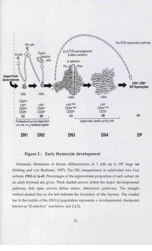
F ig u re 2 : E arly th y m o c y te d e v e lo p m e n t
The m ost im m ature DN subset (D N l) is c-kit(+)CD44(+)CD25(-). These cells tend to express a small am ount of CD4 on their cell surface and are therefore not strickly DN thym ocytes (Wu et al., 1991a). Although they are lymphoid-restricted, D N l cells are not yet fully comm itted to the T lineage, as they still can develop into B, NK or thymic dendritic cells (DC) (Ardavin et al., 1993; W u et al., 1991b). In this regard, it is im portant to stress the im portance of c-kit as a m arker for D N l cells, as the m ajority of CD44(+)CD25(-) thymocytes are not T cell precursors. CD44 is a hom ing molecule for cells that colonise the thym us (Wu et al., 1993), w hether or not they are T cell progenitors. Indeed, only about 3% of all CD44(+)CD25(-) thym ocytes express high levels of c-kit and are, therefore, "'true" D N l. This problem can be also avoided by pre-gating on Thyl(+) cells, which excludes the m ajority of CD44(+) ""contaminants". (Note: after the D N l stage, the expression of c-kit becomes very similar to that of CD44, which justifies the use of only one of these m arkers in the analysis of the DN compartment.)
U pon up-regulation of CD25, thymocytes progress to the c- kit(+)CD44(+)CD25(+), DN2 stage of developm ent. The first detectable DN2 cells during m ouse foetal developm ent occur at E13.5. DN2 cells can no longer generate B lymphocytes, b u t some still retain the potential to produce DC (Wu et al., 1996) or NK cells (Moore and Zlotnik, 1995). DN2 cells that commit to the T lineage (pro-T cells) begin to rearrange their TCR y, ô and P loci (Capone et al., 1998). This is possible due to the initiation of expression of RAG-1/2 enzym es (Capone et al., 1998; W ilson et al., 1994). TCRP rearrangem ents occur only in the D (diversity) and J (junctional) regions; no full V-DJ rearrangem ents have been detected at this stage of developm ent (Godfrey et al., 1994; Capone et al., 1998). DN2 is a population of actively proliferating cells (approximately five-fold m ore so than D N l (Moore and Zlotnik, 1995)), suggesting the existence of a cell cycle activator mechanism. Signals from IL-7 receptor and c-kit (stem cell factor receptor) have been suggested to play a role in this m echanism (Rodewald et al., 1997).
can only give rise either to yô or a p T cells. Full V-DJ rearrangem ents of TCRP genes are now detectable in this population, and if in frame they enable the synthesis of a TCRP protein. Cells that successfully rearrange and express a functional TCRp chain are selected for further m aturation (survival, proliferation and differentiation), a process term ed "p-selection" (Dudley et al., 1994; Mallick et al., 1993). The basis of this selection event is the emergence of a new signalling receptor, the pre-TCR (Saint-Ruf et al., 1994), a heterodim er consisting of a TCRP chain and an invariant chain, p T a (Bruno et al., 1995). As is the case of the TCR complex (see 1.3), the pre-TCR also associates w ith CD3 molecules, which are the active signalling components of the complex. Signals triggered by the pre-TCR are fundam ental for m aturation of aP cells beyond the DN3 stage; in contrast, y5 cell developm ent seems to be largely pre-TCR independent (Fehling et al., 1995a).
The final DN subset is DN4, characterised by a c-kit(-)CD44(-)CD25(-) surface phenotype. These cells, like DN3 thymocytes, first appear at E14.5 of foetal thymic developm ent. DN4 cells start to express CD4 and CDS at the mRNA level, and if cultured for 24 hours in m edium w ithout added differentiation factors they spontaneously develop into DP thymocytes. Their m ost strinking feature is a high rate of proliferation, a consequence of P-selection (Hoffman et al., 1996). Indeed, this population is highly enriched for productive TCRP rearrangem ents, as show n by RFLP-PCR (restriction fragm ent length polym orphism -PCR) (Dudley et al., 1994), and shows relatively high levels of surface TCRP protein expression, as assayed by liposome staining (Bruno et al., 1999). Nevertheless, except for DN4 expressing the highest levels of pre-TCR on the surface, these pre-T cells can still generate cells of both y5 and aP lineages (Bruno et al., 1999). In fact, Wilson and MacDonald have identified a subset (8%) of DN4 cells that is intracellular TCRy6(+), although extracellular TCRyô(-), and have suggested that these are yô precursors in the DN4 population (Wilson et al., 1999).
cells themselves, will be the focus of part 3 of this introduction; for the rem aining of p art 2 w e will concentrate on «P T cell development.
The vast majority of cells in the thymic cortex, and about 80% of all thymocytes, are CD4(+)CD8(+), DP ("double positive"), progressing from the DN4 stage via CD8(+) im m ature single positive (ISP) intermediates. D uring foetal m urine development, DP are first detectable at E15.5, one day later than DN4 thymocytes. In contrast, in the adult thymus, differentiation from the late DN to DP stages takes 2-3 days. This difference suggests different kinetics betw een foetal and adult thym ocyte developm ent (Manley, 2000). TCRa rearrangem ents are completed at the DP stage, which allows the expression of a TCRaP complex. Most DP cells express TCRaP at a low level, but around 30% have no detectable surface expression, while another 5 % express it at a m axim um level, identical to the one of SP thym ocytes and peripheral T cells. DP cells are less proliferative that DN4 cells, 20% being in cycle. This subset is also very susceptible to cell death, either b y neglect (lack of survival signal) or by apoptosis (program m ed cell death). Only a m inority of DP cells are able to survive both negative and positive selection (see 2.5.1).
DP cells which are positively selected (and survive negative selection) go through a lineage com m itm ent decision, betw een CD4+ and CD8+ "single positive" (SP) lineages (see 2.5.2). These constitute the m ost m ature thymic subsets. Approxim ately 10% of all thymocytes are CD4+ SP, w hereas CD8+ SP account for 5% of the thymocytes.
2 .4 The role of pre-TCR in thymocyte development
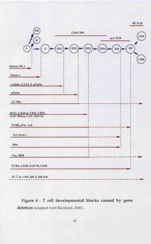
The im portance of a functional TCRP chain for early thym ocyte differentiation became obvious through the analysis of mice deprived of such entity. The first inform ative m ouse m utant was the naturally occuring SCID (severe-combined immunodeficiency), which cannot efficiently rearrange its TCR (and BCR) genes due to a defect in a DNA-dependent protein kinase that participates in somatic recom bination (Blunt et al., 1995). Further evidence was gathered from the analysis of RAG (recombination activation gene)-dehcient mice (Mombaerts et al., 1992a) and, decisively, from TCRp 'knockout' mice (M ombaerts et al., 1992b). In these m urine models, T cell differentiation is severely im paired, w ith complete (SCID, RAG^ ) or partial (TCRP'^') developm ental blocks at the DN3 stage, and therefore none (SCID, RAG'^') or very few (TCRP'^') DP thym ocytes produced. Since it was accepted that the TCR w ould not be expressed in im m ature DN thymocytes, an active search for a "pre-TCR" w as initiated.
2.4.1 p T a a s p a rt o f pre-TCR
By 1992, TCRP^ and TCRa^ mice had been generated, and it was obvious that the tw o chains that composed the TCR had distinct roles in T cell developm ent. W hereas TCRP^ differentiation was severely blocked at the DN3 stage, TCRoc^ thymocytes were able to m ature m uch further, producing a large DP com partm ent (but no SP cells) (Mombaerts et al., 1992b). This led to a puzzling question, "how could a TCRP chain prom ote T cell developm ent at the im m ature DN3 stage, in the absence of a functional TCRa chain?"
protein that w as covalently bound to TCRp; it w as nam ed pre-TCRa (pTa) (Saint- Ruf et al., 1994).
p T a belongs to the imm unoglobulin superfam ily and is encoded by a n on rearranging gene (Fehling et al., 1995b). Its expression pattern has been analysed in detail using RT-PCR (Bruno et al., 1995) and the results suggest that expression is confined to im m ature cells of the T cell lineage. Interestingly, p T a message has also been detected in m ouse bone m arrow , although B cells (including pre-B cells) and all other hematopoietic lineages do not express the gene. This m ight suggest that bone m arrow harbours very early T lineage-committed precursors, and that p T a could be used as a m arker for their identification (Fehling and von Boehmer, 1997).
In the thym us, p T a is expressed in all DN subsets (although at a very low level in D N l) and also in DP cells, b u t not in SP thymocytes. At the DP stage, p T a is competitively displaced by TCRa, thus allowing m ature TCR to take the place of pre-TCR at the cell surface (Trop et al., 2000).
The generation and analysis of pTa'^' mice (Fehling et al., 1995a) has provided conclusive evidence for p T a being the partner of TCRp in im m ature DN thymocytes. The similarity betw een the phenotypes of pTa'^' and TCRP'^' mice is striking. In both cases, a 10-foId reduction in thymic cellularity is observed, due to severely decreased num bers of DP and SP thymocytes. The DN com partm ent is devoid of DN4 cells, whereas DN3 thymocytes are twice as abundant as in norm al mice; this results in a developmental block at the DN3 stage, w here pre-TCR expression and function is required for differentiation. Interestingly, developm ent of y8 cells is not im paired in these mice (in fact, their absolute num bers are higher than in norm al mice), suggesting that the yô lineage does not depend on "p- selection" for m aturation (Fehling et al., 1995a).
2.4.2 “P -se le c tio n ”
As a consequence, there is intense proliferation and rapid progression to the DP stage. Cells that are not selected, because they fail to generate a pre-TCR complex, will die (unless they become yô cells) (Fehling and von Boehmer, 1997).
Cell survival, proliferation and differentiation at the DN3 DN4 (—> DP) transition are consequences of signals triggered by the pre-TCR. As w ith the m ature TCR, the pre-TCR also relies on association w ith CD3 - y, 5, e, Ç - molecules for signal transduction. In fact, pre-TCR and TCR complexes have very comparable subunit compositions, the major difference being p T a in the place of TCRa (reviewed in Kruisbeek, 2000). H ow ever, unlike the TCR, the pre-TCR does not seem to require a ligand for initiation of signalling. In particular, the extracellular dom ains of p T a and TCRp are not required for signalling (Irving et al., 1998).
It is im portant to note that pT a ^' and TCRP^ thymocytes, which express partial CD3 complexes on their cell surfaces, fail to signal and to differentiate, unless triggered by antibody-m ediated cross-linking (Levelt et al., 1993). Also, it seems that pre-TCR complexes do not simply serve to increase the density of CD3 subunits at the cell surface, since transgenes used to restore developm ent in pre- TCR deficient mice do not cause any detectable change in CD3 expression (Irving et al., 1998).
Therefore, pre-TCR seems to have an unique capacity to transduce P-selection signals, even w hen expressed at low levels and w hen not engaged by a surface ligand. Such properties of the pre-TCR have led to the suggestion that it acts as a constitutively active signalling complex. This ability could be conferred upon the pre-TCR by some unique property of pT a, a m atter still under investigation (reviewed in Kruisbeek, 2000).
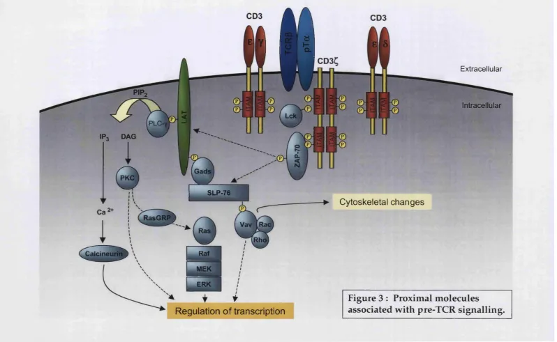
2.4.3 C o m p o n en ts o f pre-TCR s ig n a llin g p a th w a y (s)
Pre-TCR
ÏÏ
1SLP-76
Calcineurin
Cytoskeletal changes
Extracellular
Intracellular
^ i f
Regulation of transcription
Regardless of how pre-TCR signalling is initiated, its association w ith CD3 subunits (y, S, b, Q guarantees efficient signal transduction. As previously m entioned (1.3.1), CD3 molecules contain im m uno-receptor tyrosine-based activation motifs (ITAM) within their cytoplasmic domain. As in the TCR, these ITAM are phosphorylated by activated Src-family protein tyrosine kinases (PTK). Based on the phenotype of individual "knockout' mice for each of the four chains (y, Ô, e, Q, the m ost im portant for pre-TCR signalling seem to be CD3e and CD3y. Both CD3e^ (Malissen et al., 1995) and CD3y^' (Haks et al., 1998) mice display a severe block at the DN3 stage of development. In contrast, CD30^ (Dave et al., 1997) and CD3Ç'^' (Love et al., 1993) mice are m ore perm issive to thym ocyte developm ent. In CD38^ mice, for example, differentiation is only blocked at the late DP stage, beyond the influence of the pre-TCR complex. (For a thorough review on CD3 molecules, see Malissen, 1999)
Several lines of evidence docum ent the involvem ent of lymphocyte-specific tyrosine kinase p56Lck in the transmission of pre-TCR signals. Both p56Lck- deficient (Molina et al., 1992) and dom inant negative p56Lck transgenic (Levin et al., 1993) mice have a profound block in thymocyte developm ent, similar (although less severe) to that of pre-TCR-deficient mice (TCR|3’^’, pTa'^'). In addition, a constitutively active Lck transgene is capable of rescuing DP production in RAG- defident (Mombaerts et al., 1994) and in pTa-deficient mice (Fehling et al., 1997b). The proximal prom oter of p56Lck is one of two prom oters (together w ith CD2 prom oter) used for T cell-restricted expression of particular transgenes. It seems to be active from the D N l stage of developm ent, and mRNA levels for Lck are similar (varying by less than 2-fold) in all DN subsets (Buckland et al., 2000a). Lck protein, though, is dramatically (20-fold) up-regulated in DN2 —> DN3 transition, exposing a post-transcriptional mechanism that regulates Lck in accordance w ith pre-TCR expression (Buckland et al., 2000a).
'knockout' mouse (Groves et al., 1996; van Oers et al., 1996), suggesting that Fyn plays a largely redundant role in pre-TCR signalling.
Src-PTKs are activated by the phosphatase CD45, which dephosphorylates their C-terminal regulatory tyrosine residues (Src-PTKs are inactive in the phosphorylated state). CD45-deficient mice show an accumulation of DN3 cells and a reduction in DP and SP num bers (Byth et al., 1996; Kishihara et al., 1993).
Src-kinase activity is negatively regulated by carboxy-terminal Syk kinase, Csk. Consistent w ith this, Csk-deficient mice have an "opposite" phenotype to Lck- deficient animals. Csk'^' mice generate TCR(-) DP and SP cells, thus bypassing the need for pre-TCR and TCR in T cell developm ent (Schmedt et al., 1998).
Phosphorylation of ITAMs of CD3 subunits by Src-PTKs creates docking sites for SH2-domain containing PTKs ZAP-70 and Syk. Similar to Lck and Fyn, there seems to be a m arked redundancy in the function of ZAP-70 and Syk dow nstream of the pre-TCR. W hereas single 'knock-out' mice exhibit none (Syk*^ ) or only very small (ZAP-70'^ ) defects in T cell differentiation (Cheng et al., 1995; Negishi et al., 1995; Turner et al., 1995), double 'knockout' animals show a complete arrest at the DN3 stage of developm ent (Cheng et al., 1997a).
Several adaptors, exchange factors and GTPases previously know n to be involved in m ature TCR signalling have recently been implicated in transducing pre-TCR signals. A daptor proteins SLP-76 and LAT (linker for activation of T cells), which are substrates for TCR-induced PTK activity, are crucial for pre-TCR signalling. In both m ouse 'knockout' models, thym ocyte developm ent is completely blocked at the DN3 stage (Clements et al., 1998; Pivniouk et al., 1998; Zhang et al., 1999). The fact that this block is not rescued by anti-CD3e Ab cross- linking dem onstrates the importance of these adaptor proteins as "com m on platform s" for the recruitm ent of effectors of pre-TCR signalling.
Mice deficient for Vav-1 display a partial defect at the DN DP transition, with an accumulation of DN3 thymocytes (Fischer et al., 1995; Tarakhovsky et al., 1995; Turner et al., 1997; Zhang et al., 1995). Vav activates GTPases of the Rho family. The inactivation of Rho in the thym us caused a severe reduction in survival of pro-T cells and in cell cycle progression of pre-T cells (Galandrini et al., 1997; H enning et al., 1997). The pro-T cell defect m ay be related to a role in IL-7R signalling (see 2.6.2), b u t in the case of pre-T cells, Rho seems to control a p53-dependent survival checkpoint dow nstream of the pre-TCR (Costello et al., 2000). Besides Rho, another small GTPase, Rac, has been implicated in p-selection. A constitutively active Rac-1 m utant can partially substitute for the pre-TCR complex, and can fully com plem ent defects of Vav-deficient pre-T cells (Gomez et al., 2000).
2.4.4 T r a n sc r ip tio n fa c to r s in v o lv e d in “P -se le c tio n ”
The nuclear targets of the signalling pathw ays triggered by the pre-TCR are still largely unknow n. Nevertheless, gene targeting and transgenic studies, and gene m anipulation experiments in foetal thymic organ culture, have suggested crucial roles for a few transcription factors in p-selection and D N —> DP transition. It has to be said, though, that the putative targets of these TPs in this process rem ain to be defined.
EGR-1 (early grow th response gene -1) is a zinc-finger-containing TF, w hose transcription correlates w ith p-selection. M oreover, its enforced expression (via a transgene) in a RAG-deficient background rescues thymocytes from developm ental arrest at DN3, allowing m aturation to proceed to the ISP (im m ature single positive, CD8^) stage (Miyazaki, 1997). H ow ever, developm ent to the DP stage requires irradiation of the EGR-1 Tg / RAG KO mice. Thus, these data provided evidence for a two-step progression from DN3 to DP: the first step, DN3 to ISP, being prom oted by EGR-1; and the second step, ISP to DP, relying on transcriptional induction of additional genes (Miyazaki, 1997). An independent group has also show n a similar rescue (to the ISP stage) of CD3y-deficient foetal thymocytes retrovirally transduced w ith any of the EGR family members, -1, -2 or - 3 (Carleton et al., 2002). They also show ed that dom inant negative versions of Egr- 1 interfered w ith the developm ent of wild type foetal thymocytes, causing an accumulation of DN3 cells. The authors further dem onstrated that ectopic expression of EGR-1 in a SCID cell line caused dow n-regulation of p T a and up- regulation of TCRa messages, a pattern associated w ith p-selection (Carleton et al.,
2002).
thymocytes. In addition, this developm ental arrest is not rescued by CD3 cross- linking. It is therefore still not clear w hether HEB functions dow nstream of the pre- TCR, or if is p art of an unidentified pathw ay required for ISP DP progression.
E2A proteins (E47 and E12) are initially expressed at the DN2 stage, before the pre-TCR. However, although E2A "knockout' mice show a developm ental block at that stage, stimuli that mimic pre-TCR signalling (anti-CD3e mAh) lead to a severe reduction of E2A proteins activity (Engel et al., 2001). Such stimuli also induce the bH LH inhibitor ld3 through a MAPK-dependent pathw ay. Strikingly, crossing E2A-defident w ith RAG-deficient mice rescued the RAG-specific block at DN3 stage (Engel et al., 2001). Therefore, E2A proteins seem to initiate T cell differentiation at the DN2 stage b u t then inhibit further developm ent in the absence of pre-TCR expression at DN3. In the later case, E2A proteins w ould be essential com ponents of the "p-selection m achinery", although not direct targets of pre-TCR. Efficient pre-TCR signalling w ould inhibit E2A activity possibly through a sequential effect on the following transcription factors: EGR-1 (induction) ld3 (induction) —> E2A (inhibition) (Engel et al., 2001).
A recent report has suggested the importance of E2A-HEB heterodim ers in T cell developm ent. These dimers are abundant in thym ocyte extracts, and their role m ight have not been exposed in the single 'knockout' mice due to compensation b y hom odim ers of the other (not disrupted) bHLH protein. A dom inant negative allele of HEB was show n to form non-functional heterodim ers w ith E2A proteins, and mice carrying this m utation displayed a stronger and earlier block in T cell differentiation than HEB mice: cells accumulated at the DN2 stage, before pre-TCR expression, and they could not be rescued by a functional TCR trcinsgene (Barndt et al., 2000). This phenotype is similar to that of E2A-deficient mice, and could therefore be due to sequestration of E2A.


![μ Oxo bis{dichlorido[η5 2,3,4,5 tetramethyl 1 (4 methylphenyl)cyclopentadienyl]titanium(IV)}](data:image/gif;base64,R0lGODlhAQABAIAAAP///wAAACH5BAEAAAAALAAAAAABAAEAAAICRAEAOw==)