2884
Structural Analysis and Hydrogen Bonding
Interactions on Pirimor and Water Mixture by
Density Functional Theory
L.S. Anju, V.K. Suma, D. Aruldhas
Abstract-The geometrical parameters, different inter and intra molecular interactions of pirimor (PRM) and its water complex (PRM.3H2O) have been performed using density functional theory (DFT) method with 6-31G(d) basis set. Charge analysis and Hirshfeld analysis reveals the charge transfer within the molecule. HOMO-LUMO energy gap, local softness and electrophlicity indices for selected atomic sites of the PRM and its water-complex were determined.
Key Words- Bioactivity, Hirshfeld analysis, Pirimor, Water mixture.
—————————— ——————————
1.
INTRODUCTION
Pyrimidine ring and its derivatives are known for
their biological and pharmaceutical importance. Their
properties are determined by hydrogen bonding and π –
bonding systems. They belong to the family of nucleic acid.
Pyrimidine and its derivatives have immense importance as
antibiotics, and as crucial parts of many vitamins, and
coenzymes. Pyrimidine-derived biomolecules have received
much attention from spectroscopists, drug, clinical, and
industrial researchers because of their therapeutic importance
[1]. The present work gives a detailed structural analysis on
Pirimor (PRM). It is used specifically to target insects and is
applied as a foliar spray to infested plant material. The mode
of action of Pirimor is that it inhibits acetylcholinesterase
(AChE), thereby disrupting the neutral pathways of the insect.
Pirimor is also found in formulation with many other
insecticides. In this study, the structures of PRM.3H2O
complex formed by the hydrogen bonding interaction between
PRM and three water molecules were studied. The energetic,
vibrational frequencies of H-bonds were investigated. The
natural bond orbital (NBO) analysis has been carried out to
interpret hydrogen bonding, hyperconjugative interaction and
intramolecular charge transfer (ICT). The calculated value of
HOMO-LUMO energy gap is used to interpret the biological
activity of the molecule [2]. Hydrogen bonding and
hyperconjucative interactions have received much attention
from both experimental and theoretical perspectives as they
can determine the structures and biological activity of
molecules.
2. COMPUTATIONAL DETAILS
The structural analysis and spectroscopic studies of
PRM and PRM.3H2O were performed using
Beck3-Lee-Yang-Parr (B3LYP) with 6-31G(d) basis set using GAUSSIAN 09
program package [3] without any constraint on the geometry. The
optimized geometry corresponding to the minimum potential
surface has been obtained by solving self-consistent field equation
iteratively. The natural charges analysis interpret Atomic charges,
donor-acceptor NBO hyperconjugative interactions, dipole
moment, HOMO-LUMO energy gap were also computed [4].
Hirshfeld surface analysis of PRM has been constructed from CIF
files in order to identify the interactions using crystal explorer
3.1[5].Gaussview.5.0.8 visualization program has been utilized to
the shape of highest occupied molecular orbital (HOMO) and
lowest unoccupied molecular orbital (LUMO).
3. RESULT AND DISCUSSION
3.1 OPTIMIZED GEOMETRY
The optimized geometrical parameters of Pirimor (PRM) and
its water complex (PRM.3H2O) were calculated by B3LYP
with 6-31G(d) basis set. The results of the calculated
geometrical parameters (bond length, bond angle and dihedral
angle) are compared with the experimental values and are ————————————————
L.S Anju-aResearch Scholar, Register Number: 12049, Manonmaniam Sundaranar University, Abishekapatti, Tirunelveli - 627 012, Tamil Nadu, India. E-mail:[email protected]
V.K. Suma- aResearch Scholar, Register Number:11808, Manonmaniam Sundaranar University, Abishekapatti, Tirunelveli - 627 012, Tamil Nadu
D.Aruldhas- a, a*Department of Physics& Research Centre, Nesamony Memorial Christian College, Marthandam 629 165, Tamilnadu, India.
listed in table 1. The optimized molecular structures with
atom numbering scheme adopted in the computation is
shown in fig.1
TABLE 1: STRUCTURAL PARAMETERS OF HYDROGEN BONDS IN PRM.3H2O COMPLEX
In PRM , the bond length of C6-H8 in methyl group
2 decreases while comparing with other methyl groups , which
indicates the presence of C6-H8...O11 hydrogen bonding.
Similarly, C1-H4 bond length in methyl group 1 is decreased
due to the presence of C1-H4...O12 hydrogen bonding . The
C-N bond length C-N16-C17 (1.350) is increased due to the presence of
weak intermolecular hydrogen bonding C28-H31...N16. The
existence of methyl group 5 and 6 provide the additional
negative charge to the amino nitrogen atom resulting the
contraction of bond length C17-N19. Comparing with other
three compounds the C15-C20 bond length increases due to
steric hindrance of H23…H26 (2.518). The bond length of
C14-C13 increases when compared with other three
compounds shows the substitution of methyl group 4. The
dihedral angle C10-O12-C13-C14 shows the crabamate group is
non-planar with the pyrimidine ring . N18-C17-N19-C32 and
N16-C17-N19-C28 (-1.7⁰ and 6.8⁰) showing that the dimethyl
amino group in nonplanar with the pyrimidine ring. Thus the
compound pyrimidine is nonplanar in nature.
In PRM.3H2O the C1-H3 bond length increased
(0.004A0) increased due to C1-H2…O42 hydrogen bonding
interaction. In PRM.3H2O complex C6-H7 bond length is
increased when compared with PRM due to the influence of
C7-H8…O42w
hydrogen bonding interaction. Due to C32-H34…N19w
hydrogen bonding interaction the C32-C34 bond length is
decreased in PRM.3H2O complex compared with PRM.
3.2 VIBRATIONAL FREQUENCIES
The harmonic vibrational wave numbers and their shifts
calculated at the B3LYP/6-31G(d) level listed in table 2. The red
shift in the X-H stretching vibrational frequency has been widely
used important intermolecular hydrogen bonding interaction. A
shift to lower frequency relative to reference state is called a red
shift. Contrarily, a shift to higher frequency is called a blue shift
[6]. The larger shift value indicates the stronger H-bond
interactions. The shifting of X-H stretching vibrational modes
mix with other vibrational modes.
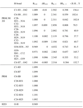
H-bond RX-H ∆RX-H RH...Y δRH…Y <X-H…Y
C1-H3…O42 1.089 -0.01 2.502 0.398 156.4
C6-H8…O11 1.089 0 2.341 0.559 102.6
PRM.3H 2O
C28-H31…N16 1.088 0 2.311 0.042 102.8
C32-H33…N18 1.097 0.009 2.858 0.808 70.5
C28-H30…N19 1.096 0 2.092 0.781 40.9
C28-H29…N19 1.100 0.002 2.119 0.796 37.7
C32-H34…N19 1.092 -0.009 2.104 0.821 40.7
O36-H38…N5 0.969 0 4.032 0.743 81.5
O39-H41…C12 0.971 0.002 2.063 0.657 165.7
C24-H25…O39 1.098 0.006 2.545 0.355 53.2
C6-H7…O42 1.094 -0.005 2.516 0.384 152.7
C1-H3 1.099
C6-H7 1.099
PRM C6-H8 1.089
C28-H31 1.088
C32-H33 1.088
C28-H29 1.098
C32-H34 1.101
C24-H25 1.092
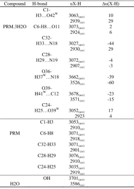
2886 Table 2: THE VIBRATIONAL WAVE NUMBER OF PRM
and PRM.3H2O COMPLEX CORRESPONDING HYDROGEN BONDS
Compound H-bond υX-H ∆υ(X-H)
C1-H3…O42W
3063(asy) 10 2939(sy) 29 PRM.3H2O C6-H8…O11 3073(asy) 2
2924(sy) 6
C32-H33…N18 3027(asy) -44 2930(sy) 29
C28-H29…N19 3072(asy) -4 2907(sy) -3
O36-H37W…N18 3662(asy) -39 3526(sy) -60
O39-H41W…C12 3678(asy) -23 3571(sy) -15
C24-H25…O39W
3052(asy) 17 2923 4 C1-H3 3053(asy)
2910(sy) PRM C6-H8 3071(asy) 2918(sy) C32-H33 3071(asy) 2901(sy) C28-H29 3076(asy) 2910(sy) C24-H25 3035(asy) 2919(sy) OH 3701(asy)
H2O 3586(sy)
The intramolecular C8-H15…N18 hydrogen bonding
interaction in PRM leads to the mixture of C-H stretching with
pyrimidine ring stretching modes. In PRM.3H2O complex the ∆υX-H value of intermolecular hydrogen bonding interaction red
shifted about 44 cm-1 due to the strong O36-H37…N18
intermolecular hydrogen bonding interaction. The C1-H3…O11
hydrogen bonding interaction have blue shifted about 10 cm-1. In
other red shifted hydrogen bonding interaction are C28-H29…N19, O36-H37…N18 andO39-H41…C12 was supported by NBO and
structural analysis.
3.3 NATURAL BOND ANALYSIS
The natural bond orbitals (NBO) calculations were
performed using NBO 3.1 program as implemented in the
Gaussian 09 package at the B3LYP 631-G(d) level in order to
understand various second-order interactions between the filled
orbitals of one subsystem and vacant orbitals of another
subsystem, which is a measure of the intramolecular
delocalization or hyper conjugation.
The second-order Fock-matrix was carried out to
evaluate the donor-acceptor interactions in the NBO basis .
The interactions result in a loss of occupancy from the
localized NBO of the idealized Lewis structure into an
empty non-Lewis orbital. For each donor (i) and acceptor (j)
the stabilization energy E(2) associated with the delocalization i → j is determined as
where, qi - donor orbital occupancy
Ei, Ej - diagonal elements
Fij - the off diagonal NBO Fock matrix element.
NBO analysis provides the most accurate possible 'natural Lewis structure' picture of ‘j’ because all orbital details
are mathematically chosen to include the highest possible
percentage of the electron density. A useful aspect of the NBO
method is that it gives information about interactions of both
filled and virtual orbital spaces that could enhance the analysis of
intra and inter molecular interactions. The electron density of
conjugated single as well as double bond of pyrimidine ring
about 1.976 - 1.987e clearly demonstrate strong
delocalization for Pirimor. The intermolecular interaction are
formed by the orbital overlap between σ(N-C)→σ*(O-C), σ(C-C)→σ*(C-C)σ(C-N)→σ*(C-N) and σ(C-N)→σ*(N-C)
bond orbital which orbital which results intramolecular
charge transfer (ICT) causing stabilization of the system as
seen from the table 3. The stabilization energy contributions from the σ(C13-C14)→σ*(C10-O12) interaction is 8.24kJ/mol.
The hydrogen bonding interaction between the oxygen cone
pair and (C-H) antibonding.ie, σ (O11)→σ*(C6-H8) increases
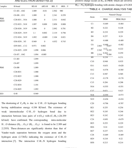
TABLE 3: SECOND ORDER PERTURBATION
THEORY ANALYSIS OF FOCK MATRIX IN NBO BASIS
CORRESPONDING TO THE INTRA MOLECULAR
HYDROGEN BONDS AND HYPERCONJUGATION IN
PRM AND PRM.3H2O COMPLEX INTRACTION
ENERGIES (E2) IN KJ MOL-1 WITH HYBRID ORBITALS
AND ELECTRON DENSITY(E.D)
Complex H-bond RX-H ∆RX-H RH...Y δRH…Y
C1-H3…O42 1.089 -0.01 2.502 0.30.980
C6-H8…O11 1.089 0 2.341 0.559
PRM.
3H2O C28-H31…N16 1.088 0 2.311 0.042
C32-H33…N18 1.097 0.009 2.858 0.808
C28-H30…N19 1.096 0 2.092 0.781
C28-H29…N19 1.1 0.002 2.119 0.796
C32-H34…N19 1.092 -0.009 2.104 0.821
O36-H38…N5 0.969 0 4.032 0.743
O39-H41…C12 0.971 0.002 2.063 0.657
C24-H25…O39 1.098 0.006 2.545 0.355
C6-H7…O42 1.094 -0.005 2.516 0.384
C1-H3 1.099
C6-H7 1.099
PRM C6-H8 1.089
C28-H31 1.088
C32-H33 1.088
C28-H29 1.098
C32-H34 1.101
C24-H25 1.092
H2O O-H 0.969
The shortening of C6-H8 is due to C-H…O hydrogen bonding
having stabilization energy 4.184 KJ/mol. The existence of
weak intermolecular C-H…O hydrogen bond due to
interaction between lone pairs of σ (O12) with (C1-H4 ) (26.359
kJ/mol) have confirmed. The corresponding intra-molecular
H…O distance (H8….O11)/ (H4….O12) is found to be 2.309 and
2.235Å. These distances are significantly shorten than that of
Vander waals seperation between the oxygen atom and the
hydrogen atom (2.720Å) indicating the existence of C-H...O
interaction [7]. The interaction C-H...N hydrogen bonding
between cone pairs (N16) to antibonding σ*(C28-H31) (8.242
kJ/mol) have been confirmed by the results of NBO analysis.
3.4 CHARGE ANALYSIS
All hydrogen atoms have positive charge. Hydrogen in
methyl group is more positive than hydrogen in ring. The largest
positive charge occurs at H33 in PRM, which are involved in C32-H33…N18 hydrogen bonding with atomic charges of 0.263e.
TABLE 4: CHARGE ANALYSIS TABLE
Atom
Natural Charge
PRM PRM.3H2O
C1 -0.469 -0.464
H2 0.222 0.228
H3 0.219 0.229
H4 0.257 0.24
N5 -0.488 -0.489
C6 -0.469 -0.482
H7 0.22 0.222
H8 0.263 0.259
H9 0.218 0.231
C10 0.948 0.935
O11 -0.631 -0.628
O12 -0.578 -0.585
C13 0.587 0.585
C14 -0.178 -0.176
C15 0.284 0.288
N16 -0.555 -0.551
C17 0.621 0.623
N18 -0.559 -0.573
N19 -0.436 -0.434
C20 -0.706 -0.707
H21 0.255 0.256
H22 0.245 0.246
H23 0.242 0.245
C24 -0.691 -0.679
H25 0.253 0.229
H26 0.24 0.243
H27 0.237 0.251
C28 -0.469 -0.469
H29 0.218 0.22
2888
H31 0.262 0.263
C32 -0.469 -0.459
H33 0.264 0.245
H34 0.212 0.216
H35 0.22 0.226
In PRM.3H2O largest positive charge occurs at H31 due
to C28-H31…N16 hydrogen bonding interaction. Charge of N18 is decreased by the influence of C25-H26…N18 hyperconjugative
interaction. It is worthy to mention that C10, C13, C17 and C15
atoms exhibit positive charge while other carbon atoms exhibit
negative charge. The more negative values on C20 and C24 of CH3
group leads to a redistribution of electron density and also
influenced by H23…H27 steric repulsion. The negative charges
located at atoms O11 and O12 will interact with C10 and C13 atoms
due to inductive effect the bond distance N5-C10 (1.364A˚)
increases. The presence of two large electronegative oxygn atom
and one nitrogen atom impose very high charge on carbon atom
(C10) in the carbamate group.
3.5 HIRSHFELD SURFACE ANALYSIS
The Hirshfeld surface analysis, a unique new method of
visualizing the intra molecular interaction, was performed in
order to explore the properties of all inter contacts within the
crystal structure of PRM. Additionally corresponding
two-dimensional (2-D) fingerprint plots [8] provide rapid quantitative
summary of them (the percentage of each contact), which is
important for understanding of the contribution of intra molecular
interactions to the crystal packing. This plot provides information
not only about close contacts, but also about more distant contacts
and areas where the interactions are the weakest. Hirshfeld
surface display all of the inter contacts within the crystal at once
and are therefore ideal for analyzing the crystal packing. The
molecular Hirshfeld surface were constructed on both de (external
distance that is the distance between the Hirshfeld Surface and the
nearest atom of an adjacent molecule.), di (internal distance that is
the distance from the nearest nucleus internal to the calculated
Hirshfeld surface and the vanderWaals radii of the atom, enabling
identification based on the electron distribution calculated as the
sum of spherical atom electron densities [9].The Hirshfeld
surfaces of PRM crystal structure have been mapped over. The de
1.000 to 2.650 A0, the di is 0.961 to 2.650 A0, dnorm is -0.271 to
1.226 A0 for PRM. Fig represents the dnorm, di, de, shape index and
curvedness of PRM. The shape index and curvedness surfaces
have been shown to give the information about each
donor-acceptor pair and to measure how much effectively divides the
surfaces into set of patches respectively.is shown in fig 2.
Fingerprint plots give an idea about interactions with in the
molecule.
3.6 HOMO LUMO ANALYSIS
According to the frontier molecular orbital theory
(FMO) of chemical reactivity, transition of electron is due to
interaction between highest occupied molecular orbital (HOMO)
and lowest unoccupied molecular orbital (LUMO) of reacting
species [10]. EHOMO is a quantum chemical parameter which is
often associated with the electron donating ability of the
molecule. High value of EHOMO is likely to a tendency of the
molecule to donate electrons to appropriate acceptor molecule of
low empty molecular orbital energy [11].
The molecule interacts with other species; hence, they are called the Frontier molecular orbitals (FMO’s). HOMO, which
can be thought the outermost orbital containing electrons, tends to
act as electron donor. On the other hand, LUMO can be thought
the innermost orbital containing free places to accept electrons.
To explain several types of reactions and for predicting the most
reactive position in conjugated systems, molecular orbital and
their properties are used. A molecule having a small frontier
orbital gap is more polarizable and is generally associated with a
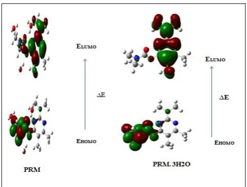
The plots of MO’s (HOMO and LUMO) are shown in
fig.3 and its energy values are given in table 5. All the HOMO
and LUMO have nodes. The nodes in each HOMO and LUMO
are placed symmetrically. The positive phase is red and the
negative is green. In the title compound, the HOMO is
delocalized over the carbamate group. By contrast, the LUMO is
located over the dimethyl amino pyrimidine.
Global Reactivity Descriptors
By using HOMO and LUMO energy values of a
molecule, the global chemical reactivity descriptor of molecules
such as hardness, chemical potential, softness, electronegativity
and electrophilicity index as well as local reactivity have been
defined [13]. The HOMO and LUMO energies, the energy gap
(E), ionization potential (I), electron affinity (A), absolute
electronegativity (χ) absolute hardness (η),and softness of title
compound and its related compounds computed by
DFT/B3LYP/6-31G(d) level.
The chemical potential provide a global reactivity
index and related to charge transfer from a system of higher
chemical potential to lower chemical potential. The reactivity
index is the measure of stabilization in energy when the system
acquires an additional electronic charge (N). A molecule or atom
that has a positive electron affinity is often called an electron
acceptor and may undergo charge transfer reactions. The electron
donating power of a donor molecule is measured by its ionization
potential which is the energy required to remove an electron from
the highest occupied molecular orbital. The overall energy
balance (ΔE), i.e., energy gained or lost, in an electron donor
acceptor transfer is determined by the difference between the
acceptor's electron affinity (EA) and the ionization potential (IP)
as ΔE=EA-IP. Electronegativity is a chemical property that
describes the ability of an atom or a functional group to attract
electrons or electron density towards itself. Parr et al. have
proposed electrophilicity index (ω) as a measure of energy
lowering due to maximal electron flow between donor and
acceptor [14]. The usefulness of this new reactivity quantity has
been recently demonstrated understanding the toxicity of various
pollutants in terms of their reactivity and site selectivity. The
electrophilicity index is positive, definite quantity and direction of
the charge transfer is fully determined by the chemical potential
(V) of the molecule. Because an electrophile is a chemical
species, it has an electron accepting capability from the
environment and its energy must decrease upon accepting
electronic charge, therefore, its electronic chemical potential must be negative. Using Koopman’s theorem for closed shell
compounds the electronegativity and chemical hardness can be
calculated as follows:
Electronegativity
Chemical reactivity,
Softness, σ=1/η
Electrophilicity index, ω = μ2 / 2η
where I and A are Ionisation Potential and Electron Affinity.
Large HOMO-LUMO gap means a hard molecule and small
HOMO-LUMO gap means a soft molecule. One can also relate
the stability of the molecule to hardness, which means that the
molecule with least HOMO-LUMO gap means it is more reactive.
The usefulness of this new reactivity quantitivity has
been recently demonstrated in understanding the toxicity of
various pollutants in terms of their reactivity and site selectivity
[15]. The calculated parameters are shown in Table.5. Global
softness 0.271 shows more chemical active nature of PRM, and
electrophilicity index 11.744, which shows more bio active nature
of PRM.3H2O complex while comparing with PRM while
comparing with PRM.3H2O.
2890
PRM PRM.3H2O
Ionization Potential (I) 8.453 8.42
Electron Affinity (A) 4.705 4.736
Electro negativity (χ) -6.579 -6.578 Chemical potential (η) 6.579 6.578 Hardness 1.874 1.842
Softness 0.267 0.271
electrophilicity index (ω) 11.547 11.744
CONCLUSION
The present study has been performed on Pirimor its
water complex (PRM.3H2O) to investigate the intermolecular
hydrogen bonding interaction to show the insecticidal activity.
The theoretically predicted optimized geometry of Pirimor and its
related compounds by DFT method suggests the possibility of
intermolecular C-H...O and C-H...N hydrogen bonding. The
stretching vibration of dimethyl amino group shows a blue shift
due to the C-H...N hydrogen bonding. The lowering of
Homo-Lumo energy gap of Pirimor while compairing with PRM.3H2O
indicate the charge transfer. The carbon atom in carbamate group
(C10) shows more positive charge which shows charge transfer
from carbamate to dimethyl amino pyrimidine group. NBO
analysis confirms the possibility of C-H...O and C-H...N
hydrogen bonding. The experimental and theoretical vibarational,
NBO, HOMO-LUMO energy gap, global softness and
electrophilicity index shows more insecticidal activity of
PRM.3H2O in comparison with Pirimor compound.
REFERENCES
[1] V. Balachandran, A. Lakshmi, A. Janaki, Spectrochimica Acta Part A: Molecular and Biomolecular Spectroscopy, 81 (2011) 1-7.
[2] Mariana Rocha, Alejandro Di Santo, Juan Marcelo Arias, Diego M. Gil,Aída Ben Altabef, Spectrochimica Acta Part A: Molecular and Biomolecular Spectroscopy, S1386-1425(14)01435-8doi:10.1016/j.saa.2014.09.077.
[3] M.Amalanathan, I.Hubrt Joe,V.K.Rastogi, Journal of Molecular Structure 985(2011) 48-56.
[4] Rong Zhang, Haoran Li, Yi Lei, Shijun Han, Journal of Molecular Structure 693 (2004) 17–25.
[5] Hojin Yang,Tae Ho Kim, Yong Woon ShinKi-Min, Park and Jineun Kim, Acta Cryst. (2010). E66, o1998.
[6] P. Hobza, Z. Havlas, Chem. Rev. 100 (2000) 4253.
[7] V.Santhana Krishnan,S.Sampath Krishnan,S.Muthu, Spectrochimica Acta Part A: Molecular and Biomolecular Spectroscopy 115 (2013) 191–201.
[8] Jack D.Dunitz, Angelo Gavezzotti, Chem.Soc.Rev. 2009,38,2622-2633.
[9] Saikat Kumar Singh, Journal of molecular structure 1064 (2014) 70-75.
[10]C.Ravikumar,L.Padmaja,I.Hubert Joe, Spectrochimica Acta Part A 75(2010) 859-866.
[11]Lynnette Joseph,D.Sajan,K.Chaitanya, Jayakumary Isac, Spectrochimica Acta Part A: Molecular and Biomolecular Spectroscopy 122(2014)375-380.
[12]A.Natraj,V.Balachandran,T.Karthick, Journal of molecular structure 1022 (2012)94-108.
[13]Wajdi.M.Zoghaib,John Husband,Usama A.Soliman, Ibrahim A Shaaban, Tarek A.Mohemed, Spectrochimica Acta Part A: Molecular and Biomolecular Spectroscopy 105(2013) 446-455.
[14]Sameh Guidara, Habib Feki, Younes Abid, , Spectrochimica Acta Part A: Molecular and Biomolecular Spectroscopy 133 (2014) 856-866.