Scholarship@Western
Scholarship@Western
Electronic Thesis and Dissertation Repository
5-7-2013 12:00 AM
A C-terminus Mitochondrial-localization Region and BH3 Domain
A C-terminus Mitochondrial-localization Region and BH3 Domain
of Puma are Required for Apoptotic Function
of Puma are Required for Apoptotic Function
Liz A. Merwin
The University of Western Ontario
Supervisor Dr. Sean Cregan
The University of Western Ontario Graduate Program in Physiology
A thesis submitted in partial fulfillment of the requirements for the degree in Master of Science © Liz A. Merwin 2013
Follow this and additional works at: https://ir.lib.uwo.ca/etd
Part of the Cellular and Molecular Physiology Commons
Recommended Citation Recommended Citation
Merwin, Liz A., "A C-terminus Mitochondrial-localization Region and BH3 Domain of Puma are Required for Apoptotic Function" (2013). Electronic Thesis and Dissertation Repository. 1283.
https://ir.lib.uwo.ca/etd/1283
This Dissertation/Thesis is brought to you for free and open access by Scholarship@Western. It has been accepted for inclusion in Electronic Thesis and Dissertation Repository by an authorized administrator of
OF PUMA ARE REQUIRED FOR APOPTOTIC FUNCTION
(Thesis format: Monograph)
by
Elizabeth A. Merwin
Graduate Program in Physiology
A thesis submitted in partial fulfillment of the requirements for the degree of
Master of Science
The School of Graduate and Postdoctoral Studies The University of Western Ontario
London, Ontario, Canada
Abstract
Apoptosis is known to contribute to the loss of neurological function in brain injury
and several neurodegenerative diseases. The Bcl-2 family of proteins consist of pro-apoptotic
and anti-apoptotic members that interact physically and functionally to regulate apoptosis in
a cell type and stimulus specific manner. We have previously demonstrated that the Bcl-2
family member Puma plays a key role in triggering neuronal apoptosis under diverse stress
conditions. However, the mechanism by which Puma mediates apoptosis remains unclear.
Here we found that Puma contains two domains required for its apoptotic function: the BH3
domain and a region in the c-terminus that regulates its localization to the mitochondria. We
found that the BH3 domain is required to bind both the anti-apoptotic protein, Bcl-xl, and the
pro-apoptotic protein, Bax. However, we found that the BH3 domain of the protein is not
required for its mitochondrial-localization. Instead, we identified a region within the
c-terminus of Puma that is essential for both mitochondrial-localization and apoptotic function
of the protein. Although we found that the c-terminus of Puma is not required to bind Bcl-xl
or Bax in vitro, it is required for co-localization and likely interaction with these proteins in
the cellular context. In summary, we determined that Puma requires both its c-terminal
mitochondrial-localization sequence and its BH3 domain to co-localize with and bind to
Bcl-2 family members, and to induce neuronal cell death.
Keywords
Apoptosis, Bax, Bcl-2, Bcl-xl, BH3-only, BH3 domain, c-terminus, Mitochondrial localization, Protein interaction, Puma
Acknowledgements
I would like to thank Dr. Sean Cregan, for his supervision and guidance throughout this project. I would also like to express appreciation to my committee: Dr. Stephen Pasternak, Dr. Caroline Schild-Poulter, and Dr. Timothy Regnault, for their advice and support.
Thanks also to my lab members: Kristin Ambacher, Lee-Anne Fochesato, Lisa Foris, Jenn Guadagno, Sarah Humphrey, Meera Karajgikar, and Pat Swan, as well as to Fabiana Caetano, Lianne Dale, and Stephanie Kulhawy, for your patient instruction and kind assistance.
Thank you, Sile, for your constant love and support, especially on the nights I was in the lab at 3a.m. Thank you, Jericho and Roen, for reminding me to find joy and excitement in science. Much gratitude to all my friends and family, for encouraging me throughout this process.
Table of Contents
Abstract...ii
Acknowledgements...iii
Table of Contents...iv
List of Tables...vi
List of Figures...vii
List of Abbreviations...viii
Preface...ix
Chapter 1 – Introduction...1
1.1 – Apoptosis ...1
1.11 - Intrinsic apoptotic pathway ...2
1.2 - Bcl-2 Family Members ...5
1.21 - Pro-apoptotic Bcl-2 Family Members ...8
1.211 - BH3-only Bcl-2 family members ...8
1.212 - Multi-BH domain Bcl-2 family members ...10
1.22 - Anti-apoptotic Bcl-2 Family Members ...11
1.3 – Bcl-2 family members in neuronal apoptosis ...12
1.4 - p53 Up-regulated Mediator of Apoptosis (Puma) ...15
1.5 – Bcl-2-associated x protein (Bax) ...18
1.6 - B-cell lymphoma extra-long (Bcl-xl) ...28
1.7 – Rationale ...30
1.8 - Hypothesis and Objectives ...31
Chapter 2 – Materials and Methods...32
2.1 – Cell Culture...32
2.2 – Plasmid constructs...33
2.3 - Transfection...37
2.5 - Peptide treatment...38
2.6 – Cell death assesment by Hoechst 33342 and Propidium Iodide staining ...38
2.7 - Mitotracker ® Red CMXRos staining...39
2.8 - Immunofluorescence...40
2.9 – Co-immunoprecipitation ...41
2.10 - Western Blot...42
2.11 - Data Analysis...43
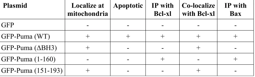
Chapter 3 - Results...44
3.1 – Interaction with other BCL-2 family proteins is required for Puma's apoptotic activity but not its localization... 3.2 - Identification of a potential membrane targeting sequence in the Puma protein..48
3.3 - Analysis of protein expression confirms that GFP plasmids express proteins at equal levels and expected molecular weights. ...51
3.4 – An element of the Puma c-terminus is required and sufficient for cellular localization of Puma...54
3.5 – Puma c-terminus is required and sufficient for mitochondrial-localization...57
3.6 – Both the BH3 domain and a distinct c-terminal domain of Puma are required for Puma-induced apoptosis...64
3.7 – Puma BH3 domain, but not Puma c-terminus, is required to bind to Bcl-xl in vitro...70
...74
3.9 - Puma BH3-domain, but not Puma c-terminus, is required to bind to Bax in vitro. ...78
Chapter 4 - Discussion...81
4.1 - Identification of two important regions in the Puma protein ...81
4.2 – Puma mitochondrial localization is regulated by a c-terminal targeting region but not its BH3 domain or Bcl-2 family interactions ...84
4.3 – Both the BH3 domain and c-terminal mitochondrial-localization region of Puma are required for Puma-mediated cell death and apoptosis ...88
Chapter 5 – Summary and Conclusions...94
References...95
Appendices ...106
Curriculum Vitae ...111
List of Tables
Table 1 - Restriction Enzyme Digest ...100
Table 2 - Ligation...101
Table 3 - Polymerase Chain Reaction Cloning ...102
Table 4 - Summary of plasmid properties ...103
Table 5 – Peptide Sequences...104
List of Figures
Figure 1 – The Intrinsic Apoptotic Pathway ...4
Figure 2 – Bcl-2 Family Members ...7
Figure 3 – Bax Activation Models ...27
Figure 5 – GFP-Puma plasmids ...36
Figure 6 – Hα1 tat-peptide interferes with Puma-induced apoptosis but not its mitochondrial-localization ...47
Figure 7 - Regions of interest in the Puma protein ...50
Figure 8 – Expression of GFP-Puma plasmids ...53
Figure 9 - Puma c-terminus is required for cellular localization ...56
Figure 10 - Co-localization of different GFP-Puma proteins with Mitotracker ® Red CMXRos ...59
Figure 11 - Co-localization of different GFP-Puma proteins with Tomm20 ...63
Figure 12 - Differing levels of apoptosis induced by various GFP-Puma proteins ...66
Figure 13 - Differing levels of cell death induced by various GFP-Puma proteins ...69
Figure 14 - Puma BH3 domain, but not Puma c-terminus, is required to bind to Bcl-xl in vitro ...73
Figure 15 - Puma c-terminus is required for co-localization with Bcl-xl in N2A cells in situ ...77
Figure 16 - Puma BH3-domain, but not Puma c-terminus, is required to bind to Bax in vitro ...80
List of Abbreviations
APAF-1 - Apoptosis protease-activating factor 1
ART - Apoptosis regulating target
Bax – Bcl-2 associated X-protein
Bcl-2 – B-cell lymphoma type 2
Bcl-xl – B-cell lymphoma extra-large
BH – Bcl-2 Homology
BH3 - Bcl-2 homology domain three
CLAC - Cytochrome-c Liberation Associated Conformation
CLIC - Cytosolic Locked In Conformation
co-IP - co-immunoprecipitation
D33A – aspartate 33 to alanine
ER – endoplasmic reticulum
Hα1 – helix alpha 1
MESSA - MEta Server for protein Sequence Analysis
MOM – Mitochondrial outer membrane
N2A – Neuro2A
PCR – polymerase chain reaction
PI - Propidium Iodide
Puma – p53 up-regulated mediator of apoptosis
UV - ultra-violet
Preface
Apoptosis plays an crucial role in normal development and maintenance of tissue
homeostasis. It is required to eliminate unnecessary tissue during development, and later on
in an organism's lifespan it is necessary to destroy damaged or mutated cells (Kerr et al.
1972; Oppenheim 1991; Yoshida et al. 1998). One of the reasons that apoptosis is utilized
for these functions is that unlike other modes of cell death, such as necrosis, apoptosis
eliminates cells without eliciting an inflammatory response or causing damage to the
surrounding tissue (Kerr et al. 1972). Disruption of the normal apoptotic process in animal
models has been shown to cause significant and often lethal defects (Ashkenazi & Dixit
1998; Li et al. 2000; Motoyama et al. 1995; Strasser et al. 1990; Yoshida et al. 1998).
Perhaps due to its vital role in normal tissue, dysregulation of the apoptotic process
also plays a major role in many diseases. Many cancer cells contain mutations in the
apoptotic machinery that allow them to continue to grow when they would otherwise die
(Adams & Cory 2007; Cory et al. 1999; Strasser et al. 1990; Yu & Zhang 2009; Vaux et al.
1988). Activation of apoptosis by acute trauma, as in ischemic injury or spinal cord injury,
can result in significant neuronal loss (Engel et al. 2011; Kotipatruni et al. 2011; Nitatori et
al. 1995; Steckley et al. 2007). Apoptotic cell death also plays a significant role in many
neurodegenerative conditions, including Alzheimer's disease, Parkinson's disease, and
Huntington's disease (Higgins et al. 2010; Portera-Cailliau et al. 1995; Vila & Przedborski
2003). Because it plays such a crucial role in both healthy and diseased tissue, a more
complete understanding of the key proteins involved in the regulation of apoptosis will be
important for the development of new treatments applicable to a variety of injury and disease
conditions.
Chapter 1 – Introduction
1.1 – Apoptosis
Apoptosis, or programmed cell death, is perhaps the most prevalent and
well-regulated means of cell death. The cell must precisely coordinate the actions of many
proteins in order to destroy itself without causing damage to surrounding cells. This
precise coordination presents a distinctly identifiable cellular morphology. In the initial
stages of apoptosis, the cellular membrane shrinks and develops a rounded appearance
(Kerr et al. 1972). At the same time, the chromatin condenses in the now shrinking
nucleus, in a process termed pyknosis (Kerr et al. 1972). The cellular membrane then
takes on the blebbed appearance characteristic of apoptotic cells (Kerr et al. 1972). In the
final stage of apoptosis, the cell packages its remaining contents into small vesicles,
known as apoptotic bodies, that can be easily engulfed by phagocytic cells for
degradation (Kerr et al. 1972).
This carefully contained process can be triggered by both extracellular (extrinsic
pathway) and intracellular (intrinsic pathway) cues. In the extrinsic apoptotic pathway,
extracellular ligands bind to death receptors on the cellular surface in order to activate
apoptosis (Ashkenazi & Dixit 1998). In the intrinsic apoptotic pathway, cellular stress
causes expression or activation of apoptosis-inducing factors (Ashkenazi & Dixit 1998;
Cory & Adams 2002). These factors activate pro-apoptotic members of the B-cell
membrane (Cory & Adams 2002). The primary focus of this thesis is the intrinsic
apoptotic pathway.
1.11 - Intrinsic apoptotic pathway
The intrinsic apoptotic pathway is activated by many different cellular stressors,
including but not limited to: genotoxic, endoplasmic reticulum (ER), and oxidative stress
(Engel et al. 2011; Galehdar et al. 2010; Reimertz et al 2003;Steckley et al. 2007). The
B-cell lymphoma 2 (Bcl-2) family of proteins plays a key role in the regulation of the
intrinsic apoptotic pathway, and downstream caspases are primarily responsible for the
execution of the apoptotic process (Cory & Adams 2002; Salvesen & Dixit 1997).
Activation of the pro-apoptotic Bcl-2 family members results in mitochondrial outer
membrane permeabilization and releases pro-death factors, such as cytochrome-c, into
the cytoplasm (Figure 1; Cory & Adams 2002; Li et al. 1997; Yoshida et al. 1998). Once
released into the cytoplasm, cytochrome-c forms a complex with apoptosis
protease-activating factor 1 (APAF-1) and caspase-9 known as the apoptosome (Figure 1; Li et al.
1997; Liu et al. 1996). The apoptosome cleaves and thereby activates caspase-9, which
then cleaves and activates the downstream effector caspases, caspase-3 and caspase-7
(Figure 1; Li et al. 1997). This caspase cascade is responsible for the characteristic
apoptotic events, including: chromatin condensation and fragmentation, membrane
blebbing, and packaging of cellular remains into apoptotic bodies (Cory & Adams 2002;
Multi-domain family members (e.g. Bax) Anti-apoptotic
Bcl-2 family members
BH3-only family members Cellular
stressor
Bax
Bax Pro-death factors
(e.g. cytochrome-c, APAF-1)
Mitochondrion
Apoptosome Caspase-9
Caspase-9 Caspase-3
Caspase-7
Caspase-7
1.2 - Bcl-2 Family Members
The B-cell lymphoma 2 (Bcl-2) family of proteins are key regulators of the
intrinsic apoptotic pathway (Cory & Adams 2002; Engel et al. 2011). These proteins
share one or more Bcl-2 homology (BH) domains with their namesake, Bcl-2. Although
they have different amino acid sequences and contain different BH domains, their
three-dimensional fold structures are strikingly similar. Most Bcl-2 family members have two
central hydrophobic alpha-helices surrounded by six or seven amphipathic alpha-helices
(Petros et al. 2004). This similarity in structure may help to explain their ability to form
homo and hetero-dimers with other family members (Petros et al. 2004).
The Bcl-2 family members are divided based on their functions into two main
categories: the pro-apoptotic family members and the anti-apoptotic family members
(Cory & Adams 2002). The pro-apoptotic family members are further sub-divided into
Bcl-2 homology domain three (BH3) only proteins and multi-BH domain proteins (Cory
& Adams 2002). Altering the expression or activation of one of the family members can
kill cells or render them resistant to apoptotic cell death (Cory et al. 1999; Motoyama et
BH3
BH3 BH3
BH3-only: Bid, Puma, Bim, Bik, Bad, Bmf, Hrk & Noxa
pro-apoptotic anti-apoptotic:
2, xl, Bcl-w, Mcl-1 & A1
multi-BH domain: Bax, Bak & Bok BH2
BH1 BH4
1.21 - Pro-apoptotic Bcl-2 Family Members
As suggested by their name, the pro-apoptotic members of the Bcl-2 family of
proteins act to promote apoptosis. They are subdivided into BH3-only proteins and
multi-domain proteins. The BH3-only proteins have only their BH3 domain in common
with Bcl-2 and are the first response to cellular stress (Engel et al. 2011). Expression and
activation of the BH3-only proteins results in the activation of the multi-domain Bcl-2
family members (Engel et al. 2011). The multi-domain Bcl-2 proteins share between one
and three of the Bcl-2 homology domains and act by forming channels in the
mitochondrial outer membrane and allowing the release of of pro-death factors, such as
cytochrome-c (Cory & Adams 2002; Lalier et al. 2007; Li et al. 1997).
1.211 - BH3-only Bcl-2 family members
The BH3-only members of the pro-apoptotic Bcl-2 proteins consist of: Puma,
Noxa, Bid, Bim, Bad, Bmf, and Hrk (Cory & Adams 2002; Engel et al. 2011). Although
they are structurally similar, the BH3-only proteins are regulated by diverse mechanisms.
Some, such as Puma, are regulated mainly through transcription; others, such as Bid, are
regulated mainly post-transcriptionally via proteolytic cleavage (Cory & Adams 2002;
Engel et al. 2011). Their affinity for binding to the anti-apoptotic proteins is also quite
diverse; some BH3-only proteins, such as Noxa and Bid, are able to bind to only some of
the anti-apoptotic proteins, whereas others, such as Puma, are able to bind to all of the
anti-apoptotic proteins (Kuwana et al. 2005; Uren et al. 2007). The BH3-only proteins
protein that is highly efficacious and of almost universal importance is Puma (Cory &
Adams 2002; Engel et al. 2011; Hikisz et al. 2012).
In accordance with their name, the BH3-only proteins contain only the third of the
Bcl-2 homology domains. It is a short, alpha-helical, amphipathic domain approximately
9-16 amino acids long (Adams & Cory 1998; Chittenden et al. 1995; Cory & Adams
2002; Engel et al 2011). The BH3 domain is the site of protein interactions (the binding
site for either anti-apoptotic or multi-domain proapoptotic Bcl-2 family members) and is
required for the apoptotic activity of BH3-only proteins (Chittenden et al. 1995; Engel et
al. 2011; Kazi et al. 2011; Kelekar & Thompson 1998; Liu et al. 2009).
Two main mechanisms of action have been proposed for these proteins: the direct
activation model and the indirect activation model. In the direct activation model, one of
the BH3-only proteins directly binds to and activates one of the multi-BH domain
pro-apoptotic Bcl-2 family members (Chipuk & Green, 2008; Cory & Adams, 2002; Engel et
al. 2011). In the indirect activation model, a BH3-only protein binds to one of the
anti-apoptotic family members and displaces a multi-BH domain pro-anti-apoptotic Bcl-2 family
member (Chipuk & Green 2008; Cory & Adams, 2002; Engel et al. 2011). Once the
multi-BH domain pro-apoptotic Bcl-2 family member is freed from the anti-apoptotic
protein, a few methods of activation have been suggested. Some believe that once
released, the proteins can bind to membrane-bound, otherwise activated multi-BH
domain pro-apoptotic Bcl-2 family members and form oligomers (Kazi et al. 2011;
bind directly to the multi-BH domain pro-apoptotic Bcl-2 family members once they are
released from the anti-apoptotic proteins (Cory & Adams 2002). It has also been
suggested that the binding and release of multi-BH domain pro-apoptotic Bcl-2 family
members from anti-apoptotic proteins may in itself be sufficient to activate them (Gautier
et al. 2011; Moreau et al. 2003).
There is evidence for both models, and it seems most likely that each of the
BH3-only proteins functions slightly differently, allowing the cell to precisely regulate
apoptosis. Some BH3 proteins, such as Bid, are widely accepted as direct activators of
multi-BH domain pro-apoptotic Bcl-2 family members (Desagher et al. 1999; Eskes et
al. 2000; Lovell et al. 2008). Others, such as Bad, appear to function only through the
indirect model (Lalier et al. 2007). The conflicting evidence for other BH3-only proteins,
such as Puma (Gallenne et al 2009; Gautier et al. 2011; Ming et al. 2006; Willis et al.
2007), may indicate that they function via a combination of both the indirect and direct
activation models (Du et al. 2011; Engel et al. 2011; Kuwana et al. 2005; Steckley et al.
2007; Zhang et al. 2009).
1.212 - Multi-BH domain Bcl-2 family members
There are three multi-BH domain members of the pro-apoptotic Bcl-2 proteins:
Bax, Bak, and Bok (Adams & Cory 1998; Cory & Adams, 2002). They contain the first
three BH domains: BH1, BH2, and BH3 (Cory & Adams, 2002; Petros et al. 2004).
amphipathic alpha-helices (Petros et al. 2004). This structure allows them to exist in one
conformation in the cytosol and to form oligomeric channels in the mitochondrial outer
membrane when activated (Chipuk & Green 2008; Lalier et al. 2007).
Bax, Bak, and Bok are typically activated by the BH3-only proteins, either
through a direct or an indirect interaction (Chipuk & Green 2008). Once activated, they
oligomerize and form channels in the outer mitochondrial membrane (Cory & Adams
2002; Lalier et al., 2007). These channels permeabilize the outer mitochondrial
membrane and allow the release of pro-apoptotic factors, such as cytochrome-c, into the
cytoplasm. When released into the cytoplasm, these pro-death factors are free to cleave
and activate caspase-9 (Cory & Adams 2002; Li et al. 1997). Once activated, caspase-9
activates downstream caspases, resulting in apoptotic cell death (Li et al. 1997; Liu et al.
1996).
1.22 - Anti-apoptotic Bcl-2 Family Members
The anti-apoptotic Bcl-2 family members consist of: Bcl-xl, Bcl-2, Bcl-w, A1,
and Mcl-1 (Adams & Cory 1998; Cory & Adams 2002). They contain all four of the BH
domains as well as a c-terminal membrane-localization sequence (Lindsay et al. 2011;
Petros et al. 2004; Reed et al. 1996). Their three-dimensional structure consists of two
hydrophobic alpha-helices surrounded by five amphipathic alpha-helices (Petros et al.
2004). The first three BH domains form a hydrophobic binding pocket that is responsible
(Petros et al. 2004; Sattler et al. 1997). Some of the anti-apoptotic family members have
also been shown to form soluble homodimers in the cytoplasm by sequestering their
c-terminal membrane-localization sequence in this hydrophobic pocket until activated
(Jeong et al. 2004; Petros et al. 2004).
Anti-apoptotic Bcl-2 family proteins function primarily by binding to and
restricting both the BH3-only and multi-domain pro-apoptotic Bcl-2 family members.
When anti-apoptotic proteins bind to the BH3-only proteins, it prevents them from
binding to and/or activating the multi-domain pro-apoptotic Bcl-2 family members
(Nakano & Vousden 2001; Steckley et al. 2007; Yu et al. 2001). When anti-apoptotic
Bcl-2 proteins bind to the multi-domain pro-apoptotic Bcl-2 family members, it prevents
the activation and/or oligimerization of the multi-BH domain pro-apoptotic proteins
(Gallenne et al. 2009; Gautier et al. 2011; Jeong et al. 2004; Sattler et al. 1997).
1.3 – Bcl-2 family members in neuronal apoptosis
The wide variety of apoptotic regulators seems to suggest that each protein may
be of differing importance in various cell types and for various cell-death stimuli. There
is growing evidence to indicate that different cell types may regulate apoptosis in distinct
ways (Chen et al. 2005; Chipuk et al. 2010; Cory & Adams 2002; Engel et al. 2011;
Hikisz et al. 2012; Kim et al. 2006; Kuwana et al. 2005; Letai et al. 2002). For this
reason it is increasingly important to determine the roles of the Bcl-2 family proteins in
important role in a number of neurodegenerative disorders including Alzheimer's and
Huntington's disease, as well as in acute conditions, such as stroke and traumatic brain
injury (Engel et al. 2011; Higgins et al. 2010; Portera-Cailliau 1995; Vila & Przedborski
2003). The Bcl-2 family of proteins appears to play an important role in the regulation of
neuronal apoptosis under these conditions (Boise et al. 1993; Frankowski et al. 1995;
Galehdar et al. 2010; Kotipatruni et al. 2011; Rubin et al. 1994; Steckley et al. 2007).
Anti-apoptotic members of the Bcl-2 family have been shown to inhibit neuronal
cell death induced by a number of different stimuli, including: neurotrophic factor
withdrawal, reactive oxygen species, and ischemia (Allsopp et al. 1993; Garcia et al.
1992; Martinou et al. 1994; Zhong et al. 1993). Of the anti-apoptotic Bcl-2 family
members, Bcl-xl appears to play a particularly important role in preventing neuronal
apoptosis (Boise et al. 1993; Frankowski et al. 1995; Rubin et al. 1994).
Of course, the role of the pro-apoptotic members of the Bcl-2 family in neuronal
apoptosis cannot be neglected. The multi-domain pro-apoptotic Bcl-2 family member,
Bax, has been shown to mediate neuronal apoptosis in a rat model of spinal cord injury
(Kotipatruni et al. 2011), and neuronal apoptosis induced by oxidative stress (Steckley et
al. 2007). The pro-apoptotic BH3-only protein, Puma, has been shown to be of
particular importance in neuronal death induced by a number of factors, including:
trophic factor deprivation, endoplasmic reticulum stress, ischemia, and oxidative stress
(Ambacher et al. 2012; Galehdar et al. 2010; Niizuma et al. 2009; Reimertz et al. 2003;
regulators of neuronal apoptosis in a number of conditions, both chronic (as in the case of
neurodegenerative disorders) and acute (as with stroke or spinal cord injury).
The Bcl-2 family of proteins has also been shown to play a crucial role in the
regulation of neuronal apoptosis during development. Anti-apoptotic Bcl-2 family
members are highly expressed in developing neurons (Abe-Dohmae 1993; Frankowski et
al. 1995). Bcl-xl knockout mice show increased neuronal apoptosis (Motoyama et
al.1995) that can be mitigated by also knocking out the pro-apoptotic protein, Bax
(Shindler et al. 1997). The pro-apoptotic members of the Bcl-2 family also play an
important role in normal neuronal development. Bax/Bak double knockout mice have a
number of neurological deficits, including apparent deafness and seizures (Lindsten et al.
2000). Histologically, the brains of these mice display accumulations of small,
undifferentiated neuronal cells, most notably in the periventricular area, the
hippocampus, the cerebellum, and the olfactory bulb; all regions where neural stem cells
are common (Lindsten et al. 2000). Bax knockout mice show decreased sympathetic and
neuronal apoptosis during development and are resistant to neuronal apoptosis induced
by trophic factor deprivation (Deckwerth et al. 1996). The Bcl-2 family of proteins is
also critical in maintaining tissue homeostasis by preventing the accumulation of
damaged or dysfunctional cells (Adams & Cory 2007; Cory et al. 1999; Cory & Adams
2002). The pro-apoptotic members of the Bcl-2 family, most notably Puma, are activated
by oncogenic stress in order to safely eliminate cancerous and potentially cancerous cells
(Dudgeon et al. 2010; Ming et al. 2006; Yu et al. 2001; Yu et al. 2003). The
regulation of neuronal apoptosis, both during development and for maintenance of
homeostasis.
1.4 - p53 Up-regulated Mediator of Apoptosis (Puma)
p53 up-regulated mediator of apoptosis (Puma) is perhaps the most prevalent and
effective of the BH3-only Bcl-2 family members. Puma has been shown to play an
essential role in apoptosis induced by many different factors, including: oxidative stress,
genotoxic stress, endoplasmic reticulum (ER) stress, trophic-factor withdrawal, and
oncogenesis (Ambacher et al. 2012; Engel, T et al. 2011; Galehdar et al. 2010; Niizuma
et al 2009; Reimertz et al. 2003; Steckley et al. 2007). Although other BH3-only
proteins appear to contribute to neuronal apoptosis in some of these conditions, none of
them are as universally essential as Puma. Even under conditions where other BH3-only
proteins are expressed, Puma still seems to be the essential factor for apoptosis to occur
(Ambacher et al. 2012; Galehdar et al. 2010; Niizuma et al 2009; Reimertz et al. 2003;
Steckley et al. 2007).
Puma was identified by several independent groups at almost the same time (Han
et al. 2001; Nakano & Vousden 2001; Yu et al. 2001). As suggested by its name, Puma
is transcriptionally up-regulated by the apoptotic protein p53 (Cregan et al 2004; Nakano
& Vousden 2001; Yu et al. 2001), although it has also been shown to be induced
independently of p53 expression (Wu et al. 2007; Yu & Zhang 2009). Other
Ray et al. 2011), FoxO3a (Dudgeon et al. 2010; You et al. 2006), CHOP (Galehdar 2010;
Wu et al. 2007), E2F1 (Hershko & Ginsberg 2004; Ray et al. 2011), and c-Myc
(Fernandez et al. 2003). This variety of regulators allows Puma expression to be
controlled specifically in different cell types and under different forms of cell stress.
Puma is present at very low levels in healthy cells and is transcribed when the cell
experiences a wide variety of stressors (Nakano & Vousden 2001; Puthalakath & Strasser
2002; Yu et al. 2001). The transcriptional up-regulation of Puma has been widely
accepted as its main form of regulation, but recent evidence suggests that it may also be
post-transcriptionally regulated by phosphorylation (Fricker et al. 2010; Lam et al. 2009;
Puthalakath & Strasser 2002; Sandow et al. 2012). Phosphorylation of Puma at
serine-ten has been shown to target the protein for degradation and reduce Puma-induced
apoptosis (Fricker et al.2010; Sandow et al. 2012). Although other phosphorylation sites
have been identified, their function in regulating Puma has not yet been determined
(Hikisz & Kilianska 2012; Fricker et al. 2010; Lam et al. 2009; Puthalakath & Strasser
2002; Sandow et al. 2012).
There are two main splice-variants of Puma produced by cells: Puma-α and
Puma-β (Nakano & Vousden 2001; Yu et al. 2001). Initially, these variants were thought
to produce proteins that were similar in size and structure, both of which had full
pro-apoptotic activity (Nakano & Vousden 2001; Yu et al. 2001). However, recent evidence
has emerged to suggest that Puma-β may be smaller than Puma-α and lack full apoptotic
transcribe proteins without the BH3 domain or the full length c-terminus (Nakano &
Vousden 2001; Yu et al. 2001). These proteins lack the apoptotic activity of full-length
Puma and their cellular function is unknown, though perhaps also negligible because they
are produced at such low levels (Nakano & Vousden 2001; Yu et al. 2001).
Like the other BH3-only proteins, Puma contains a BH3 domain that is required
for its interaction with other Bcl-2 family members (Day et al. 2008; Nakano & Vousden
2001; Yu et al. 2001). It is amphipathic and alpha-helical in structure, spanning amino
acids 137 to 150 of the protein (Day et al. 2008). The BH3 domain of Puma also has a
similar function to the BH3 domains of the other BH3-only proteins; it is required to bind
to the anti-apoptotic Bcl-2 proteins, Bcl-xl, Bcl-2, Bcl-w, A1, and Mcl-1 (Day et al.
2008; Gautier et al. 2011; Nakano & Vousden 2001; Yu et al. 2001). The BH3 domain
of Puma is the site of Puma's putative direct interaction with the multi-BH domain
pro-apoptotic Bcl-2 family members. The BH3 domains of Bid and Bim, two of the other
BH3-only proteins, have been shown to interact directly with the multi-BH domain
pro-apoptotic protein, Bax (Kuwana et al. 2005; Lovell et al. 2008). Many groups have
found evidence that the BH3 domain of Puma can directly bind to and activate Bax in the
same manner (Cartron et al. 2004; Du et al. 2011; Gallenne et al. 2009; Steckley et al.
2007; Yee & Vousden 2008; Zhang et al. 2009). However, unlike the other members of
the Bcl-2 family, Puma does not contain a classic c-terminal membrane localization
sequence (Nakano & Vousden 2001; Yee & Vousden 2008; Yu et al. 2001; Yu et al.
involved and their exact function have not been well-defined (Hikisz & Kilianska 2012;
Nakano & Vousden 2001; Yee & Vousden 2008; Yu et al. 2001).
The reasons for the importance of Puma in cellular apoptosis are currently
unclear. It has been suggested that the reason for Puma's efficacy is its ability to bind all
of the anti-apoptotic family members, or that Puma may bind with greater strength than
the other Bcl-2 family members (Chen et al. 2005; Kuwana et al. 2005; Yu & Zhang
2009). One important difference between Puma and the other BH3-only proteins is that
Puma can bind directly to all of the anti-apoptotic Bcl-2 family members with high
affinity (Chen et al. 2005; Kuwana 2005). This is likely part of the reason that Puma is
such an efficacious and important protein in neuronal apoptosis. However, even these
ideas fail to explain what it is about Puma that enables it to be such an effective and
essential mediator of apoptosis.
1.5 – Bcl-2-associated x protein (Bax)
Bcl-2-associated x protein (Bax) is a multi-domain pro-apoptotic Bcl-2 family
member. Similar in structure to the other Bcl-2 family members, Bax consists of two
hydrophobic alpha-helices surrounded by seven amphipathic alpha-helices (Lalier et al.
2007; Petros et al. 2004; Suzuki et al. 2000). Like the other multi-domain pro-apoptotic
members of the Bcl-2 family, Bax contains three BH domains: BH1, BH2, and BH3
(Petros et al. 2004; Suzuki et al. 2000). Several regions of Bax have been shown to be
central helices, Hα5 and Hα6; the n-terminus; and the c-terminus (Carton et al. 2004;
Lalier et al. 2007; Reed et al.1996; Suzuki et al. 2000; Wolter et al. 1997).
The number of important domains in Bax is unsurprising, considering the overall
significance of the protein in apoptosis. Our lab and others have shown that Bax plays a
particularly important role in Puma-mediated neuronal apoptosis (Engel et al, 2011;
Shindler et al. 1997; Steckley et al. 2007). Under normal conditions, Bax is present in
the cellular cytoplasm in an inactive conformation. Upon activation, Bax translocates to
the mitochondrial outer membrane (MOM) and forms homo- or hetero-oligomers with
other Bax proteins or with Bak. These oligomers act as channels in the MOM and allow
the release of cytochrome-c and other death factors into the cytoplasm.
Like most of the BH3 domains found in Bcl-2 family members, the BH3 domain
of Bax has been implicated in its interactions with other Bcl-2 family proteins. A peptide
modeled on the BH3 domain of Bax has been shown to interact with the hydrophobic
binding pocket on the anti-apoptotic proteins, Bcl-xl and Bcl-2; the same peptide also
interferes with Bax binding to Bcl-xl and Bcl-2 (Moreau et al. 2003). Mutations of the
BH3 domain of Bax appear to interfere with its ability to bind Bcl-xl, Bcl-2, and Mcl-1
(Cartron et al. 2004; Wang et al. 1998). The Bax BH3 region also seems to be necessary
for its interaction with and activation by the pro-apoptotic BH3-only protein, Bad
(Cartron et al. 2004). However, the BH3 region of Bax does not appear to be responsible
Indeed, the BH3-only proteins Puma and Bid appear to interact with Bax via its
Hα1 domain rather than its BH3 domain (Cartron et al. 2004). The first alpha helix at the
n-terminus of Bax, Hα1, is one of the amphipathic helices sequestered in the unactivated,
cytoplasmic conformation of the protein. It has been suggested that the exposure of the
Hα1 domain may be a critical step in the activation and conformational change of Bax
(Lalier et al. 2007). The aspartate at position 33 of Bax is required for Bax to interact
with the BH3 regions of Bid and Puma; mutation of this acidic residue into a neutral
residue, alanine, abrogates these interactions (Cartron et al. 2004). However, the Hα1
domain of Bax is not required for its interactions with Bcl-xl and Bcl-2, and mutation of
the Hα1 domain does not affect the ability of Bax to bind the anti-apoptotic proteins
Bcl-xl and Bcl-2 (Cartron et al. 2004).
Although they do not appear to play a role in protein interactions, the central
helices of Bax, Hα5 and Hα6, are important for the pro-apoptotic function of the protein.
These central hydrophobic alpha-helices are tightly-sequestered in the inactive
cytoplasmic conformation of Bax, but are exposed upon its activation (Lalier et al. 2007;
Suzuki et al. 2000). Their resemblance to the pore-forming domains of diptheria toxin
and colicins inspired research into their potential for a similar function in Bax (Nouraini
et al. 2000). Structurally, Hα5 and Hα6 of Bax appear to be trans-membrane domains;
peptides based on these helices have been shown to form pores in a synthetic lipid
membrane (Annis et al. 2005; Garcia-Saez et al. 2006). Although Hα5 and Hα6 are
required to form the channel in the mitochondrial outer membrane that allows activated
localization to the mitochondria (Carton et al. 2005; Lalier et al. 2007). Instead, the
localization of Bax appears to be regulated by both its c-terminus and its n-terminus.
At the c-terminus of Bax, its ninth alpha-helix, Hα9, occupies the hydrophobic
binding pocket formed by the first three BH domains of Bax in its inactive conformation
(Suzuki et al. 2000). Activation of Bax exposes this helix, which resembles the
c-terminal membrane-localization domains in Bcl-xl and Bcl-2 (Lalier et al. 2007). The
c-terminus of Bax has been shown to play an important role in the localization and function
of the protein (Gardia et al. 2004; Nechustan et al. 1999). However, some studies have
demonstrated that Bax retains its ability to translocate to the mitochondria despite
c-terminus deletion (Antonsson et al. 2000; Carton et al. 2003). Perhaps this is because the
n-terminus of Bax also appears to play a role in the mitochondrial-localization of the
protein (Carton et al. 2003). The n-terminus of Bax has been shown to contain a
mitochondrial-localization sequence, and deletion of this sequence has been shown to
abrogate its localization (Carton et al. 2003). Interestingly, the first 20 amino acids of
Bax appear to inhibit its mitochondrial-localization, and have been termed the Apoptosis
Regulating Target (ART) domain (Goping et al. 1998). Deletion of this domain results in
a protein constitutively localized to the mitochondria (Goping et al. 1998). However, the
complete n-terminus alone is unable to insert into the mitochondrial outer membrane
(Carton et al. 2003). It appears that the n-terminus and the c-terminus of Bax co-operate
Mitochondrial localization is of particular importance in the apoptotic function of
Bax. Without its ability to localize to the mitochondrial outer membrane, Bax cannot
induce apoptosis. The precise reasons for the importance of membrane-localization in
the apoptotic function of Bax are still a subject of investigation, but some recent studies
have suggested a role for the membrane in protein interactions (Bogner et al. 2010;
Lalier, L. et al. 2007, Lovell, J.F. et al. 2008; Roucou et al. 2002; Yethon et al. 2003).
The BH3-only protein, tBid, requires the presence of a membrane in order to bind to and
activate Bax (Lovell, J.F. et al. 2008). It has been suggested that contact with the
membrane may open the conformation of Bax and encourage protein interactions
(Yethon et al. 2003), or that mitochondrial proteins may play a role in Bax
oligomerization (Roucou et al. 2002). Of course, in order for Bax to form the channels in
the mitochondrial outer membrane that are required for release of pro-apoptotic factors
into the cytoplasm, it must be in the mitochondrial outer membrane. The
mitochondrial-localization of Bax plays an essential role in the apoptotic function of the protein.
Bax is constitutively expressed in cells in its inactive form and is regulated
post-transcriptionally by interactions with the pro-apoptotic BH3-only proteins as well as the
anti-apoptotic members of the Bcl-2 family (Lalier et al. 2007). Bax has two major
conformations: a cytosolic, inactive conformation and a membrane-bound, active
conformation (Lalier et al. 2007; Wolter et al. 1997). These two conformations have
been dubbed CLIC (for Cytosolic Locked In Conformation) and CLAC (for
In its cytosolic conformation, Bax is tightly-packed and the regions responsible
for protein interaction and membrane-localization are sequestered within the structure of
the protein. The three BH domains of Bax form a hydrophobic binding pocket, similar to
the one seen in the anti-apoptotic proteins Bcl-2 and Bcl-xl, that facilitates the binding of
other Bcl-2 family members via the BH3-domain (Jeong et al. 2004; Suzuki et al. 2000).
When in its cytosolic conformation, the ninth alpha-helix of Bax (Hα9), at the c-terminus
of the protein, occupies the hydrophobic binding pocket formed by the three BH3
domains (Suzuki et al. 2000). This interaction effectively sequesters two important
regions of Bax: the BH3 domain and the c-terminus. Occupation of the hydrophobic
binding pocket, and specifically of the BH3 domain, deters protein interactions;
sequestration of the c-terminus prevents Bax from anchoring in the mitochondrial outer
membrane (Suzuki et al. 2000; Lalier et al. 2007). The third region of importance for the
function of Bax, the Hα1 helix at the n-terminus of the protein, forms bonds with the
hydrophobic helices at the centre of the protein and acts to stabilize the protein structure
in its cytoplasmic conformation (Suzuki et al. 2000; Lalier et al. 2007). CLIC Bax is a
closely-packed protein unlikely to interact with other proteins or membranes. In order for
Bax to promote apoptosis, it must be changed into its active conformation so that it is
able to both insert into membranes and bind to other proteins.
There are two proposed models of Bax activation by BH3-only pro-apoptotic
proteins: the indirect and the direct model of Bax activation. In the direct activation
model, BH3-only proteins directly interact with Bax in order to alter its conformation and
been shown to occur between the BH3 domain of some BH3-only proteins and the Hα1
domain of Bax (Carton et al. 2004). It is believed that a BH3-only protein binding to the
Hα1 domain of Bax may open up the structure of the protein and encourage the switch to
its active conformation (Carton et al. 2004). There is an extensive body of evidence to
show that Bid, one of the BH3-only proteins, can directly interact with and activate Bax
in order to promote apoptosis (Billen et al. 2009; Eskes et al. 2000; Lovell et al. 2008).
Bid appears to require a membrane in order to bind to and activate Bax (Lovell et al.
2008; Veresov & Davidovskii 2009). Upon binding to the mitochondrial outer
membrane, Bid is able to bind and activate Bax, which then inserts into the mitochondrial
outer membrane and forms oligomeric channels (Lovell et al. 2008; Veresov &
Davidovskii 2009). Many studies have also shown that Puma can directly activate Bax in
a manner similar to Bid (Cartron et al. 2004; Du et al. 2011; Melino et al. 2004; Ren et
al. 2010). In the indirect model, BH3-only proteins activate Bax through a variety of
proposed mechanisms, such as the inhibition of anti-apoptotic Bcl-2 family proteins
(Kazi et al. 2010; Willis et al.2007). The indirect activation of Bax by BH3-only proteins
has been explained by three slightly different sub-models: the sensitizer model, the
derepressor model, and the neutralization model (Figure 3; Chipuk et al. 2010; Kazi et al.
2011; Kuwana et al. 2005; Ming et al. 2006; Uren et al. 2007; Willis et al. 2007). In
each of these models, the indirect activators interact directly with anti-apoptotic proteins
in order to activate Bax. Evidence has been given for both the indirect and the direct
activation models; it has also been suggested that the activation of Bax may be regulated
different models (Juin et al. 2005; Kim et al. 2006; Kuwana et al. 2005; Letai et al.
2002). The ability of the various BH3-only proteins to induce apoptosis via the different
Bax indirect
activator anti-apoptotic
direct activator
Bax indirect
activator anti-apoptotic
direct activator
anti-apoptotic
direct activator
inactive Bax indirect
activator
anti-apoptotic anti-apoptotic
active Bax
inactive Bax Hα1 active Bax
BH3
Bax BH3-only
(b) (a)
(c)
Figure 3 - Bax Activation Models. (a) Direct activation model: Direct activator BH3-only proteins, such as Bid and perhaps Puma, bind directly to the Hα1 domain of Bax via their BH3 domain; this switches Bax into its active conformation (Cartron et al. 2004; Du et al. 2011; Eskes et al. 2000; Lalier et al. 2007). (b) Sensitizer model: Indirect activator BH3-only proteins sequester anti-apoptotic proteins so that they cannot inhibit direct activator BH3-only proteins; this leaves direct activator BH3-only proteins free to activate Bax (Chipuk et al. 2010; Willis et al. 2007). (c) De-repressor model: Direct activator proteins are bound to anti-apoptotic proteins in cytoplasm. Indirect activator BH3-only proteins compete with direct activator BH3-only proteins for binding to anti-apoptotic proteins; this releases direct activator BH3-only proteins so they are free to activate Bax (Chipuk et al. 2010; Chipuk et al. 2008; Kuwana et al. 2005). (d)
1.6 - B-cell lymphoma extra-long (Bcl-xl)
Bcl-xl is a 233 amino acid protein, consisting of seven alpha-helices (Boise et al.
1993; Muchmore et al 1997). Like the other anti-apoptotic proteins, Bcl-xl contains
multiple BH domains. Specifically, Bcl-xl contains a BH1, a BH2, a BH3, and a BH4
domain. In the normal configuration of the protein, the first three BH domains are in
close proximity and appear to form a hydrophobic binding pocket (Muchmore et al
1997). All three of these BH domains seem to be essential for the anti-apoptotic function
of Bcl-xl, as mutation or deletion of any one of them abrogates its anti-apoptotic activity
(Muchmore et al 1997). The BH3 domain is the most common site for the interaction of
Bcl-2 family members; the proximity of the other BH domains in the hydrophobic
binding pocket likely enhances the ability of other Bcl-2 family members to bind to this
region. The BH3 domain of Bcl-xl has been shown to interact with both multi-domain
and BH3-only pro-apoptotic proteins (Kazi et al. 2011; Kelekar & Thompson 1998;
Muchmore et al 1997).
Under normal conditions, Bcl-xl is found in both the cytosol and the
mitochondrial outer membrane (Hsu et al.1997; Wolter et al. 1997). Free cytosolic
Bcl-xl appears to exist mainly in homodimers. These dimers are formed by one Bcl-Bcl-xl
protein inserting its hydrophobic c-terminus into the pocket formed by the BH domains
of another Bcl-xl protein (Jeong et al 2004). This sequesters the c-terminal
membrane-localization domain and allows the protein to remain cytosolic, while also blocking the
BH3 domain. However, the bonds of the Bcl-xl homodimers are easily broken when the
Bad, has been shown to break apart the Bcl-xl homodimers and allow Bcl-xl to
translocate to the mitochondrial outer membrane (Jeong et al 2004). When Bcl-xl inserts
its c-terminal membrane domain into the mitochondrial outer membrane, the hydrophobic
binding pocket is free to bind to other Bcl-2 family members (Jeong et al 2004). In fact,
the c-terminus of Bcl-xl appears to be necessary to bind to and sequester the
pro-apoptotic multi-BH domain protein Bax (Jeong et al 2004).
Bcl-xl has been shown to be of particular importance in neuronal apoptosis (Boise
et al. 1993; Frankowski et al. 1995; Motoyama et al. 1995; Rubin et al. 1994) and is
considered by some to be the most effective anti-apoptotic protein (Ming et al. 2006).
Part of the reason for its efficacy and crucial role in apoptosis may be its ability to
specifically target the mitochondrial outer membrane, a crucial site for the regulation of
apoptotic events. Unlike the other anti-apoptotic Bcl-2 family members, Bcl-xl contains
a membrane localization sequence specific to the mitochondrial outer membrane
(Kaufmann et al. 2003; Lindsay et al. 2011). Bcl-xl contains a classic c-terminal
membrane targeting sequence flanked by groups of basic amino acids (Kaufmann et al.
2003; Lindsay et al. 2011). This sequence allows Bcl-xl to localize specifically to the
mitochondrial membrane, while Bcl-2, which contains a similar membrane localization
sequence without the flanking groups of basic amino acids, does not specifically target
mitochondria (Kaufmann et al. 2003; Lindsay et al. 2011). Because of its overall
importance and its specific importance in neuronal cells, we will focus on Bcl-xl as our
1.7 – Rationale
Neuronal apoptosis plays a crucial role in normal development and in a number of
diseases. In normal development, the Bcl-2 family of proteins are important regulators of
neuronal apoptosis, and alteration of these proteins can result in significant abnormalities
(Lindsten et al. 2000; Motoyama et al. 1995; Shindler et al. 1997). Bcl-2 family
members have also been shown to play a key role in the regulation of neuronal apoptosis
in both chronic conditions, such as Alzheimer's disease (Higgins et al. 2010) and
Parkinson's disease (Vila & Przedborski, 2003), and in acute trauma, such as ischemia
(Niizuma et al. 2009; Steckley et al. 2007) and spinal cord injury (Kotipatruni et al.
2011).
We have previously shown that the BH3-only Bcl-2 family member, Puma, plays
an important role in the mediation of neuronal apoptosis due to a number of cell stressors
(Ambacher et al. 2012; Galehdar et al. 2010; Steckley et al. 2007). The BH3 domain has
been shown to be the site of Puma's interactions with both multi-domain (e.g. Bax) and
anti-apoptotic (e.g. Bcl-xl) members of the Bcl-2 family (Cartron et al. 2004; Day et al.
2008; Du et al. 2011; Gautier et al. 2011; Nakano & Vousden 2001; Steckley et al. 2007;
Yu et al. 2001 Gallenne et al. 2009; Yee & Vousden 2008; Zhang et al. 2009). Both Bax
and Bcl-xl have been shown to localize at the mitochondria when the cell is undergoing
apoptosis (Carton et al. 2003; Gardia et al. 2004; Goping et al. 1998; Kaufmann et al.
2003; Lalier et al. 2007; Lindsay et al. 2011; Nechustan et al. 1999). We and others
mechanism of this membrane targeting and whether it is important for apoptotic function
of the protein remain unclear. However, our preliminary studies indicate that the
c-terminus of Puma contains a motif that may be involved in targeting the protein to the
mitochondrial membrane. In this work, we will attempt to identify the region of Puma
responsible for its mitochondrial-localization. We will also examine the importance of
this region and the BH3 domain of Puma in its interactions with other Bcl-2 family
members and its apoptotic activity.
1.8 - Hypothesis and Objectives
We hypothesize that both the BH3 domain and a distinct mitochondrial
localization region of p53-upregulated mediator of apoptosis (Puma) are required for
Puma-mediated neuronal apoptosis.
Objectives:
1. To determine whether BH3 domain mediated BCL-2 family interactions are required for mitochondrial localization of Puma
2. To determine whether the c-terminus is required for mitochondrial targeting of Puma
3. To determine whether the c-terminus is required for Puma-induced neuronal apoptosis
4. To determine whether the c-terminus is required for Puma interaction with Bcl-xl and/or Bax
Chapter 2 – Materials and Methods
2.1 – Cell Culture
Neuro-2A (N2A) cells are an adherent neuronal cell line, derived from a
neuroblastoma in a strain A albino mouse (Augusti-Tocco & Sato 1969; Klebe & Ruddle
1969). Differentiation of the cell line into a more neuronal cell-type via
serum-withdrawal has been well-documented (Amano et al. 1972; Evangelopoulos et al. 2005).
Upon differentiation, cells develop extensive neurites and produce levels of caspase-3
comparable to primary neurons (Amano et al. 1972; Evangelopoulos et al. 2005). Early
studies demonstrated that N2A cells express choline acetyltransferase and tyrosine
hydroxylase, enzymes required for neurotransmitter synthesis (Amano et al. 1972;
Augusti-Tocco & Sato 1969). They have also been shown to contain glutamate (NMDA)
receptors (LePage et al. 2005; Van der Valk JB & Vijverberg 1990) and voltage-gated
sodium channels (LePage et al. 2005). In cell-death studies, these cells have been shown
to behave in a similar manner to primary neurons (Klebe et al. 1969; Gotoh et al. 2012).
Because these cells are easy to transfect and exhibit neuronal behaviour, we chose
differentiated N2As as our preferred cell-type for these experiments.
Neuro-2A (N2A) cells (ATCC #CCL131) were cultured in complete media
(Minimum Essential Medium (MEM) (Invitrogen #11095), 10% Fetal Bovine Serum
(FBS) (Invitrogen #10437-028), 10 μg/ml gentamycin (Invitrogen #15710), and 1%
penicillin/streptomycin (Invitrogen #15070063)) at 37 C in 5% CO⁰ 2. To plate, cells were
0.25% Trypsin-EDTA (Invitrogen #25200) at 37 C in 5% CO⁰ 2 (humidified incubator) for
five minutes. Cells were then suspended in complete media at an appropriate density for
plating. For live-cell microscopy experiments, cells were plated at a density of 15000
cells per glass-bottomed 35mm dish. For fixed-cell microscopy experiments, cells were
plated at 5000 cells per well on glass cover slips (Fisher Scientific #12-545-83) in 4-well
dishes. For co-immunoprecipitation (co-IP) experiments, cells were plated at a density of
45000 cells per 60mm dish. The day after plating, cells for all experiments were
differentiated by serum-withdrawal (Amano et al. 1972; Evangelopoulos et al. 2005). In
summary, cells were washed once in 1x PBS then incubated in serum-free media
(Minimum Essential Medium (MEM) (Invitrogen #11095)) at 37 C in 5% CO⁰ 2
(humidified incubator) for 24 hours to induce differentiation. Cells were then maintained
in complete media for the duration of treatment.
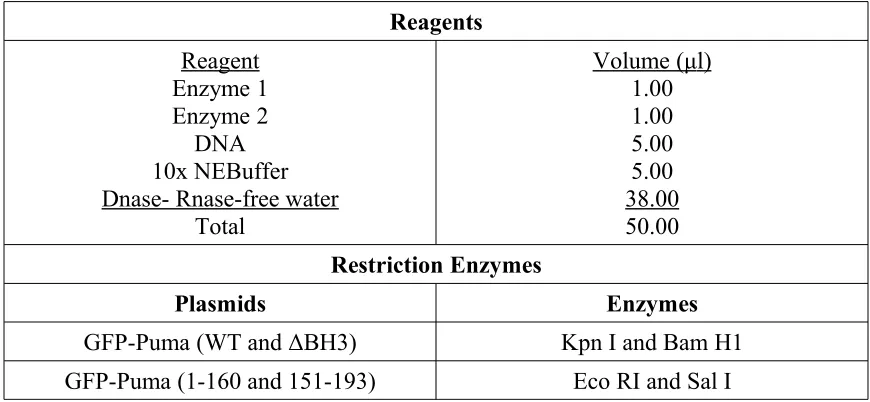
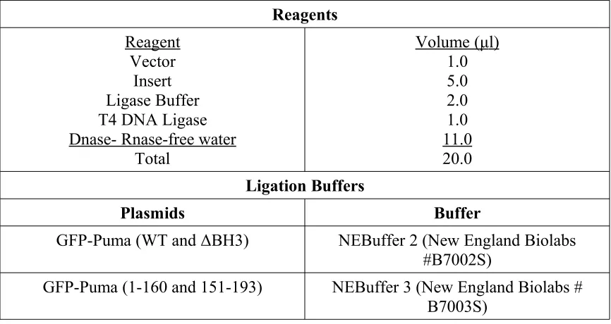
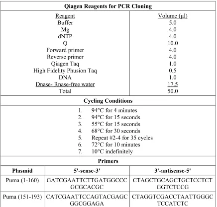
2.2 – Plasmid constructs
HA-Puma and HA-Puma (ΔBH3) cloned in pCEP4 vector were generously
donated by Dr. Vogelstein (Yu et al. 2001). The plasmids and the desired vector, GFP
C1 (Clontech #6084-1), were digested with Kpn I (New England Biolabs #R0142L) and
BamH 1 (New England Biolabs #R0136L) restriction enzymes (Table 1). The digested
DNA was then ligated into the digested GFP C1 vector with T4 DNA Ligase (New
The other GFP-Puma plasmids, GFP-Puma (1-160) and GFP-Puma (151-93) were
generated by PCR amplification (Table 3) and cloned into GFP C2 vector between Eco
RI and Sal I. The DNA template for Puma was diluted to 10ng/μl and and a PCR was run
using Qiagen Taq (Qiagen #201203) and High Fidelity Taq (Qiagen #202602) (Table 3).
Ten μl of the PCR product was then run on a 1.5% agarose gel and purified with the
Qiagen Gel Purification kit as per manufacturer's instructions (Qiagen #28704). The
purified product and 5μg of the GFP C2 vector were then digested with Eco RI (New
England Biolabs #R0101L) and Sal I (New England Biolabs #R0138L) restriction
enzymes (Table 1). The digested vector and DNA were then ligated with T4 DNA
Ligase (New England Biolabs #M0202S) (Table 2) overnight at 4ºC. All plasmids are
GFP-Puma (WT)
c-terminus
BH3 c-terminus
BH3
c-terminus GFP
GFP GFP
GFP GFP-Puma (ΔBH3)
GFP-Puma (1-160)
2.3 - Transfection
Differentiated N2As were transfected using calcium/phosphate. For live-cell
confocal microscopy, cells in 35mm glass-bottomed dishes were transfected using 1ug of
plasmid DNA (GFP, Puma (WT), Puma (∆BH3), Puma (1-160), or
GFP-Puma (151-193)) per dish. Plasmid DNA was diluted in 45µl ddH20 and then mixed
with 5µl of 2.5M CaCl2. Fifty microlitres of 2xHBS (0.28M NaCl, 0.05M HEPES,
1.5mM Na2HPO4, at pH 7.1) was added drop-wise and the resulting mixture was
carefully inverted two to three times. To each 35mm dish, 100µl of this solution was
added drop-wise.
For fixed-cell confocal microscopy and live-cell microscopy, cells in 4-well
dishes were transfected using 0.25ug of plasmid DNA (GFP, Puma (WT),
GFP-Puma (∆BH3), GFP-GFP-Puma (1-160), GFP-GFP-Puma (151-193) and/or 3x Flag-Bcl-xl) per
well. One microgram of plasmid DNA was diluted in 60ul of ddH20 and then mixed
with 6.7ul of 2.5M CaCl2. To this solution, 66.7ul of 2xHBS (0.28M NaCl, 0.05M
HEPES, and 1.5mM Na2HPO4, at pH 7.1) was added drop-wise and the resulting
mixture was carefully inverted two to three times. Thirty-three microlitres of this
solution was added drop-wise to each well in a 4-well plate.
For co-immunoprecipitation experiments, cells in 60mm dishes were transfected
using 3ug of plasmid DNA (GFP, GFP-Puma (WT), GFP-Puma (∆BH3), GFP-Puma
(1-160), GFP-Puma (151-193), 3x Flag-Bcl-xl, and/or 3xFlag) per dish. Three micrograms
CaCl2. To this solution, 334ul of 2xHBS (0.28M NaCl, 0.05M HEPES, 1.5mM
Na2HPO4, at pH 7.1) was added drop-wise and the resulting mixture was carefully
inverted two to three times. The entirety of this solution was added drop-wise to each
60mm dish.
2.5 - Peptide treatment
For experiments with GFP or GFP-Puma (WT) transfected cells, cells were
treated with 1μM of either Bax-Hα1 (CanPeptide #CP08640) or Bax-Hα1(D33A)
(CanPeptide #CP08641) Tat-fused peptide per well 12 hours after transfection. Tat is a
short sequence of basic amino acids derived from human immunodeficiency virus that
allows the transport of proteins across cell membranes (Fawell et al. 1994; Ruben et al.
1989; Vives et al. 1997). See Table 5 for complete peptide sequences. Peptide treatment
was repeated every six hours thereafter, for a total of 28 hours post-transfection, at a
concentration of 0.2μM of either Bax-Hα1 or Bax-Hα1(D33A) tat-peptide per well.
2.6 – Cell death assesment by Hoechst 33342 and Propidium Iodide staining
Hoechst 33342 (Sigma #B2261) was added to cells at a final concentration of
1μg/ml and samples were incubated for 20 minutes at 37 C in 5% CO⁰ 2 (humidified
incubator). For staining with Propidium Iodide, Propidium Iodide (Sigma # P1470) was
added to cells at a final concentration of 1μg/ml and samples were incubated for 20
To examine apoptosis and cell death after transfection with GFP-Puma plasmids,
cells were stained with Hoechst 33342 and/or Propidium Iodide at 28 hours
post-transfection or 36 hours after the initial drug treatment. Nuclear morphology was
visualized with a 40x objective lens under ultra-violet (UV) light (for Hoechst 3342)
and/or with a rhodamine (TRITC) filter (for Propidium Iodide) using a Zeiss Axiovert
200 fluorescent microscope. At least five representative images per well were captured
using Zeiss Axiovision 4.8 software and at least 500 nuclei were counted for each
treatment. For analysis of apoptosis by Hoechst staining, nuclei were categorized as
either healthy or apoptotic (blebbed, shrunken, and/or fragmented) as previously
described (Steckley et al. 2007). For each treatment, the percentage of apoptotic cells
was calculated as the number of GFP-positive apoptotic cells out of the total number of
cells. For analysis of cell death by Propidium Iodide and Hoechst staining,
Hoechst-stained nuclei were categorized as either live (Propidium Iodide-negative) or dead
(Propidium Iodide-positive). Percent cell death was calculated as the number of
Propidium Iodide-positive (dead cells) out of the total number of cells for each treatment.
2.7 - Mitotracker ® Red CMXRos staining
Neuro2A cells were transfected with GFP, GFP-Puma (WT), GFP-Puma (∆BH3),
GFP-Puma (1-160), or GFP-Puma (151-193) plasmids (see Transfection). Twenty-four
hours post-transfection, cells were stained with Mitotracker ® Red CMXRos (Invitrogen
#M-7512). at 1ug/ml in complete MEM culture media to identify mitochondria and
Mitotracker ® Red CMXRos were added to the cells simultaneously and samples were
incubated for 20 minutes at 37 C in 5% CO⁰ 2 (humidified incubator). Samples were
visualized with a 63x objective lens on an LSM 510 Meta Zeiss confocal microscope and
images were captured using Zeiss software. To detect Hoechst 33342 staining, an
excitation wavelength of 405nm at 3% intensity was used with a bandpass filter for
420-480nm. For GFP, the fluorophore was excited at 3% intensity at 488nm with a bandpass
filter for 505-530nm. Mitotracker ® Red CMXRos fluorescence was excited at a
wavelength of 633nm at 10% intensity and viewed through a 650nm longpass filter.
2.8 - Immunofluorescence
Cells were incubated at 37 C in 5% CO⁰ 2 (humidified incubator) for 20 minutes
with Hoechst 33342 (Sigma #B2261) diluted to 1ug/ml in complete media. Cells were
then fixed with Lana’s fixative (4% paraformaldehyde (Sigma-#P6148), 160mM dibasic
and monobasic sodium phosphate, and 0.2% picric acid (Sigma-#80456)) for 20 minutes
at room temperature, then washed twice with 1xPBS. Cells were incubated at room
temperature for 20 minutes with permeabilizing solution (0.2% Triton-X in 1xPBS), and
then in blocking solution (0.2% Triton-X and 2% BSA in 1x PBS) for 45 minutes at room
temperature. Cells were then incubated overnight at 4 C with primary antibody at a ⁰
1:500 dilution in blocking solution (1x PBS, 0.2% Triton-X, 2% BSA). Primary
antibodies used for immunofluorescent staining were Anti-Tomm-20 (Abcam #ab56783),
or Anti-Flag M2 (Sigma #F-3165). The following day, cells were washed twice in 1x
at a 1:500 dilution. Secondary antibody used in this experiment was Alexa Fluor® 594
goat anti-mouse IgG (Invitrogen #A11005). Cells were washed twice in 1x PBS and then
the glass coverslips were affixed to slides using Immu-mount (Thermo Scientific
#9990402). Samples were visualized with a 63x objective lens on an LSM 510 Meta
Zeiss confocal microscope and images were captured using Zeiss software. To detect
Hoechst 33342 staining, an excitation wavelength of 405nm at 3% intensity was used
with a bandpass filter for 420-480nm. For GFP, the fluorophore was excited at 3%
intensity at 488nm with a bandpass filter for 505-530nm. Alexa Fluor® 594
fluorescence was excited at a wavelength of 543nm at 10% intensity and viewed through
a 585nm longpass filter.
2.9 – Co-immunoprecipitation
Anti-Flag M2 agarose beads (Sigma #A2220) were aliquoted into a 15ml falcon
tube and washed with 1xPBS, then centrifuged at 500xg for five minutes. The PBS was
then carefully aspirated off and the rinse was repeated twice. Flag-bead/CHAPS slurry
was prepared by re-suspending the rinsed Flag-beads in an equal volume of 1% CHAPS
buffer (10mM HEPES, 150mM NaCl, 0.5mM EDTA, 1% CHAPS (Sigma #C9426),
0.5mM phenylmethyl sulfonyl fluoride (PMSF) (Bioshop #PMS123), 10mg/ml
Aprotonin, 2mg/ml Leupeptin).
Twenty-four hours after transfection, cells were rinsed twice with ice-cold 1x PBS
centrifuged at 14000xg for 12 minutes at 4 C to pellet cell debris. Protein concentration ⁰
in the supernatant was determined using the Pierce © BCA protein determination assay
(Thermo Fisher Scientific © #22325). Five hundred micrograms of protein from each
sample was added to 50µl of the Flag-bead/CHAPS slurry and rotated at 4 C f⁰ or one
hour. Twenty-five micrograms of protein from each sample was not exposed to beads
and reserved for Western blotting to assess protein expression.
After incubation, beads were rinsed three times with 1x PBS, centrifuged at
14000xg for 20 seconds and aspirated with a syringe. Beads were resuspended in 50µl
1% CHAPS and 10µl of 6x sodium dodecyl sulfate (SDS) (Bioshop #SDS101). The
resulting mixture was boiled at 100 C for 10 minutes to denature the protein and release it⁰
from the beads. Twenty-five microlitres of each of the bead-incubated samples and the
entirety of the 25µg lysate samples were analysed by Western blot (see Western Blot).
2.10 - Western Blot
Protein samples were run on a 12.5% polyacrylamide gel and transferred to a
nitrocellulose membrane by semi-dry electroblotting. The membrane was washed three
times in 1x TBS-T (150mM NaCl, 10mM Tris, 0.05% Tween-20 (Fisher Biotech
#BP1605)) and blocked for an hour in blocking buffer (10% milk (w/v) in 1x TBS-T) at
room temperature on a rocker. The membrane was again washed three times in 1x