1
The Dual Role of Oat Bran Water Extract in Bone Homeostasis Through the
Regulation of Osteoclastogenesis and Osteoblast Differentiation
Shin-Hye Kim1,2,†, Kwang-Jin Kim3,†, Hyeon Jung Kang1, Young-Jin Son3, Sik-Won
Choi4,*, Mi-Ja Lee1,*
1Division of Crop Foundation, National Institute of Crop Science (NICS), Rural
Development Administration (RDA), Wanju 55365, Korea; 2Department of Biological
Sciences, College of Natural Science, Chonbuk National University, Jeonju 54896, Korea; 3Department of Pharmacy, Sunchon National University, Suncheon, Jeonnam
57922, Korea; 4Forest Biomaterials Research Center, National Institute of Forest
Science (NIFS), Jinju, Gyeongnam 52817
†SHK and KJK contributed equally to this study
*Co-corresponding authors:
Sik-Won Choi, Ph.D.; Forest Biomaterials Research Center, National Institute of Forest Science (NIFS), Jinju, Gyeongnam 52817, Republic of Korea; Tel: +82-55-760-5093; Fax: +82-55-759-8432; E-mail: [email protected]
Mi-Ja Lee, Ph.D.; Laboratory of Crop Resource Development, Division of Crop Foundation, National Institute of Crop Science, Rural Development Administration, Wanju 55365, Republic of Korea; Tel: +82-63-238-5332; Fax: +82-63-238-5305; E-mail: [email protected]
2
Abstract
The number of patients with bone metabolic disorders including osteoporosis is increasing worldwide. These disorders often facilitate bone fractures, which seriously impact the patient’s quality of life and could lead to further health complications. Bone homeostasis is tightly regulated to balance bone resorption and formation. However, many osteoporotic agents are broadly categorized as either bone forming or anti-resorptive, and their therapeutic use is often limited due to unwanted side effects. Therefore, safe and effective therapeutic agents are needed for osteoporosis. This study aims to clarify the bone protecting effects of oat bran water extract (OBWE) and its mode of action. OBWE inhibited RANKL-induced osteoclast differentiation by blocking c-Fos/NFATc1 through the alteration of I-κB. Furthermore, we found that OBWE enhanced BMP-2-stimulated osteoblast differentiation by the induction of Runx2 via Smad signaling molecules. In addition, the anti-osteoporotic activity of OBWE was also evaluated using an in vivo model. OBWE significantly restored ovariectomy-induced bone loss. These in vitro and in vivo results showed that OBWE has the potential to combat bone metabolic disorders including osteoporosis.
3
1. Introduction
Fractures caused by bone metabolic disorders including osteoporosis are recognized as a serious public health issue worldwide [1]. Osteoporotic fragility fractures can cause considerable pain and severe disability, which can reduce the quality of life. Therefore, protection against bone fragility is an important means of improving the quality of life. The maintenance of bone homeostasis depends on both the number and stability of the activity of bone-resorbing osteoclastic and bone-forming osteoblastogenic cells [2]. The imbalance between bone formation and resorption leads to pathological bone disorders resulting in osteoporosis, rheumatoid arthritis, Paget’s disease, and periodontal disease [3-5]. Numerous pharmaceutical agents, which are broadly categorized as either anabolic or anti-resorptive, have been developed to treat bone metabolic diseases including osteoporosis. Although several effective agents are currently available for the treatment of osteoporosis, their use is often limited due to safety issues associated with side effects and long-term use. Accordingly, new therapies that have both bone forming and anti-resorptive effects with satisfactory safety assessments would be valuable to mitigate osteoporotic bone loss.
4
necessary for osteoclast differentiation [6]. In response to RANKL, these signaling molecules contribute to the regulation of the AP-1 transcription factor member, c-Fos, and the nuclear factor of activated T cells, cytoplasmic 1 (NFATc1), which are known to be key regulators for osteoclast differentiation, fusion, and maturation [7-9]. NFATc1, which requires c-Fos for its induction, regulates the process of osteogenesis by controlling osteoclast-related genes including tartrate-resistant acid phosphatase (TRAP, also known as ACP5), dendritic cell-specific transmembrane protein (DC-STAMP), and cathepsin K [10, 11]. Apparently, transcription factors such as c-Fos and NFATc1 play a critical role in the regulation of molecules for osteoclast differentiation. Therefore, the pharmacological inhibition of osteoclast differentiation-mediated transcription factors is effective to overcome bone resorbing-mediated disorders [3, 12].
5
phosphoprotein 1 (OPN; osteopontin, also known as Spp1), and bone gamma-carboxyglutamate protein (OCL; osteocalcin, also known as Bglap) [19, 20]. Consequently, Runx2 has significant potential to be a target for new therapies that prevent bone loss by mitigating bone formation.
6
2. Results and Discussion
2.1 OBWE inhibits RANKL-induced osteoclast differentiation
7
Figure 1. OBWE impairs RANKL-mediated osteoclast differentiation. (A) The BMMs were cultured for 4 d in the presence of RANKL (10 ng/ml) and M-CSF (30 ng/ml) with either the vehicle (water) or the indicated concentration of OBWE. Multinucleated osteoclasts were visualized using TRAP staining. (B) TRAP+ MNCs were counted (left panel) and TRAP activity was measured (right panel). ###, p < 0.001 (versus the control);
**, p < 0.01; ***, p < 0.001 (versus the RANKL-treated group). (C) The effect of BSE on the viability of BMMs was evaluated using the CCK-8 assay. Data are expressed as mean ± SD and are representative of at least three experiments.
2.2 OBWE inhibits RANKL-related expression of c-Fos and NFATc1 through the
modulation of NF-κB/I-κB signaling molecules
8
9
10
The indicated densitometric values were obtained using Multi Gauge version 3 software. One representative result obtained from three independent experiments yielding similar results is shown.
To gain insight into the mechanism by which OBWE inhibits osteoclast differentiation by attenuating c-Fos/NFATc1 expression, we investigated whether OBWE could affect the activation of the RANKL-mediated several signaling molecules that are associated with the regulation of master transcription factors. RANKL signaling during osteoclast differentiation activates various signaling pathways including NF-κB, PI3K/AKT, and MAP kinases [30, 35]. In particular, the expression of c-Fos and NFATc1 requires the assembly of NF-κB signaling pathways [36]. NF-κB p50/p52 double deficiency mice show acute osteopetrosis and weaknesses in osteoclastogensis because c-Fos and NFATc1 are not expressed by RANKL stimulation [37]. The function of NF-κB proteins is regulated by I-κB signaling molecules. I-κB inhibits NF-κB by preventing the nuclear localization signals of NF-κB molecules, thus segregating and maintaining them in an inactive state in the cytoplasm [38]. As shown in Figure 2C, RANKL stimulated the degradation of I-κB and the phosphorylation of RAC-Alpha Serine/Threonine-Protein Kinase (AKT), but the treatment of OBWE only prevented the RANKL-induced degradation of I-κB. These results suggest that the inhibition of I-κB degradation could be attributed to the anti-osteoclastogenic action of OBWE.
2.3 OBWE enhances BMP-2-mediated osteoblast differentiation in C2C12 cells
11
is important for the effective treatment of bone metabolic disorders. Therefore, we examined whether OBWE could induce the bipotential of mesenchymal C2C12 cells to commit to osteoblast differentiation. BMP-2 enhances osteoblast differentiation by the induction of ALP activity and expression in C2C12 cells [39]. Treatment with OBWE dose-dependently enhanced BMP-2-mediated ALP expression, which was deduced by ALP staining (Figure 3A). Consistent with this result, OBWE significantly enhanced BMP-2-induced ALP activity in a dose-dependent manner (Figure 3B). However, no cytotoxicity of OBWE was observed at the doses used to assess its effects (Figure 3C). These results indicate that OBWE may possess osteogenic activity without apparent cytotoxicity.
12
concentration of OBWE. Osteoblast differentiation was visualized by ALP staining. (B) ALP activity was monitored by measuring absorbance at 405 nm. ### p < 0.001 (versus
control); *** p < 0.001 (versus BMP-2–treated group). (C) Effects of OBWE on the viability of C2C12 cells were evaluated using the CCK-8 assay. Data are expressed as mean ± SD and are representative of at least three experiments.
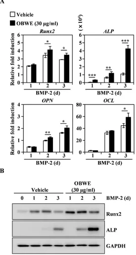
2.4 OBWE contributes to the BMP-2-stimulated expression of Runx2 through the
activation of Smad signaling pathways
13
14
real-time PCR. GAPDH was used as the internal control. * p < 0.05; ** p < 0.01; *** p < 0.001 (versus vehicle control). (B) Effects of OBWE on the levels of Runx2 and ALP were evaluated by immune blot analysis. GAPDH was used as the internal control. One representative result from three independent experiments yielding similar results is shown.
15
Figure 5. OBWE induces BMP-2–mediated phosphorylation of Smad signaling molecules. Following serum starvation for 1 d, C2C12 cells were pre-treated with vehicle or OBWE (30 µg/ml) for 1 h prior to BMP-2 stimulation (100 ng/ml) for the indicated times. The expression levels of the signaling molecules were evaluated by western blotting. Actin was used as the internal control.
2.5 OBWE prevents ovariectomy-induced bone loss in vivo
The in vitro anti-osteoporotic effect of OBWE prompted us to explore its putative in vivo effects using an osteoporosis model with ovariectomy (OVX). OVX-induced bone
16
substantially protected by OBWE administration (Figure 6B). These results demonstrate that it is likely that OBWE can prevent and improve postmenopausal osteoporosis.
Figure 6. OBWE prevents OVX-induced bone loss in vivo. The effect of OBWE on OVX-induced bone loss was investigated in mice.(A) Micro-CT analysis-based images show transverse and longitudinal images of sham and OVX bone at 1 mg, 10 mg, and 100 mg/kg from left to right, and (B) bone mineral density (BMD), percent bone volume ratio (BV/TV), trabecular separation (Tb. Sp) of femur measurements by 3D images analyzer. ### p < 0.001 (versus the sham group); * p < 0.05; ** p < 0.01 (versus
17
3. Materials and Methods
3.1 Preparation of the oat bran water extract
The oat bran used in this study was grounded in a laboratory test mill (Brabender Technologie, Germany). The flour (100 g) was defatted three times with hexane (1 L) for 24 h at room temperature. After filtration with filter paper (Whatman No. 3), the residual oat bran was extracted three times with prethanol (1 L) and filtrated by means of a Buechner funnel lined with filter paper (Carl Roth, Germany, 111A, Ø100 mm). The filtrates were combined and concentrated in a rotary evaporator. The residuals from the prethanol extraction were extracted twice with water (1 L) for 24 h at room temperature and combined and dried with a freeze dryer.
3.2 Reagents and antibodies
Mouse soluble RANKL, M-CSF, recombinant human bone morphogenetic protein-2 (rhBMP-2), and ALP antibodies were purchased from R & D Systems (Minneapolis, MN). Penicillin, streptomycin, cell culture medium and foetal bovine serum (FBS) were purchased from Invitrogen Life Technologies (Carlsbad, CA). Antibodies against c-Fos, NFATc1, actin, IκB, Smad, and the secondary antibody conjugated to horseradish peroxidase (HRP) were purchased from Santa Cruz Biotechnology (Dallas, TX). All other antibodies were obtained from Cell Signaling Technology (Beverly, MA).
3.3 Preparations of osteoclast precursor cells
18
Institutional Animal Care and Use Committee (IACUC) of Yonsei University College of Medicine. Every effort was made to minimise the number of animals used in the study and minimise their suffering and stress/discomfort.
All experiments were carried out as described in a previous study, with modifications [48]. Five-week-old male imprinting control region (ICR) mice (Damul Science Co., Deajeon, Korea) were maintained in a room illuminated daily from 07:00 to 19:00 (12:12 h light to dark cycle), with controlled temperature (23 ± 1C) and ventilation (10–12 times per h). Humidity was maintained at 55% ± 5% and the animals had free access to a standard animal diet and tap water. Bone marrow cells were obtained from the five-week-old male ICR mice by flushing their femurs and tibias with alpha minimum essential medium (α-MEM) supplemented with antibiotics (100 units/ml penicillin, 100 g/ml streptomycin). The bone marrow cells were cultured on culture dishes for 1 d in α-MEM containing 10% FBS and M-CSF (10 ng/ml). The non-adherent bone marrow cells were plated onto Petri dishes and cultured for 3 d in the presence of M-CSF (30 ng/ml). After the non-adherent cells were washed out, the adherent cells were used as bone marrow-derived macrophages (BMMs).
3.4 Osteoclast cell culture and osteoclast differentiation
The BMMs were maintained in α-MEM supplemented with 10% FBS, 100 units/ml penicillin, and 100 g/ml streptomycin. The medium was changed every 3 d in a humidified atmosphere of 5% CO2 at 37C. To differentiate the osteoclasts from the
BMMs, the BMMs (1 104 cells/well in a 96-well plate or 3 105 cells/well in a 6-well
19 3.5 TRAP staining and activity assay
The mature osteoclasts were visualized using TRAP staining, a biomarker of osteoclast differentiation. Briefly, the multinucleated osteoclasts were fixed with 3.7% formalin for 10 min, permeabilized with 0.1% Triton X-100 for 10 min and then stained with TRAP solution (Sigma-Aldrich, Saint Louis, MO). The TRAP-positive multinucleated osteoclasts (MNC; nuclear ≥ 3) were counted. To measure TRAP activity, the multinucleated osteoclasts were fixed in 3.7% formalin for 5 min, permeabilized with 0.1% Triton X-100 for 10 min and then treated with TRAP buffer (100 mM sodium citrate, pH 5.0, 50 mM sodium tartrate) containing 3 mM p-nitrophenyl phosphate (Sigma-Aldrich) at 37°C for 5 min. The reaction mixtures in the wells were transferred to new plates containing an equal volume of 0.1 N NaOH, and the optical density values were determined at 405 nm.
3.6 Cell viability assay
The BMMs and C2C12 cells were plated on 96-well plates (three replicate plates) at a density of 1 104 cells/well (BMMs) or 2.5 × 10³ cells/well (C2C12 cells). After
treatment with the indicated concentrations of OBWE, the cells were incubated for 3 d, and cell viability was measured using the Cell Counting Kit 8 (CCK-8) according to the manufacturer’s protocol. The CCK-8 assay kit was purchased from Dojindo Molecular Technologies (Rockville, MD).
3.7 RNA isolation and real-time polymerase chain reaction analysis
20
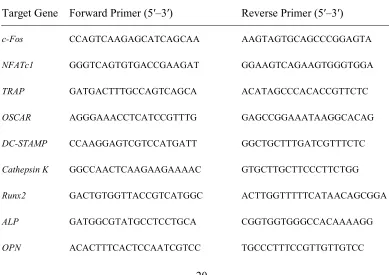
The primers were chosen using the online Primer3 design program [49]. The primer sets used in this study are shown in Table 1. Briefly, total RNA was isolated with TRIzol reagent, and the first-strand of the cDNA was synthesized with the RevertAid First Strand cDNA Synthesis Kit (Thermo Scientific, Waltham, MA) according to the manufacturer’s recommended protocol. SYBR green-based quantitative PCR (qPCR) was performed using the Bio-Rad CFX96 Real-Time PCR Detection System (Hercules, CA) and Topreal qPCR 2 PreMIX (Enzynomics, Daejeon, Korea). All reactions were run in triplicates, and the data were analyzed using the 2−ΔΔCT method [50].
Hypoxanthine phosphoribosyltransferase 1 (HPRT1) and glyceraldehyde 3-phosphate dehydrogenase (GAPDH) were used as the internal standard genes. The statistical significance was determined using a Student’s t-test with HPRT1/GAPDH-normalized 2−ΔΔCT values; the differences were considered significant at p < 0.05.
Table 1. The primer sequences used in this study.
Target Gene Forward Primer (5′–3′) Reverse Primer (5′–3′)
c-Fos CCAGTCAAGAGCATCAGCAA AAGTAGTGCAGCCCGGAGTA
NFATc1 GGGTCAGTGTGACCGAAGAT GGAAGTCAGAAGTGGGTGGA
TRAP GATGACTTTGCCAGTCAGCA ACATAGCCCACACCGTTCTC
OSCAR AGGGAAACCTCATCCGTTTG GAGCCGGAAATAAGGCACAG
DC-STAMP CCAAGGAGTCGTCCATGATT GGCTGCTTTGATCGTTTCTC
Cathepsin K GGCCAACTCAAGAAGAAAAC GTGCTTGCTTCCCTTCTGG
Runx2 GACTGTGGTTACCGTCATGGC ACTTGGTTTTTCATAACAGCGGA
ALP GATGGCGTATGCCTCCTGCA CGGTGGTGGGCCACAAAAGG
21
OCL AGGGAAACCTCATCCGTTG GAGCCGGAAATAAGGCACAG
GAPDH ACCACAGTCCATGCCATCAC TCCACCACCCTGTTGCTGTA
HPRT1 TGCTCGAGATGTCATGAAGG AGAGGTCCTTTTCACCAGCA
3.8 Western blot analysis
The western blot analysis was performed as previously described [51]. Briefly, the cultured cells were washed with ice-cold phosphate-buffered saline (PBS) and lysed in lysis buffer (50 mM Tris-HCl, 150 mM NaCl, 5 mM EDTA, 1% Triton X-100, 1 mM sodium fluoride, 1 mM sodium vanadate, and 1% deoxycholate) supplemented with protease inhibitors. After centrifugation at 15,000 g for 15 min, the protein quantification of the supernatant was determined using the detergent compatible (DC) protein assay kit (Bio-Rad). The quantified proteins were denatured, separated on sodium dodecyl sulphate-polyacrylamide gel electrophoresis (SDS-PAGE) gels, and transferred onto a polyvinylidene difluoride (PVDF) membrane (Merck Millipore, Darmstadt, Germany). After incubation with an antibody, the membranes were developed using SuperSignal West Femto Maximum Sensitivity Substrate (Thermo Scientific) and visualised with the LAS-4000 luminescent image analyser (GE Healthcare Life Sciences, Little Chalfont, UK). Actin and GAPDH were used as a loading control.
3.9 Osteoblast differentiation
22
alpha minimum essential medium (α-MEM) with 10% FBS, 100 units/ml penicillin, and 100 g/ml streptomycin. Cells were seeded in 96-well plates at 2.5 × 10³ cells/well or in
6-well plates at 2.5 × 10⁵ cells/well. After 1 d, cells were differentiated by replacing the
medium with α-MEM containing 5% FBS and rhBMP-2 (100 ng/ml) with OBWE at the indicated dose. Osteoblastic bone formation was observed by ALP staining.
3.10 Alkaline phosphatase staining and activity assays
ALP is an early biomarker of osteoblast differentiation. After differentiation for 3 d, cells were washed twice with PBS, fixed with 10% formalin in PBS for 5 min, rinsed with deionized water, and stained using an ALP Kit (Sigma-Aldrich). To measure ALP activity, differentiated cells were washed twice with PBS, fixed with 10% formalin in PBS for 5 min, rinsed with PBS, and measured using a one-step PNPP substrate solution (Thermo Scientific, MA) according to the manufacturer’s protocol.
3.11 Ovariectomy-induced bone erosion
23
were fixed in 3.5% formaldehyde for 1 d. The fixed femurs were scanned and analyzed with the SkyScan 1272 micro-CT imaging system provided by Bruker (Billerica, MA).
3.12 Statistical analysis
24
4. Conclusions
25
Acknowledgments
This work was conducted with the support of the Cooperative Research Program for Agriculture Science & Technology Development (Project No. PJ012107032018) of the Rural Development Administration (RDA), Korea
Author Contributions
SHK and KJK contributed equally to this work. SHK contributed to all experiments and the manuscript preparation. KJK performed the in vivo study. HJK performed in vitro experiments. YJS prepared part of the manuscript and supervised the in vivo research. SWC and MJL designed and supervised all experiments and wrote and finalized the manuscript.
Conflicts of Interest
26
References
1. Klibanski, A.; Adams-Campbell, L.; Bassford, T.; Blair, S. N.; Boden, S. D.; Dickersin, K.; Gifford, D. R.; Glasse, L.; Goldring, S. R.; Hruska, K.; Johnson, S. R.; McCauley, L. K.; Russell, W. E.; Osteopor, N. C. D. P., Osteoporosis prevention, diagnosis, and therapy. Jama-Journal of the American Medical Association 2001, 285, (6), 785-795.
2. Karsenty, G.; Wagner, E. F., Reaching a genetic and molecular understanding of skeletal development. Developmental Cell 2002, 2, (4), 389-406.
3. Rodan, G. A.; Martin, T. J., Therapeutic approaches to bone diseases. Science 2000,
289, (5484), 1508-1514.
4. Khosla, S.; Riggs, B. L., Pathophysiology of age-related bone loss and osteoporosis. Endocrinology and Metabolism Clinics of North America 2005, 34, (4), 1015-1030.
5. Manolagas, S. C.; Parfitt, A. M., What old means to bone. Trends in Endocrinology and Metabolism 2010, 21, (6), 369-374.
6. Lee, Z. H.; Kim, H. H., Signal transduction by receptor activator of nuclear factor kappa B in osteoclasts. Biochemical and Biophysical Research Communications
2003, 305, (2), 211-214.
7. Feng, X., RANKing intracellular signaling in osteoclasts. Iubmb Life 2005, 57, (6), 389-395.
8. Takayanagi, H., Osteoimmunology: shared mechanisms and crosstalk between the immune and bone systems. Nature Reviews Immunology 2007, 7, (4), 292-304. 9. Takayanagi, H., Inflammatory bone destruction and osteoimmunology. Journal of
27
10. Grigoriadis, A. E.; Wang, Z. Q.; Cecchini, M. G.; Hofstetter, W.; Felix, R.; Fleisch, H. A.; Wagner, E. F., C-Fos - a Key Regulator of Osteoclast-Macrophage Lineage Determination and Bone Remodeling. Science 1994, 266, (5184), 443-448.
11. Takayanagi, H.; Kim, S.; Koga, T.; Nishina, H.; Isshiki, M.; Yoshida, H.; Saiura, A.; Isobe, M.; Yokochi, T.; Inoue, J.; Wagner, E. F.; Mak, T. W.; Kodama, T.; Taniguchi, T., Induction and activation of the transcription factor NFATc1 (NFAT2) integrate RANKL signaling in terminal differentiation of osteoclasts. Developmental Cell 2002, 3, (6), 889-901.
12. Teitelbaum, S. L., Bone resorption by osteoclasts. Science 2000, 289, (5484), 1504-1508.
13. Komori, T., Regulation of osteoblast differentiation by transcription factors. Journal of Cellular Biochemistry 2006, 99, (5), 1233-1239.
14. Almalki, S. G.; Agrawal, D. K., Key transcription factors in the differentiation of mesenchymal stem cells. Differentiation 2016, 92, (1-2), 41-51.
15. Hollinger, J. O.; Schmitt, J. M.; Buck, D. C.; Shannon, R.; Joh, S. P.; Zegzula, H. D.; Wozney, J., Recombinant human bone morphogenetic protein-2 and collagen for bone regeneration. Journal of Biomedical Materials Research 1998, 43, (4), 356-364.
16. Cheng, H. W.; Jiang, W.; Phillips, F. M.; Haydon, R. C.; Peng, Y.; Zhou, L.; Luu, H. H.; An, N. L.; Breyer, B.; Vanichakarn, P.; Szatkowski, J. P.; Park, J. Y.; He, T. C., Osteogenic activity of the fourteen types of human bone morphogenetic proteins (BMPs). Journal of Bone and Joint Surgery-American Volume 2003, 85A, (8), 1544-1552.
28
pathway and other major signaling pathways results in tightly regulated cell-specific outcomes. Febs Journal 2007, 274, (12), 2977-2985.
18. Lian, J. B.; Stein, G. S.; Javed, A.; van Wijnen, A. J.; Stein, J. L.; Montecino, M.; Hassan, M. Q.; Gaur, T.; Lengner, C. J.; Young, D. W., Networks and hubs for the transcriptional control of osteoblastogenesis. Reviews in Endocrine & Metabolic Disorders 2006, 7, (1-2), 1-16.
19. Kahn, H. S.; Tatham, L. M.; Rodriguez, C.; Calle, E. E.; Thun, M. J.; Heath, C. W., Stable behaviors associated with adults' 10-year change in body mass index and likelihood of gain at the waist. American Journal of Public Health 1997, 87, (5), 747-754.
20. Bruderer, M.; Richards, R. G.; Alini, M.; Stoddart, M. J., Role and Regulation of Runx2 in Osteogenesis. European Cells & Materials 2014, 28, 269-286.
21. Bryngelsson, S.; Mannerstedt-Fogelfors, B.; Kamal-Eldin, A.; Andersson, R.; Dimberg, L. H., Lipids and antioxidants in groats and hulls of Swedish oats (Avena sativa L). Journal of the Science of Food and Agriculture 2002, 82, (6), 606-614. 22. Dimberg, L. H.; Gissen, C.; Nilsson, J., Phenolic compounds in oat grains (Avena
sativa L.) grown in conventional and organic systems. Ambio 2005, 34, (4-5), 331-337.
23. Emmons, C. L.; Peterson, D. M.; Paul, G. L., Antioxidant capacity of oat (Avena sativa L.) extracts. 2. In vitro antioxidant activity and contents of phenolic and tocol antioxidants. Journal of Agricultural and Food Chemistry 1999, 47, (12), 4894-4898.
29
(Avena sativa L.). Journal of Phytopathology 2008, 156, (1), 1-7.
25. Tapola, N.; Karvonen, H.; Niskanen, L.; Mikola, M.; Sarkkinen, E., Glycemic responses of oat bran products in type 2 diabetic patients. Nutrition Metabolism and Cardiovascular Diseases 2005, 15, (4), 255-261.
26. Guo, W. M.; Nie, L.; Wu, D. Y.; Wise, M. L.; Collins, F. W.; Meydani, S. N.; Meydani, M., Avenanthramides Inhibit Proliferation of Human Colon Cancer Cell Lines In Vitro. Nutrition and Cancer-an International Journal 2010, 62, (8), 1007-1016.
27. Ozkaya, H.; Ozkaya, B.; Duman, B.; Turksoy, S., Effect of Dephytinization by Fermentation and Hydrothermal Autoclaving Treatments on the Antioxidant Activity, Dietary Fiber, and Phenolic Content of Oat Bran. Journal of Agricultural and Food Chemistry 2017, 65, (28), 5713-5719.
28. Robitaille, J.; Fontaine-Bisson, B.; Couture, P.; Tchernof, A.; Vohl, M. C., Effect of an oat bran-rich supplement on the metabolic profile of overweight premenopausal women. Annals of Nutrition and Metabolism 2005, 49, (3), 141-148.
29. Varga, N.; Vereb, Z.; Rajnavolgyi, E.; Nemet, K.; Uher, F.; Sarkadi, B.; Apati, A., Mesenchymal stem cell like (MSCl) cells generated from human embryonic stem cells support pluripotent cell growth. Biochemical and Biophysical Research Communications 2011, 414, (3), 474-480.
30. Boyle, W. J.; Simonet, W. S.; Lacey, D. L., Osteoclast differentiation and activation. Nature 2003, 423, (6937), 337-342.
31. Raisz, L. G., Pathogenesis of osteoporosis: concepts, conflicts, and prospects. Journal of Clinical Investigation 2005, 115, (12), 3318-3325.
30 differentiation. Bone 2007, 40, (2), 251-264.
33. Ishida, N.; Hayashi, K.; Hoshijima, M.; Ogawa, T.; Koga, S.; Miyatake, Y.; Kumegawa, M.; Kimura, T.; Takeya, T., Large scale gene expression analysis of osteoclastogenesis in vitro and elucidation of NFAT2 as a key regulator. Journal of Biological Chemistry 2002, 277, (43), 41147-41156.
34. Wang, Z. Q.; Ovitt, C.; Grigoriadis, A. E.; Mohlesteinlein, U.; Ruther, U.; Wagner, E. F., Bone and Hematopoietic Defects in Mice Lacking C-Fos. Nature 1992, 360, (6406), 741-745.
35. Blair, H. C.; Robinson, L. J.; Zaidi, M., Osteoclast signalling pathways. Biochemical and Biophysical Research Communications 2005, 328, (3), 728-738. 36. Kobayashi, N.; Kadono, Y.; Naito, A.; Matsumoto, K.; Yamamoto, T.; Tanaka, S.;
Inoue, J., Segregation of TRAF6-mediated signaling pathways clarifies its role in osteoclastogenesis. Embo Journal 2001, 20, (6), 1271-1280.
37. Iotsova, V.; Caamano, J.; Loy, J.; Yang, Y.; Lewin, A.; Bravo, R., Osteopetrosis in mice lacking NF-kappaB1 and NF-kappaB2. Nat Med 1997, 3, (11), 1285-9. 38. Jacobs, M. D.; Harrison, S. C., Structure of an IkappaBalpha/NF-kappaB complex.
Cell 1998, 95, (6), 749-58.
39. Jang, W. G.; Kim, E. J.; Kim, D. K.; Ryoo, H. M.; Lee, K. B.; Kim, S. H.; Choi, H. S.; Koh, J. T., BMP2 protein regulates osteocalcin expression via Runx2-mediated Atf6 gene transcription. Journal of Biological Chemistry 2012, 287, (2), 905-15. 40. Gersbach, C. A.; Byers, B. A.; Pavlath, G. K.; Garcia, A. J., Runx2/Cbfa1
31
Quarles, L. D., Selective Runx2-II deficiency leads to low-turnover osteopenia in adult mice. Dev Biol 2005, 283, (2), 345-56.
42. Harada, H.; Tagashira, S.; Fujiwara, M.; Ogawa, S.; Katsumata, T.; Yamaguchi, A.; Komori, T.; Nakatsuka, M., Cbfa1 isoforms exert functional differences in osteoblast differentiation. Journal of Biological Chemistry 1999, 274, (11), 6972-8. 43. Chen, G.; Deng, C.; Li, Y. P., TGF-beta and BMP signaling in osteoblast
differentiation and bone formation. Int J Biol Sci 2012, 8, (2), 272-88.
44. Chen, D.; Zhao, M.; Mundy, G. R., Bone morphogenetic proteins. Growth Factors
2004, 22, (4), 233-41.
45. Feng, X. H.; Derynck, R., Specificity and versatility in tgf-beta signaling through Smads. Annu Rev Cell Dev Biol 2005, 21, 659-93.
46. Chen, D.; Harris, M. A.; Rossini, G.; Dunstan, C. R.; Dallas, S. L.; Feng, J. Q.; Mundy, G. R.; Harris, S. E., Bone morphogenetic protein 2 (BMP-2) enhances BMP-3, BMP-4, and bone cell differentiation marker gene expression during the induction of mineralized bone matrix formation in cultures of fetal rat calvarial osteoblasts. Calcif Tissue Int 1997, 60, (3), 283-90.
47. Li, J. Z.; Li, H.; Sasaki, T.; Holman, D.; Beres, B.; Dumont, R. J.; Pittman, D. D.; Hankins, G. R.; Helm, G. A., Osteogenic potential of five different recombinant human bone morphogenetic protein adenoviral vectors in the rat. Gene Ther 2003,
10, (20), 1735-43.
32
49. Rozen, S.; Skaletsky, H., Primer3 on the WWW for general users and for biologist programmers. Methods Mol Biol 2000, 132, 365-86.
50. Livak, K. J.; Schmittgen, T. D., Analysis of relative gene expression data using real-time quantitative PCR and the 2(T)(-Delta Delta C) method. Methods 2001,
25, (4), 402-408.
51. Choi, S. W.; Lee, K. S.; Lee, J. H.; Kang, H. J.; Lee, M. J.; Kim, H. Y.; Park, K. I.; Kim, S. L.; Shin, H. K.; Seo, W. D., Suppression of Akt-HIF-1alpha signaling axis by diacetyl atractylodiol inhibits hypoxia-induced angiogenesis. BMB Rep 2016,
49, (9), 508-13.