International Journal of Nanomedicine
Dove
press
O r I g I N a l r e s e a r c h
open access to scientific and medical research
Open access Full Text article
comparison of the cellular transport mechanism
of cationic, star-shaped polymers and liposomes
in hacat cells
heng-cong luo,1,2,* Na
li,1,* li Yan,1 Kai-jin Mai,3
Kan sun,1 Wei Wang,1
guo-Juan lao,1 chuan Yang,1
li-Ming Zhang,3 Meng ren1
1Department of endocrinology, sun Yat-sen Memorial hospital, guangdong Provincial Key laboratory of Malignant Tumor epigenetics and gene regulation Medical research center, sun Yat-sen University, guangzhou, People’s republic of china; 2Department of endocrinology, The Third affiliated hospital of guangzhou Medical University, guangzhou, People’s republic of china; 3school of Materials science and engineering, school of chemistry, sun Yat-sen University, guangzhou, People’s republic of china
*These authors contributed equally to this work
Abstract: Several biological barriers must be overcome to achieve efficient nonviral gene delivery. These barriers include target cell uptake, lysosomal degradation, and dissociation from the carrier. In this study, we compared the differences in the uptake mechanism of cationic, star-shaped polymer/MMP-9siRNA complexes (β-CD-(D3)7/MMP-9siRNA complexes: polyplexes) and commercial liposome/MMP-9siRNA complexes (Lipofectamine® 2000/
MMP-9siRNA complexes: liposomes). The uptake pathway and transfection efficiency of the polyplexes and liposomes were determined by fluorescence microscopy, flow cytometry, and reverse transcriptase-polymerase chain reaction. The occurrence of intracellular processing was assessed by confocal laser scanning microscopy. Endosomal acidification inhibitors were used to explore the endosomal escape mechanisms of the polyplexes and lysosomes. We concluded that the polyplexes were internalized by non-caveolae- and non-clathrin-mediated pathways, with no lysosomal trafficking, thereby inducing successful transfection, while the majority of liposomes were internalized by clathrin-dependent endocytosis (CDE), caveolae-mediated endocytosis, and macropinocytosis, and only CDE induced successful transfection. Liposomes might escape more quickly than polyplexes, and the digestion effect of acidic organelles on liposomes was faint compared to the polyplexes, although both complexes escaped from endolysosomes via the proton sponge mechanism. This may be the key aspect that leads to the lower transfection efficiency of the β-CD-(D3)7/MMP-9siRNA complexes. The present study may offer some insights for the rational design of novel delivery systems with increased transfection efficiency but decreased toxicity.
Keywords:β-CD-(D3)7, liposome, endocytosis, endolysosomal escape, intracellular trafficking
Introduction
Diabetic foot ulcers are a complication of chronic diabetes and an important factor of morbidity and mortality in patients with diabetes. Our previous study showed that the application of small interfering RNA (siRNA) to inhibit matrix metalloproteinase
(MMP-9) expression could promote the process of cutaneous wound healing.1 However,
as siRNA is large and charged, it has difficulty in traversing the plasma membrane on its own. Thus, an appropriate gene delivery system is required for its efficient
cellular uptake.2
In our previous study, we synthesized a cationic, star-shaped polymer consisting
of a β-CD core and poly(amidoamine) dendron arms, ie, β-CD-(D3)7. The ability
of β-CD-(D3)7 to condense MMP-9siRNA into particulate structures was
con-firmed by agarose gel electrophoresis, particle size measurements, and transmission
electron microscopy observations.1 However, this previous study showed that the
correspondence: Meng ren Department of endocrinology, sun Yat-sen Memorial hospital, guangdong Provincial Key laboratory of Malignant Tumor epigenetics and gene regulation Medical research center, No 107 Yan Jiang West road, sun Yat-sen University, guangzhou 510120, People’s republic of china
Tel +86 20 8133 2286 Fax +86 20 8133 2404 email [email protected]
li-Ming Zhang
school of Materials science and engineering, school of chemistry, sun Yat-sen University, No 135, Xingang West road, guangzhou 520175, People’s republic of china
Tel +86 20 8411 2354 email [email protected]
Journal name: International Journal of Nanomedicine Article Designation: Original Research
Year: 2017 Volume: 12
Running head verso: Luo et al
Running head recto: Comparison of the cellular transport mechanism DOI: http://dx.doi.org/10.2147/IJN.S121450
International Journal of Nanomedicine downloaded from https://www.dovepress.com/ by 118.70.13.36 on 23-Aug-2020
For personal use only.
Number of times this article has been viewed
This article was published in the following Dove Press journal: International Journal of Nanomedicine
Dovepress luo et al
β-CD-(D3)7/MMP-9siRNA complexes (also denoted as
polyplexes) had a higher internalization rate but a lower interference effect compared with commercial liposome
gene vector-Lipofectamine® 2000 (Thermo Fisher Scientific,
Waltham, MA, USA)/MMP-9siRNA complexes (also denoted as lip2000/MMP-9siRNA complexes or liposomes; uptake rates of 98.4% vs 28.2% and interference rates of 44.2% vs 77.2%). We speculated that these results may be related to the extracellular and intracellular barriers. Past research has shown that to achieve successful transfection, nonviral deliv-ery systems, such as those delivering siRNA, need to over-come several cellular barriers, including being internalized by cells, escaping from endolysosomes, and dissociating the
cargo from the complex for delivery into the cytoplasm.3
Endocytosis has been established as the main mechanism
for the uptake of nonviral vectors into the cells.4 Endocytosis
can be classified into five categories: phagocytosis, clathrin-dependent endocytosis (CDE), caveolae-mediated endocytosis (Cav-ME), non-clathrin- and non-caveolae dependent endocytosis, and macropinocytosis. Phagocytosis is typically restricted to specialized mammalian cells. CDE pathway is characterized by the formation of endosomes through the constriction and fission of clathrin-coated pits triggered by the GTPase dynamin. In Cav-ME pathway, the composition of the primary endocytic vesicle was in relation to lipid rafts. Lipid rafts are freely floating plasma mem-brane microdomains that are comprised of a combination of glycosphingolipids and protein receptors where cellular processes are compartmentalized. On the contrary, vesicles formed during macropinocytosis, which result from actin-driven membrane protrusions that collapse onto the engulfed nanoparticle and fuse with the plasma membrane, can con-tain lipid rafts together with nonraft membrane domains. Non-clathrin- and non-caveolae-dependent endocytosis has not been thoroughly studied, but may involve the adenosine diphosphate-ribosylation factor 6 (ARF6)-dependent route
and the RhoA-dependent pathway, among others.5
In our study, we compared β-CD-(D3)7/MMP-9siRNA
complexes with lip2000/MMP-9siRNA complexes using endocytosis inhibitors, endosomal acidification inhibitors, and organelle fluorescent probes in terms of the follow-ing elements: cellular uptake, intracellular traffickfollow-ing and transfection efficiency after internalization by different uptake modes, and potential endolysosomal escape mecha-nisms. The aim of this work is to provide some insights for further improving the molecular design and thereby the transfection efficiency of the polycationic amphiphilic cyclodextrin vector.
Materials and methods
Materials
The β-CD-(D3)7 synthesis and β-CD-(D3)7/MMP-9siRNA
complexes preparation methods were reported in our
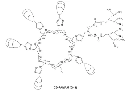
previ-ous publication.1 Figure 1 shows the chemical structure of
β-CD-(D3)7. Lipofectamine® 2000 was purchased from
Thermo Fisher Scientific. Human keratinocyte HaCat cells were purchased from the Chinese Academy of Sciences Cell
Bank (Shanghai, People’s Republic of China). α-Modified
Eagle Medium (α-MEM) and fetal bovine serum (FBS)
were purchased from GIBCO (Grand Island, NY, USA). Cell Counting Kit-8 (CCK-8) was purchased from Dojindo
(Kumamoto, Japan). Endocytosis inhibitors, such as M-β-CD,
genistein, chlorpromazine, and amiloride and the endosomal acidification inhibitors, bafilomycin A1 and chloroquine, were purchased from Sigma (Schnelldorf, Germany). The roles of the inhibitors are shown in Table 1. The fluorescent nuclear probe Hoechst 33342 and the fluorescent lysosomal probe LysoTracker DND-99 were purchased from Thermo Fisher Scientifice. The fluorescently labeled organelle fluo-rescent probes used in the study are shown in Table 2. All other reagents were of analytical grade and used without further purification. MMP-9 siRNA was purchased from GenePharma (Suzhou, People’s Republic of China). The sequences of the human MMP-9siRNA were as follows: sense,
5′-CUAUGGUCCUCGCCUGAATT-3′; antisense, 5′-UUCA
GGGCGAGGACCAUAGTT-3′.
cell culture
HaCat cells were cultured in α-MEM supplemented with 10%
FBS, penicillin (100 U/mL) and streptomycin (100 mg/mL)
(Beyotime, Haimen, People’s Republic of China) at 37°C in
a humid atmosphere with 5% CO2.
Treatment with endocytosis inhibitors
effects of different endocytosis inhibitor concentrations on hacat cell activity
The following inhibitors were used: 3–8 mM M-β-CD (Sigma
Cat No: C4555), 100–400 µM genistein (Sigma Cat No:
91955), 5–10 µg/mL chlorpromazine (Sigma Cat No: C8138),
and 25–75 µM amiloride (Sigma Cat No: A7410). Cells were
treated with endocytosis inhibitors for 1 h at 37°C.
Subse-quently, polyplexes or liposomes were added and the cells
were incubated for another 4 h. Then, 10 µL of CCK-8
reagent was added to the plate and incubated for another 3 h. After incubation, the optical density (OD) was measured at 450 and 630 nm using a plate spectrophotometer. Each group had six parallel replicates. Cell viability was calculated as
International Journal of Nanomedicine downloaded from https://www.dovepress.com/ by 118.70.13.36 on 23-Aug-2020
Dovepress comparison of the cellular transport mechanism
follows: cell viability (%) = (OD [sample] - OD [blank]/OD
[control] - OD [blank]) ×100. As a positive control, cells
were treated with β-CD-(D3)7/MMP-9siRNA complexes
or lip2000/MMP-9siRNA complexes in the absence of any inhibitors. Three independent experiments were performed using HaCat cells. The most efficient concentration of each endocytosis inhibitor with the lowest cytotoxicity was selected as described later.
effects of different endocytosis inhibitors on complex uptake efficiency
HaCat cells were seeded 24 h prior to transfection in 24-well plates (10,000 cells/well). Cells were treated with
chlorpro-mazine (8 µg/mL), genistein (100 µM), amiloride (50 µM),
or M-β-CD (3 mM) in Opti-MEM medium for 1 h at 37°C
before transfection. Subsequently, polyplexes or liposomes were added and the cells were incubated for an additional 4 h. The siRNA was labeled with 5-carboxyfluorescein (5-FAM). The cells were viewed by fluorescence microscopy before being detached by trypsinization. After detachment and cen-trifugation, the cells were resuspended and analyzed using a FACSCalibur system (BD Biosciences, San Jose, CA, USA). Ten thousand cells were measured in each sample.
Intracellular process of complex uptake by different endocytosis pathways
Eight thousand HaCat cells were seeded in dishes and cul-tured for 24 h. The cells were treated as described earlier. Then,
Figure 1 chemical structure of β-cD-(D3)7.
&'3$0$0* 1
1 1
1
1
1+
1+
1+
1+
1+
1+
1+
+1
1 1
+ 1 1 +
+&
+&
&+
+
&
1 1 1
1 2
2
1 1 1 1 1 1
1 1
1
1 1 1
1 1 1
2 2
2 2
2 2
2+
2+ 2+ 2+
2+ 2+
2+ 2+ 2+
2+
2+
+2 +22
+2
2
2 2
2
2
2
2
Table 1 Inhibitors and markers used for different endocytic pathways
Pathway Inhibitor Mechanism
clathrin-mediated endocytosis (cDe) chlorpromazine Interacts with clathrin, to block cDe pathway caveolae-mediated endocytosis
(cav-Me)
genistein a tyrosine-kinase inhibitor, to block cav-Me pathway
Methyl-β-cyclodextrin(M-β-cD) Inclusion complexes with cholesterol, simultaneous inhibition of cDe and cav-Me pathway
Macropinocytosis amiloride Blocks Na+/h+ pump, to block macropinocytosis
Acidification of endosomes chloroquine
Bafilomycin A1
Increases endosomal PH, inhibits the acidification of endosomes Blocks (h+)ATPase, inhibits the acidification of endosomes
International Journal of Nanomedicine downloaded from https://www.dovepress.com/ by 118.70.13.36 on 23-Aug-2020
Dovepress luo et al
the cells were extensively washed with phosphate-buffered saline (PBS) and treated with 0.04% trypan blue to quench extracellular fluorescence. Subsequently, the cells were
treated with 1 µM LysoTracker DND-99 for 1 h at 37°C and
the nuclei dyed with Hoechst 33342 (1 µg/mL) for 30 min
at 37°C. The samples were examined using laser scanning
confocal microscopy (Zeiss AxioPlan 2; Carl Zeiss Meditec AG, Jena, Germany) as a preliminary investigation of the intracellular processing of polyplexes and liposomes inter-nalized by different pathways.
effect of different endocytosis inhibitors on complex transfection efficiency
After treatment with endocytosis inhibitors and polyplexes
or liposomes, the cells were washed 3× with PBS, and then
α-MEM supplemented with 10% FBS was added and the
cells further incubated for another 48 h. As a positive control, cells were transfected with complexes without inhibitors. MMP-9 mRNA expression levels were detected to evaluate the effect of the endocytosis inhibitors on transfection effi-ciency. MMP-9 mRNA transcription levels were determined by quantitative reverse-transcription PCR (RT-qPCR; Takara, Beijing, People’s Republic of China) according to the manu-facturer’s instructions. In brief, total RNA was isolated from cells using RNA Plus reagent (Takara). Then, 400 ng of total RNA was used as the template for first-strand cDNA synthesis by a PrimeScript® RT Master Mix Perfect Real-Time system
(Takara). In all, 2 µL of each reverse-transcription reaction was
amplified in a 20 µL PCR system using ABI7500. The results
were normalized by the threshold cycle (CT) of GAPDH.
Imaging of intracellular fluorescently
labeled polyplexes or liposomes at
different times
Cells were seeded in dishes (8,000 cells/dish) and cultured for 24 h. Then, the cells were transfected with complexes for 2 h, extracellular fluorescence was quenched with 0.04% trypan blue, and the cells were incubated with LysoTracker DND-99 for 1 h. Subsequently, the cell nuclei were dyed with Hoechst 33342 for 30 min; the cells were then rinsed with
PBS and incubated with α-MEM supplemented with 10%
FBS for 0, 8, or 20 h, respectively. Finally, the cells were
fixed with 3% paraformaldehyde and observed by confocal microscopy. Images of cells containing fluorescently labeled polyplexes or liposomes were obtained at 4, 12, and 24 h after transfection.
endosomal release of polyplexes or
liposomes
Cell viability after treatment with endosomal acidification inhibitors was assessed by CCK-8 assays, as described in the “Effects of different endocytosis inhibitor concentra-tions on HaCat cell activity” section. The following reagents
were used: 50–150 µM chloroquine and, 50–200 nM
bafilomycin A1. The concentrations of these reagents with the lowest toxicities were selected.
Ten thousand HaCat cells were seeded in 24 well plates
for 24 h and then treated with chloroquine (50 µM) or,
bafilomycin A1 (50 nM) for 1 h at 37°C before transfection.
Polyplexes or liposomes were then added and incubation
continued for 4 h. Subsequently, the cells were washed 3× with
PBS, and α-MEM supplemented with 10% FBS was added
followed by an additional incubation of 48 h. Complexes not treated with inhibitors were used as positive controls. MMP-9 mRNA transcription levels were determined by RT-qPCR.
statistical analysis
Quantitative data are expressed as the mean ± standard
deviation. Statistical analyses were performed using unpaired Student’s t-tests, and analysis of variance was used for data subjected to multiple comparisons, followed by Bonferroni’s post hoc analysis for comparisons between two groups. SPSS16.0 software (SPSS, Chicago, IL, USA) was
used for all analyses. Values of P,0.05 were considered
statistically significant.
Results
Polyplex and liposome internalization
mechanisms
We studied the uptake of β-CD-(D3)7/MMP-9siRNA
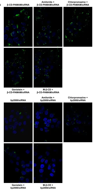
complexes and lip2000/MMP-9siRNA complexes using endocytosis inhibitors in HaCat cells. We labeled the MMP-9siRNA with 5-FAM and assessed uptake by fluorescence microscopy and flow cytometry. Cell viability in the presence
Table 2 characteristics of the dyes used
Dye/marker Application/purpose Excitationmax (nm) Emissionmax (nm)
liposome marker lysoTracker DND-99 Incorporated into liposome membrane 577 590
cell marker hoechst 33342 Nucleolus 346 460
Polyplexes or liposomes marker 5-FaM Incorporated into MMP-9sirNa 494 522
International Journal of Nanomedicine downloaded from https://www.dovepress.com/ by 118.70.13.36 on 23-Aug-2020
Dovepress comparison of the cellular transport mechanism
of different concentrations of endocytosis inhibitors was first determined to establish the optimal concentrations. Cellular toxicity was assessed using CCK-8 assays. The results are shown in Figure 2. Both complexes exerted toxic effects to a certain extent on HaCat cells, as cell viability values dropped to 88% and 74% for polyplexes and liposomes, respectively. Amiloride alone showed limited toxicity toward HaCat cells after 5 h of incubation. The highest
concentra-tion tested (100 µM) reduced viability by less than 10%
(Figure 2A). Incubation in the presence of amiloride and polyplexes or liposomes reflected an almost additive effect in cell viability. Treatment with chlorpromazine concentrations
higher than 10 µg/mL caused cytotoxicity (Figure 2B), while
combined incubation with chlorpromazine (15 µg/mL) and
polyplexes or liposomes resulted in 63.7% and 53.4% cell viability, respectively. After 5 h of incubation, HaCat cells
were observed to be sensitive to genistein up to 200 µM and to
M-β-CD up to 8 mM. On the other hand, higher concentrations
led to 50% cell death (Figure 2C and D) and were not used for
further studies. On the basis of initial study results, we selected
100 µM genistein, 50 µM amiloride, 8 µg/mL chlorpromazine
and 3 mM M-β-CD for the subsequent experiments.
HaCat cells were preincubated for 1 h with 100 µM
genistein, 50 µM amiloride, 8 µg/mL chlorpromazine, or
3 mM M-β-CD. Next, the cells were incubated for 4 h with
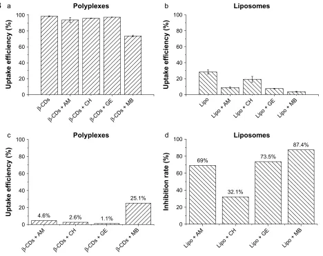
the 5-FAM-labeled polyplexes or liposomes in the presence of the corresponding inhibitor. Figure 3 shows that only 24%
of the β-CD-(D3)7/MMP-9siRNA complexes were
internal-ized into HaCat cells by the CDE and Cav-ME pathways; 70% of the lip2000/MMP-9siRNA complexes were absorbed by macropinocytosis and the Cav-ME pathway, while only 30% were absorbed by the CDE pathway into HaCat cells.
The internalization mode of complexes might affect the kinetics of their intracellular processing. The intracellular processing of polyplexes and liposomes internalized by dif-ferent pathways into HaCat cells was unclear. In this study, we used laser scanning confocal microscopy to observe the location of complexes in the cytoplasm after intervention with
Figure 2 cytotoxicity of different concentrations of endocytosis inhibitors to hacat cells.
Notes: (A): amiloride, (B): chlorpromazine, (C): genistein, (D): M-β-cD. Data presented as mean ± standard deviation.
,QKLELWRU ,QKLELWRUSRO\SOH[HV ,QKLELWRUOLSRVRPH
&HOOYLDELOLW\
'
P0
&HOOYLDELOLW\
%
JP/
&HOOYLDELOLW\
&
0
&HOOYLDELOLW\
$
0
International Journal of Nanomedicine downloaded from https://www.dovepress.com/ by 118.70.13.36 on 23-Aug-2020
Dovepress luo et al
β&'3$0$0VL51$ β&'3$0$0VL51$$PLORULGH β&KORUSURPD]LQH&'3$0$0VL51$
*HQLVWHLQ
β&'3$0$0VL51$ β&'3$0$0VL51$0β&'
OLSVL51$ OLSVL51$$PLORULGH &KORUSURPD]LQHOLSVL51$
*HQLVWHLQ
OLSVL51$ OLSVL51$0β&'
$
Figure 3 (Continued)
International Journal of Nanomedicine downloaded from https://www.dovepress.com/ by 118.70.13.36 on 23-Aug-2020
Dovepress comparison of the cellular transport mechanism
8SWDNHHIILFLHQF\
D 3RO\SOH[HV
β&'V β&'V
$0
β&'V&+β&'V*(β&'V0%
E /LSRVRPHV
/LSR $0 /LSR
/LSR&+ /LSR*( /LSR0%
8SWDNHHIILFLHQF\
8SWDNHHIILFLHQF\
F 3RO\SOH[HV
β&'V $0
β&'V&+ β&'V*( β&'V0%
G /LSRVRPHV
/LSR $0
/LSR&+ /LSR*( /LSR0%
,QKLELWLRQUDWH
%
Figure 3 The uptake of β-cD-(D3)7/MMP-9sirNa complexes or lip2000/MMP-9sirNa complexes into hacat cells after endocytosis inhibitors treatment.
Notes: (A): The internalization of 5-carboxyfluorescein (5-FAM)-loaded vesicles into HaCat cells observed by fluorescence microscopy. Punctuated green fluorescence was
observed in β-CD-(D3)7/MMP-9siRNA complexes treatment groups. Less green fluorescence was observed in lip2000/MMP-9siRNA treatment group. (B): a) The uptake rates
of β-CD-(D3)7/MMP-9siRNA complexes after different inhibitors treatment were quantified by flow cytometry; b) The uptake rates of lip2000/MMP-9siRNA complexes after
different inhibitors treatment were quantified by flow cytometry; c) The uptake inhibition rates of β-cD-(D3)7/MMP-9sirNa complexes of each inhibition group compared with
control group; d) The uptake inhibition rates of lip2000/MMP-9sirNa complexes of of each inhibition group compared with control group.
Abbreviations:β-cDs, β-cD-(D3)7/sirNa; lipo, lip2000/sirNa; aM, amiloride; ch, chlorpromazine; ge, genistein; MB, M-β-cD.
different endocytosis inhibitors. The colocalization of acidic organelle probes with polyplexes or liposomes indicated the combination of the complexes with the acidic organelles, which could by visualized as yellow particles. In the case of treatment with amiloride, chlorpromazine, or genistein, acidic organelles and polyplexes were partially colocalized, as indi-cated by few yellow particles in the cytoplasm (Figure 4). On the contrary, acidic organelles and polyplexes were not
colocalized after treatment with M-β-CD, indicating that
β-CD-(D3)7/MMP-9siRNA complexes internalized by
non-clathrin- and non-caveolae-dependent endocytosis pathways would not be translocated into acidic organelles. It is worth noting that acidic organelle and liposome colocalization was observed in each group. Acidic organelles and liposomes were completely colocalized after treatment with
chlorpromazine or M-β-CD, while partial colocalization was
observed after treatment with genistein or amiloride. The sd results suggested that the lip2000/MMP-9siRNA complexes internalized by the Cav-ME pathway and macropinocytosis would be translocated into acidic organelles.
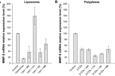
We used RT-qPCR to measure the MMP-9 mRNA transcription level, resulting from treatment with the dif-ferent endocytosis inhibitors, to explore the effect of endocytosis pathways on transfection efficiency. As shown in Figure 5, the levels of MMP-9 mRNA in the groups
transfected with β-CD-(D3)7/MMP-9siRNA complexes
were not upregulated, suggesting that no endocytosis inhibitor could completely suppress the interference effect of MMP-9siRNA. On the contrary, the levels of MMP-9 mRNA were all increased in the groups transfected with
International Journal of Nanomedicine downloaded from https://www.dovepress.com/ by 118.70.13.36 on 23-Aug-2020
Dovepress luo et al
lip2000/MMP-9siRNA complexes, and MMP-9 expression was significantly upregulated in the chlorpromazine-treated
group (with a relative value of ~8). These results indicated
that the uptake of lip2000/MMP-9siRNA complexes by the CDE pathway could induce successful transfection.
Intracellular polyplex and liposome
processing
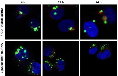
The uptake of polyplexes and liposomes at different time points was also monitored by confocal microscopy
using fluorescently labeled complexes and acidic organ-elle fluorescent probe. Images obtained at different time points are presented in Figure 6. Both the polyplexes and liposomes were partially combined with acidic organelles at 4 h after transfection, as indicated by the presence of many yellow particles, which tended to accumulate in the vicinity of the nucleus. At 12 h, the polyplexes remained colocalized with acidic organelles, while the liposomes had separated from the acidic organelles. Fewer lip2000/MMP-9siRNA complexes were observed
&RQWURO $PLORULGH &KORUSURPD]LQH *HQLVWHLQ 0β&'
β
&'3
$0$0
VL51$
/LS
003VL51$
Figure 4 colocalization of polyplexes or liposomes with acidic organelles after different internalization modes.
Notes: acidic organelles: late endosomes and lysosomal; green: 5-FaM labeled polyplexes or 5-FaM labeled liposomes; red: lysoTracker DND-99 which is incorporated into liposome membrane; Blue: Hoechst 33342 which is the probe of nucleolus. Yellow: the colocalization of red acidic organelles probe with green fluorescent polyplexes or liposomes.
003P51$
UHODWLYHH[SUHVVLRQOHYHO
3RO\SOH[HV
%
&RQWURO β&'V
β&'V
$0
β&'V&+β&'V*(β&'V0%
003P51$
UHODWLYHH[SUHVVLRQOHYHO
/LSRVRPHV
$
&RQWURO /LSRVRPHV/LSR
$0
/LSR&+/LSR*(/LSR0%
Figure 5 The rNa interference to MMP-9 messenger rNa induced by β-cD-(D3)7/MMP-9sirNa complexes or lip2000/MMP-9sirNa complexes that were internalized by different endocytosis pathways.
Notes: (A): lip2000/MMP-9sirNa complexes; (B): β-cD-(D3)7/MMP-9sirNa complexes.
Abbreviations:β-cDs, β-cD-(D3)7/sirNa; lipo, lip2000/sirNa; aM, amiloride; ch, chlorpromazine; ge, genistein; MB, M-β-cD.
International Journal of Nanomedicine downloaded from https://www.dovepress.com/ by 118.70.13.36 on 23-Aug-2020
Dovepress comparison of the cellular transport mechanism
at 12 h than at 4 h. As shown in Figure 6, the complete separation of polyplexes from acidic organelles was
observed at 24 h, and significantly fewer β-CD-(D3)7/
MMP-9siRNA complexes were observed at 24 h than at 4 h.
endosomal escape mechanisms of
polyplexes and liposomes
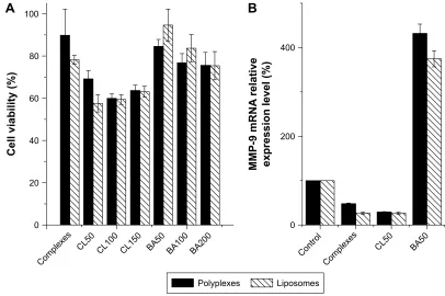
As shown in Figure 6, we found that the liposomes separated from acidic organelles faster than did the polyplexes, and we aimed to determine whether this difference was related to the endosomal escape mechanism. We applied such endosomal acidification inhibitors as chloroquine and bafilomycin A1, and cell viability in the presence of different concentrations of the inhibitors was first determined to establish the optimal protocol
for their use (Figure 7A). Finally, 50 µM chloroquine and 50 nM
bafilomycin A1 were selected for the subsequent experiments. We measured the effect of endosomal acidification on transfection efficiency by assaying the MMP-9 mRNA transcription level at 48 h after transfection (Figure 7B). In the case of chloroquine, no MMP-9 mRNA upregulation was observed in the group transfected with polyplexes or liposomes. However, in the case of bafilomycin A1, the MMP-9 mRNA levels were 8-fold and 7-fold greater than those in the polyplex and liposome positive control groups, respectively, suggesting that both complexes escaped from endolysosomes via the proton sponge mechanism.
Discussion
Our previous studies showed that β-CD-(D3)7/MMP-
9siRNA complexes had a higher internalization rate but a lower interference effect than lip2000/MMP-9siRNA complexes (uptake rate 98.4% vs 28.2%, interference rate 44.2% vs 77.2%). According to these results, we concluded
that although β-CD-(D3)7/MMP-9siRNA complexes could
be adequately absorbed, they could not effectively exert siRNA interference. This previous research confirmed that many barriers prevented gene vector–siRNA complexes from producing siRNA interference effects. Such barriers include being internalized by target cells, escaping from endolyso-somes, and dissociating the siRNA cargo from the complex to achieve successful transfection. Hence we preliminarily explored these barriers in this study.
For the development of efficient vectors, it has to be taken into account that the internalization mechanism of the vehicles will affect the whole transfection process. The entry route will first influence the uptake quantitatively, while the intracellular fate among the different compartments within the cytoplasm (late endosomes, lysosomes, etc) will eventually outline the nuclear entry and the expression of the delivered genetic material. Endocytosis has been estab-lished as the main mechanism for the uptake of nonviral
vectors into cells.4 Using selective endocytosis inhibitors
was a method to explore the internalization mechanism of
K K K
β
&'3
$0$0VL51$
/LS003VL51$
Figure 6 Uptake of polyplexes or liposomes was monitored by confocal microscopy.
Notes: Photographs were taken at 4, 12, and 24 h after transfection. green: 5-FaM labeled polyplexes or liposomes; red: lysoTracker DND-99; Blue: hoechst 33342.
International Journal of Nanomedicine downloaded from https://www.dovepress.com/ by 118.70.13.36 on 23-Aug-2020
Dovepress luo et al
nonviral gene vectors. Most studies in the field of particle uptake focus on pathways via the endocytosis of caveolae- or clathrin-coated vesicles and via macropinocytosis. We used the following four types of endocytosis inhibitors: amiloride (macropinocytosis), chlorpromazine (clathrin-mediated),
genistein (caveolae-mediated), and M-β-CD (caveolae- and
clathrin-mediated). We studied the effects of the chemical endocytosis inhibitors on the uptake of polyplexes and liposomes by HaCat cells using fluorescence microscopy and flow cytometry. As shown in Figure 3, we concluded that the uptake of polyplexes might be largely mediated by another internalization mode, such as non-caveolae- and non-clathrin-mediated pathways. However, the uptake of liposomes was diversified. Liposomes were mainly internalized by HaCat cells via macropinocytosis and the Cav-ME pathway and were marginally internalized by the clathrin-mediated endocytosis pathway.
Many factors regarding the characteristics of the carri-ers such as size, superficial charge, shape, or cell type can influence the internalization route. Size has been reported to be one of the most important influencing factors. Obser-vations of the uptake of fluorescent polymers published by
Rejman et al6 revealed that the size of particles determines the
pathway of their entry into a cell as well as their subsequent
intracellular trafficking.6,7 Cav-ME has been described as
using vesicles that are usually small (50–80 nm in diameter), while the vesicles internalized in macropinocytosis, ie,
macro-pinosomes, can have sizes of up to 5 µm. CDE is reportedly
the preferred pathway for microspheres up to 200 nm in size.
This was inconsistent with our results showing that β
-CD-(D3)7/MMP-9siRNA complexes 200 nm in diameter were not internalized by the above three endocytic pathways but by non-clathrin- and non-caveolae-mediated pathways instead. Although non-clathrin- and non-caveolae-mediated pathways are not currently well understood, a study by Lai et al also found that small polymeric particles entered live cells via a novel non-cholesterol-independent and non-clathrin- and non-caveolae-mediated mechanism, resulting in trafficking
outside the endolysosomal pathway.8 Many viruses, such
as the influenza, Sendai, and echoviruses, were also found to infect cells utilizing non-clathrin- and non-caveolae-mediated pathways for transport to the golgi apparatus and,
subsequently, to the endoplasmic reticulum.9,10
Previous research has reported that the mode of com-plex internalization might affect not only the intracellular
processing kinetics but also the transfection efficiency.11
Thus, we studied the intracellular processing of polyplexes and liposomes internalized by different endocytosis pathways
Figure 7 The effect of endosomal acidification to the transfection efficiency of β-cD-(D3)7/MMP-9sirNa complexes or lip2000/MMP-9sirNa.
Notes: (A): The HaCat cells viability after treating with different concentrations of endosomal acidification inhibitors. CL: chloroquine (concentration range: 50–150 µM);
BA: Bafilomycin A1 (concentration range: 50–200 nM). (B): The relative expression level of MMP-9 mRNA after different endosomal acidification inhibitors treatments
quantified by RT-PCR.
Abbreviation: rT-Pcr, real-time polymerase chain reaction.
3RO\SOH[HV /LSRVRPHV
&RPSOH[HV &/ &/ &/ %$ %$ %$
&HOOYLDELOLW\
$
&RPSOH[HV
&RQWURO &/ %$
%
003P51$
UHODWLYH
H[SUHVVLRQOHYHO
International Journal of Nanomedicine downloaded from https://www.dovepress.com/ by 118.70.13.36 on 23-Aug-2020
Dovepress comparison of the cellular transport mechanism
with acidic organelle fluorescent probes. The LysoTracker®
(Thermo Fisher Scientific, Waltham, MA, USA) probes are fluorescent probes with high selectivity for labeling and tracking acidic organelles in live cells. Polyplexes and liposomes were labeled with 5-FAM, and LysoTracker DND-99 labeling resulted in red fluorescence. We explored the colocalization of the two types of fluorescence using laser scanning confocal microscopy, which showed yellow particles in the cytoplasm. Through the analysis of images presented in Figure 4, we speculated that polyplexes absorbed by clathrin- or caveolae-mediated uptake pathways would be transported to acidic organelles such as lysosomes and secondary endosomes. Polyplexes internalized by macro-pinocytosis and non-clathrin- and non-caveolae-mediated pathways would avoid acidic organelles. Liposomes absorbed by macropinocytosis would be sent to acidic organelles, while liposomes internalized by clathrin-mediated endocy-tosis would avoid acidic organelles. Particles transported into acidic organelles might be digested, and lysosomal trafficking is routinely cited as a critical barrier to nonviral
gene therapy.12 Lysosomal trafficking, characterized by a
low pH and an environment rich in acidic hydrolases, poses particularly severe constraints on the efficient intracellular delivery of various chemical and biological molecules. A broad range of therapeutics, including DNA, RNA, and many proteins and chemotherapeutic agents, rapidly degrade in acidic environments. The trafficking of drug and gene car-riers in acidic vesicles is a critical bottleneck in therapeutic delivery applications.
We also measured the effect of internalization mode on transfection efficiency by assaying the MMP-9 mRNA transcription level at 48 h after transfection (Figure 5). We concluded that polyplexes internalized by these three endocytosis pathways would not induce successful transfec-tion. However, liposomes internalized by each type of endo-cytosis pathway, especially clathrin-mediated endoendo-cytosis pathways, might induce successful transfection.
As shown in Figure 4, we found that both β-CD-(D3)7/
MMP-9siRNA complexes and lip2000/MMP-9siRNA complexes could partially be transported into acidic organelles, resulting in either digestion or escape into the cytoplasm. Therefore, we loosely compared the digestion rate and endosomal escape rate of polyplexes and liposomes by confocal microscopy using fluorescently labeled com-plexes and fluorescent acidic organelle probes. As shown in Figure 6, we captured images at 4, 12, and 24 h after transfec-tion. Through the analysis of images presented in Figure 6, we proposed that the lip2000/MMP-9siRNA complexes
could escape from acidic organelles more quickly than the
β-CD-(D3)7/MMP-9siRNA complexes. Digestion by acidic
organelles affected the lip2000/MMP-9siRNA complexes
less than the β-CD-(D3)7/MMP-9siRNA complexes, which
may be the main reason for the lower transfection efficiency
of the β-CD-(D3)7/MMP-9siRNA complexes.
As the polyplexes and liposomes showed different endosomal escape rates, we explored the possible endosomal escape mechanisms of the polyplexes and liposomes. The “proton sponge effect” was proposed as one of the potential
mechanisms for the endosomal release of the polymers.13
The ATPase proton pump inhibitor bafilomycin A1 and the lysosomotropic base chloroquine were thus applied in our study, both of which would disturb endosomal acidification and increase the endosomal pH. We measured the effect of the endosomal acidification inhibitors on transfection efficiency by assaying the MMP-9 mRNA transcription level at 48 h after transfection to explore the endosomal release mechanisms (Figure 7B). The results showed that while bafilomycin A1 could inhibit the endosomal escape of complexes, thus downregulating the siRNA interference effect, chloroquine failed to inhibit the endosomal escape of complexes. These results could be attributed to the
concentra-tion used in the current study (50 µM), as chloroquine has
been shown to raise endosomal pH levels at concentrations over 0.6 M. However, it is possible that HaCat cells are more sensitive to chloroquine treatment. We observed sub-stantial cytotoxicity in HaCat cells treated with chloroquine
concentrations greater than 50 µM, which precluded further
endosomal acidification to achieve the endosomal escape of polyplexes and liposomes in the current study. We con-cluded that both polyplexes and liposomes were dependent on the proton sponge mechanism for endosomal escape. Ouyang et al, described the mode of siRNA release from
cationic polymers as the proton sponge mechanism,13 which
implies that polymers sequester the protons transported into the endosomal compartment, thus preventing endosomal acidification. Subsequently, the increased osmolarity in this compartment causes the influx of water, which eventually leads to endosome lysis and the release of its contents into the cytoplasm.
In brief, we could increase the transfection efficiency
of β-CD-(D3)7 by transforming the vector’s structure to
facilitate the interaction with the epidermal cells surface, and thus, improve the internalization by non-clathrin- and non-caveolae-dependent endocytosis pathways, and then increase the release from the endosomal compound to get successful transfection. We will apply the transformed
International Journal of Nanomedicine downloaded from https://www.dovepress.com/ by 118.70.13.36 on 23-Aug-2020
International Journal of Nanomedicine
Publish your work in this journal
Submit your manuscript here: http://www.dovepress.com/international-journal-of-nanomedicine-journal
The International Journal of Nanomedicine is an international, peer-reviewed journal focusing on the application of nanotechnology in diagnostics, therapeutics, and drug delivery systems throughout the biomedical field. This journal is indexed on PubMed Central, MedLine, CAS, SciSearch®, Current Contents®/Clinical Medicine,
Journal Citation Reports/Science Edition, EMBase, Scopus and the Elsevier Bibliographic databases. The manuscript management system is completely online and includes a very quick and fair peer-review system, which is all easy to use. Visit http://www.dovepress.com/ testimonials.php to read real quotes from published authors.
Dovepress
Dove
press
luo et al
β-CD-(D3)7vector/MMP-9siRNA complexes into diabetic
foot skin to promote wound healing in the clinic. Our group is working on related research.
Conclusion
β-CD-(D3)7/MMP-9siRNA complexes were internalized by
non-caveolae- and non-clathrin-mediated pathways and could induce successful transfection, and lip2000/MMP-9siRNA complexes internalized by CDE could induce successful transfection. Although both of these complexes escaped from endolysosomes via the proton sponge mechanism, we still found that the lip2000/MMP-9siRNA complexes escaped
more quickly from the acidic organelles than did the β
-CD-(D3)7/MMP-9siRNA complexes and that digestion by acidic organelles affected the lip2000/MMP-9siRNA complexes
less than the β-CD-(D3)7/MMP-9siRNA complexes. This
effect may be the reason for the lower transfection efficiency
of β-CD-(D3)7. Our study provided insights for further
improvements in the molecular design of the polycationic amphiphilic cyclodextrin vector, eg, the incorporation of functional elements promoting endo-lysosomal escape and restraining digestion by acidic organelles. Research in this direction is currently underway in our laboratories.
Acknowledgments
This study was supported by the National Natural Science Foundation of China (81500640, 81471034, 81270916, 81370910, 81670764), the Major Projects of People’s Liveli-hood Science and Technology in Guangzhou (201300000102); Applied Science and Technology Research and Development in Guangdong Province (2016B020238001), and the National High Technology Research and Development Program (2015AA020927). This work was also supported by the Key Laboratory of Malignant Tumor Molecular Mechanisms and Translational Medicine of the Guangzhou Bureau of Science and Information Technology and the Key Laboratory of
Malignant Tumor Gene Regulation and Target Therapy of Guangdong Higher Education Institutes.
Disclosure
The authors report no conflicts of interest in this work.
References
1. Li N, Luo HC, Yang C, et al. Cationic star-shaped polymer as an siRNA carrier for reducing MMP-9 expression in skin fibroblast cells and promoting wound healing in diabetic rats. Int J Nanomedicine. 2014;9: 3377–3387.
2. Schott JW, Morgan M, Galla M, Schambach A. Viral and synthetic RNA vector technologies and applications. Mol Ther. 2016;24(9):1513–1527. 3. Namvar A, Bolhassani A, Khairkhah N, Motevalli F. Physicochemical
properties of polymers: an important system to overcome the cell bar-riers in gene transfection. Biopolymers. 2015;103(7):363–375. 4. Perez Ruiz de Garibay A. Endocytosis in gene therapy with non-viral
vectors. Wien Med Wochenschr. 2016;166(7–8):227–235.
5. Khalil IA, Kogure K, Akita H, Harashima H. Uptake pathways and sub-sequent intracellular trafficking in nonviral gene delivery. Pharmacol
Rev. 2006;58(1):32–45.
6. Rejman J, Oberle V, Zuhorn IS, Hoekstra D. Size-dependent internaliza-tion of particles via the pathways of clathrin- and caveolae-mediated endocytosis. Biochem J. 2004;377(1):159–169.
7. Parakhonskiy B, Zyuzin MV, Yashchenok A, et al. The influence of the size and aspect ratio of anisotropic, porous CaCO3 partcles on their uptake by cells. J Nanobiotechnology. 2015;13:53.
8. Lai SK, Hida K, Man ST, et al. Privileged delivery of polymer nano-particles to the perinuclear region of live cells via a clathrin, non-degradative pathway. Biomaterials. 2007;28(18):2876–2884. 9. Gerondopoulos A, Jackson T, Monaghan P, Doyle N, Roberts LO.
Murine norovirus-1 cell entry is mediated through a clathrin-, non-caveolae-, dynamin- and cholesterol-dependent pathway. J Gen Virol. 2010;91(6):1428–1438.
10. Sieczkarski SB, Whittaker GR. Dissecting virus entry via endocytosis.
J Gen Virol. 2002;83(7):1535–1545.
11. Hwang ME, Keswani RK, Pack DW. Dependence of PEI and PAMAM gene delivery on clathrin- and caveolin-dependent trafficking pathways.
Pharm Res. 2015;32(6):2051–2059.
12. Cardarelli F, Digiacomo L, Marchini C, et al. The intracellular traf-ficking mechanism of Lipofectamine-based transfection reagents and its implication forgene delivery. Sci Rep. 2016;11(6):25879. 13. Ouyang D, Zhang H, Parekh HS, Smith SC. The effect of pH on
PAMAM dendrimer-siRNA complexation: endosomal considerations as determined by molecular dynamics simulation. Biophys Chem. 2011; 158(2–3):126–133.
International Journal of Nanomedicine downloaded from https://www.dovepress.com/ by 118.70.13.36 on 23-Aug-2020