1
Title
Artificial membrane induced by novel biodegradable nanofibrous scaffold
in the Masquelet procedure for the treatment of segmental bone defects
Authors
Yi-Hsun Yu, M.D.
Department of Orthopedic Surgery, Musculoskeletal Research Center, Chang Gung Memorial Hospital-Linkou, Taoyuan, Taiwan
Department of Mechanical Engineering, Chang Gung University, Taoyuan, Taiwan Email: [email protected]
Ren-Chin Wu, M.D.
Department of Pathology, Chang Gung Memorial Hospital-Linkou, Tao-Yuan, Taiwan Email: [email protected]
Demei Lee, Ph.D.
Department of Pathology, Chang Gung Memorial Hospital-Linkou, Tao-Yuan, Taiwan Email: [email protected]
Che-Kang Chen, M.S.
Department of Pathology, Chang Gung Memorial Hospital-Linkou, Tao-Yuan, Taiwan Email: [email protected]
Shih-Jung Liu, Ph.D.
Department of Orthopedic Surgery, Chang Gung Memorial Hospital-Linkou, Taoyuan, Taiwan
Department of Mechanical Engineering, Chang Gung University, Taoyuan, Taiwan Email: [email protected]
*YH Yu and RC Wu have equal contribution to this study and are co-first authors of this paper.
Correspondence Author: Prof. Shih-Jung Liu, Ph.D.
Biomaterials Lab, Mechanical Engineering Chang Gung University
259, Wen-Hwa 1st Road Kwei-Shan, Tao-Yuan 333 Taiwan
Tel: +886-3-2118166, Fax: +886-3-2118558 Email: [email protected]
2
Abstract
Masquelet induced-membrane technique for the treatment of segmental bone
defects includes a two-stage surgical procedure, and polymethylmethacrylate (PMMA)
plays a major role in the treatment. However, the PMMA spacer must be surgically
removed. Here, we investigated the potential of poly (lactic-co-glycolic acid) (PLGA)
nanofibers, a biodegradable material to replace PMMA spacer, allowing the bioactive
membrane to be induced, and the spacer to degrade without the additional surgery
on a rabbit femoral segmental bone defect model. PLGA nanofibers were shown to
degrade completely six weeks after implantation in the investigated animals, and a
thick membrane was found to circumferentially fold around the segmental bone
defects. Results from image studies demonstrated that, in the group without bone
graft, all studied femurs exhibited either nonunion or considerable malunion. In
contrast, the femurs in the bone graft group had a high union rate without
considerable deformities. Histological examinations suggested that the membranous
tissue in this group was rich in small blood vessels and the expression of BMP2 and
VEGF increased. Our results demonstrate that the biodegradable PLGA nanofibers may
be useful for replacing the PMMA spacer as the bioactive-membrane inducer,
facilitating the process of healing and removing the need for repeated surgeries.
3 1. Introduction
Segmental bone defects may be a result of trauma, tumor resection, or the
sequelae of osteomyelitis, and their management remains challenging for
orthopedic surgeons [1,2]. Additionally, the development of these defects is
accompanied by considerable functional disabilities in patients. Two
approaches have been commonly employed for the treatment of segmental
bone defects. First, the transplantation of vascularized autologous bone graft
[2-4] has been commonly used, however, the donor site morbidity from the
autologous fibula graft, including infection and stress fracture, remains as the
main concern. In addition, the operation must be performed by a microsurgery
specialist [4]. The second approach is the bone transport with distraction
osteogenesis by Ilizarov ring fixator, which is a standard procedure for the
management of segmental bone defect applied by the experienced surgeons
in some medical institutes [5-7]. Nevertheless, various complications, including
pin tract infection, failure of the transported bone consolidation, and nonunion
at the docking site have been reported [8,9].
An alternative approach for the segmental bone defect repair was first
proposed by Masquelet in 1980 [10], showing that, following the implantation of
a polymethylmethacrylate (PMMA) spacer for 6 to 8 weeks in the segmental
4
containing osteogenic and osteoinductive factors, can be induced. This
PMMA-induced bioactive membrane serves as an envelope, encapsulating the
autologous cancellous bone graft and promoting bone healing. Other studies,
following this two-stage Masquelet procedure, demonstrated satisfactory
results for bone union [11-17]. In spite of the promising outcomes achieved
using this technique, one major drawback pertains to be the requirement for the
surgical removal of the PMMA spacer, demanding the patients to undergo
several surgeries and thus increasing the cost and complexity of the treatment.
Since the actual mechanisms underlying the formation of induced
membrane have not been completely elucidated, we aimed to examine whether
the PMMA spacer can be replaced by a different biodegradable implant, in order
to avoid the requirement for an additional surgical intervention during the
Masquelet procedure. Therefore, we developed biodegradable nanofibrous
implants and examined their ability to induce bioactive membranes using the
segmental bone defect model. Additionally, we investigated the role of
biodegradable implants as the reservoirs for bone grafting during the formation
5 2. Materials and methods
Preparation of poly (lactic-co-glycolic acid) (PLGA) nanofibers
PLGA polymers (LA:GA=50:50, Sigma, USA) were adopted. An electrospinning
setup was employed to produce the nanofibers. A high-voltage of 17 kV was
applied to the needle that emits the solution jet. The distance of the needle to
the collecting plate and the flow rate of the syringe were 12 cm and 0.5 mL/h,
respectively. Fabricated nanofibrous membrane was incubated in a chamber
equipped with a vacuum pump at 40°C for three days to volatilize the solvent.
Scanning electron microscope (SEM) characterization
The morphological structure of the polymeric fibers was characterized by a
JEOL Model JSM-7500 F field emission SEM (Tokyo, Japan).
In vivo study and animal care
This study and all procedures used acquired approval from the Institutional
Review Board and Animal Care Center of Chang Gung University, Taiwan (IRB
number: CGU106-058). Twelve 6-month-old male New Zealand rabbits were
cared and grown using the standardized procedures of the Animal Care Center
of Chang Gung University. Rabbits were housed in individual pens with free
6
±0.2 kg).
Prior to the surgeries, oxygen was delivered to the animals via a face
mask at a flow rate of at 4 L/min for five minutes. Isoflurane was then
transmitted via the face mask till the rabbit became anesthetized and continued
during the entire surgical procedure. Rabbits were kept in the decubitus position
which allowed the surgical field upwards, and the right thighs were shaved and
disinfected. A longitudinal incision was made along the lateral aspect of the
thigh, and an internervous plane was created bluntly between the vastus
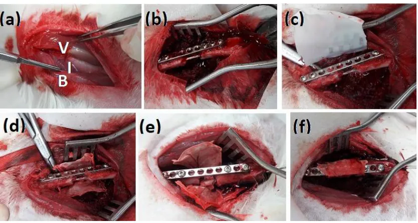
lateralis and biceps femoris to expose antero-lateral aspect of the femur (Figure
1a). Afterward, the femur was fixed with a 10-hole stainless-steel plate (Lisen
Technology Co. Ltd., New Taipei City, Taiwan) with two 2.0-mm screws at each
end, and 1.8-mm Kirschner wire was inserted intramedullary from intercondylar
notch of the femur retrogradely. After stabilization, a critical-size bone defect,
measuring 10 mm, was created in the middle of femur shaft using an osteotome
(Figure 1b).
Following the creation of the defect, the defect was wrapped
circumferentially with the PLGA nanofibers (Figure 1c), and we randomized the
operated rabbits into two groups: bone graft-free (BG-f) and bone graft (BG)
3-7
0 Vicryl (Ethicon, Johnson & Johnson, New Jersey, USA) suture to secure the
wrapping, leaving the inside of the wrapped nanofiber empty (Figure 1d), while
in the BG group, the same wrapping procedure was completed after placing the
bone chips, obtained from the osteotomized femur by chipping of the cortical
bone, inside the wrapped PLGA nanofibers (Figure 1e).
Figure 1 Surgical procedure applied for the formation and treatment of the
segmental bone defect in a rabbit model. (a) Identification the internervous
plane (I) between the vastus lateralis (V) and biceps femoris (B). (b) Creation
of a 10-mm femoral defect following the fixation procedure with stainless plate
and intramedullary nail. (c) Application of PLGA nanofibers around the bone
defect. (d) Suture-fixation of the PLGA nanofibers in BG-f group. (e) Bone
grafting (BG) inside the femoral defect before the suture-fixation of the PLGA
8
Afterward, the wound was irrigated with sterile saline and the fascia of
vastus lateralis and biceps femoris were approximated using 2-0 Vicryl suture,
while the subcutaneous tissue and skin were occluded using 3-0 Vicryl suture
(Ethicon, Johnson & Johnson, New Jersey, USA).
All animals were monitored daily for any altered behavior or
complications, and analgesics were administered for 5 days postoperatively.
They were allowed free movements and full weight bearing immediately
following the recovery from anesthesia. The rabbits were also checked twice
daily for mentation and attitude, ability to ambulate, willingness to bear weight
on the surgically treated limb, food and water consumption, respiratory rate,
and inflammation at the surgical site.
All the rabbits were euthanized 6 weeks after the surgical procedure by
a standard euthanasia procedure. The entire femur was harvested through the
plane used in the previous surgical procedures. Periosteal and fibrous tissues
surrounding the defects were preserved. The observed membranes were
excised carefully and further analyzed. Femur samples were fixed in 10%
neutral buffered formalin for 48 h and transferred to 70% ethanol, until further
9 X-ray and micro-CT examinations
The animals underwent X-ray examinations twice: immediately after the
surgical procedure and after euthanasia at six weeks. Prior to the first radiative
inspection, the animals were consoled with an intravenous injection of
zolazepam/tiletamine (Zoletil, Taipei, Taiwan). X-ray images of the
anteroposterior and lateral views were obtained. During the second X-ray
imaging, the target femora were evaluated using micro-CT as well.
Histologic analysis
Capsular tissue processing
Capsular tissue samples obtained from the investigated animals were
preserved in 10% phosphate-buffered formalin and sliced into 2-mm wide
fragments, which were processed and embedded in paraffin. Tissue sections
(4 µm) were obtained using a microtome (Sakura Finetek, Tokyo, Japan) for
histological and immunohistochemical (IHC) evaluations. Additionally, the
obtained samples were blotted with H&E and observed under a microscope
with magnification up to 400×.
IHC staining of capsular tissue
10
stainer (BOND-MAX, Leica Microsystems, Singapore). After deparaffinization,
heat-induced epitope retrieval was performed (100°C/20 min) in EDTA buffer
(pH 9). For bone morphogenic protein (BMP2) analysis, a mouse anti-BMP2
monoclonal antibody (1:200; clone 65529.111, Cat# ab6285, Abcam,
Cambridge, UK) was adopted as the primary antibody. For the characterization
of vascular endothelial growth factor (VEGF), a mouse anti-VEGF antibody
(1:400; clone VG1, Cat#: NB100-664, Novus Biologicals, Littleton CO, USA)
was used. PolyTek goat anti-mouse polymerized horseradish peroxidase (HRP;
Scytek Laboratories, Logan, UT, USA) was employed as the secondary
antibody. Bond Polymer Refine Detection Kit (DS9800, Leica Microsystems,
Singapore) was applied for the visualization of obtained signals.
3. Results
SEM analysis PLGA of nanofibers
The microscopic photos of the biodegradable nanofibers are displayed in Figure
2 (x8,000). Measured diameters of PLGA nanofibers spanned from 40 to 430
11
Figure 2 The SEM photographs of fabricated nanofibers.
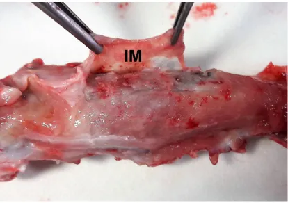
Femoral sample examination
After euthanizing the rabbits and excising the target femora, we observed that
in all specimens, a membranous layer was observed to surround the applied
PLGA nanofibers densely, immediately between the applied material and the
12
Figure 3 Induced membrane formation. Identification of the induced membrane
(IM) around the poly (lactic-co-glycolic acid) nanofibers in a representative
rabbit femoral specimen.
In the BG group, the femoral samples were shown to have continuous
hard calluses without any considerable deformities, shortening of the
osteotomized femurs, or loosened implants (Figure 4a). In contrast, in the
BG-f group, various adverse eBG-fBG-fects oBG-f the implantation were observed, such as
residual fracture gap in the calluses, loosened screws, changes in the position
of the intramedullary K wires, and considerable malunion rate and the
13
Figure 4 Representative images of the examined femoral defects after the
rabbit euthanization. (a) Representative images of the BG group femur,
showing a continuous femoral cortex with good union rate without deformity. (b)
Representative images of a malunited femur obtained from an animal included
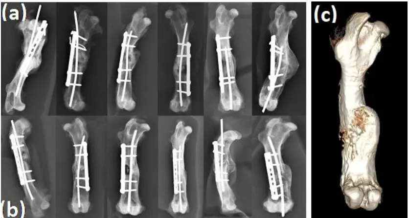
14 X-ray and micro-CT results
As observed in the follow-up series of X-ray images obtained in the BG-f group,
four femurs failed to achieve bone union, leading to the residual bony gaps,
while two femurs were malunited with considerably deformity and shortening
rates (Figure 5a). In contrast, in the BG group, only one femoral sample was
shown to have a residual gap on one side. Five tissue samples were found to
achieve bone union without serious deformities (Figure 5b), while one sample
had a loosed implant with some degree of shortening and malunion. Micro-CT
examinations revealed united bone gaps and good bone remodeling in the BG
group (Figure 5C), with good callus formation and continuous femoral cortex.
Figure 5 X-ray and micro-computed tomography (CT) evaluations of the treated
15
in the BG-f group. (b) Representative X-ray images of the treated femoral
defects in the BG group. R, a residual gap in one femur belonging to this group.
(c) Representative micro-CT images of the treated femoral defects in the BG
group.
Histologic and IHC characteristics of the capsules
The surfaces of the capsular membranes in both groups were found to be lined
by one to three layers of round to ovoid cells or short spindle cells (Figure 6a).
These cells were shown to lack the underlying basement membrane and
morphologically resembled synoviocytes. The deeper layers of the membranes
consisted of fibroblast-like spindle cells with longer cytoplasmic processes and
haphazard orientation in an extracellular matrix-rich stroma. Membranous
tissue was rich in small blood vessels. Scattered eosinophils and lymphocytes
were noted in most cases, and multinucleated giant cells were occasionally
identified.
IHC staining revealed that the membrane-lining cells and spindle cells
showed a diffuse and strong cytoplasmic expression of BMP2 (Figure 6b), with
16
exhibited intense cytoplasmic VEGF staining (Figure 6c).
Figure 6 Histologic and immunohistochemical examination of the femoral
samples. (a) Capsular membrane stained with hematoxylin and eosin. (b)
BMP2 and (c) VEGF expression in the femoral samples. Scale bars, 50 µm.
4. Discussion
In this study, we examined the effectiveness of the PLGA nanofibers used in
the Masquelet technique for the induction of periosteum-like bioactive
membrane and the reparation of the segmental bone defects. Our results
demonstrated that the bioactive membrane can be successfully induced by the
application of the biodegradable material tested here, PLGA, which was shown
to be accompanied by the expression of growth factors such as BMP2 and
VGEF. PLGA nanofibers were shown to play an important role as the bone graft
reservoirs, assisting fracture union in the segmental femoral defect model.
17
procedures since the 1970s. They have been applied in the orthopedic
surgeries, as internal fixators [18,19], drug delivery media [20-22], and bone
graft reservoirs [23,24]. PLGA has been one of the most prospective
biodegradable polymers, mainly due to its controllable degradation and
superior biocompatibility with the human tissues. This polymeric material has
received approval for clinical applications, owing to that it is innocuous, evokes
an acceptable inflammation, and can be degraded via the hydrolysis of its ester
bond [25]. These end products may induce inflammatory responses in the
surrounding tissue, recruiting fibroblasts, inflammatory cells, and stimulating
angiogenesis. Additionally, we supposed that inflammatory responses due to
the degradation of the biodegradable materials may induce tissue adhesion,
leading to the formation of an encapsulated cavity, which may play a role as a
reservoir for bone grafts. We demonstrated here that the PLGA fibers induced
the formation of a mature periosteum-like membrane circumferentially wrapped
around the fibers. These nanofibers were shown to be hydrolyzed and
degraded at the time of examination, while the cells localized in the healing
tissue expressed osteoinductive factors such as BMP2 and VGEF.
The Masquelet technique combined with a two-stage external and
18
large bone defects: the first stage consists of radical debridement, limb
stabilization, PMMA spacer implantation, and soft tissue coverage, while the
second stage consists of the clearance of infection, if infective nonunion
locations exist, removal of PMMA spacer, massive autologous cancellous bone
grafting, and a permanent internal fixation [28-30]. The implantation of the
PMMA spacer is a crucial step in this procedure, as it prevents the fibrous
tissues from invading the bone defect (mechanical role), and induces the
growth of surrounding membrane (biological role) that envelops bone grafts and
stimulates bone healing mediated by osteoinductive growth factors [26,27,31].
However, the PMMA cement spacer needs to be surgically removed, so that
the bone graft can be implanted within the “envelop” generated by the PMMA
spacer. The PLGA nanofibers examined in this study exhibited a similar ability
to induce the development of bioactive membranes, as shown by histologic and
IHC analyses. Additionally, bone healing process was shown to proceed
simultaneously with the degradation of the PLGA nanofibers in the BG group,
indicating that these grafts were securely fixed inside the PLGA nanofiber layer.
With the formation of the induced membrane and degradation of the PLGA
nanofibers, new bone formation was stimulated, suggesting that the PLGA
19
using the biodegradable nanofibers, the original two-stage Masquelet
procedure can be reduced to a single step, decreasing the time, cost, and
patient discomfort associated with the treatment, in addition to minimizing the
risk of surgical site infection.
Our study has several limitations. First, the rabbits used in the study
were all euthanized at 6 weeks, and this time point was selected based on the
timing of the standard Masquelet procedure, as in previous studies.12-16 To the
best knowledge of the authors, this research was the pioneer one to use
biodegradable material to induce bioactive membrane. Therefore, the actual
time necessary to induce membrane formation was unknown. However, we
successfully induced bioactive membrane formation, which the results of our
analyses confirmed. Furthermore, due to the loss of the fixation of one femur in
the BG group, it shape was shown to be deformed, with the bone shortening.
However, the results obtained from both groups demonstrated that the
preservation of bone grafts is crucial for bone healing. Finally, we did not
quantify the obtained micro-CT results, failing to determine the quantitative
difference between the analyzed groups. However, the union rates can be
determined from the photographs and X-ray images. In future, these differences
20 5. Conclusion
In conclusion, in this study, we successfully used the PLGA nanofibers as a
biodegradable material in Masquelet technique, which were shown to induce
the generation of bioactive membranes, envelope bone grafts, and enhance
bone union. We demonstrated additionally that this material can replace the
PMMA in the treatment of large bone defects, and it does not need to be
removed. Further studies should focus on the duration of post-operative
induction of membrane formation and the use of different biodegradable
materials with the better performance than the PLGA nanofibers.
Acknowledgements
The financial supports of the Ministry of Science and Technology, Taiwan
(Contract No. 104-2221-E-182-048-MY3) and Chang Gung Memorial
Hospital (Contract No. CMRPD2H0031) for this research are gratefully
acknowledged.
Author Contributions
Conceptualization: Y.-H.Y. and S.-J.L.; Funding acquisition S.-J.L.;
Investigation: Y.H.Y. and C.K.C.; Writing: Y.H.Y. and R.C.W.; Writing
21
Conflicts of Interest
22
References
1. Hake, M.E.; Oh, J.K.; Kim, J.W.,; Ziran, B.; Smith, W.; Hak, D.; Mauffrey,
C. Difficulties and challenges to diagnose and treat posttraumatic long
bone osteomyelitis. Eur J Orthop Surg Traumatol. 2015, 25(1), 1-3.
2. Nauth, A.; McKee, M.D.; Einhorn, T.A.; Watson, J.T.; Li R.; Schemitsch,
E.H. Managing bone defects. J Orthop Trauma. 2011, 25(8), 462-466.
3. Taylor, G.I.; Miller, G.D.; Ham, F.J. The free vascularized bone graft. A
clinical extension of microvascular techniques. Plast Reconstr Surg. 1975,
55(5), 533–544.
4. Molina, C.S.; Stinner, D.J.; Obremskey, W.T. Treatment of traumatic
segmental long-bone defects. A critical analysis review. JBJS Rev. 2014,
2(4).
5. Madhusudhan, T.R.; Ramesh, B.; Manjunath, K.; Shah H.M.; Sundaresh,
D.C.; Krishnappa, N. Outcomes of Ilizarov ring fixation in recalcitrant
infected tibial non-unions—a prospective study. J Trauma Manag
Outcomes. 2008, 2(1), 6.
23
Baek, S.G.; Jung, G.H. Bone transport with an external fixator and a
locking plate for segmental tibial defects. Bone Joint J. 2013, 95-B(12),
1667-1672.
7. Demiralp, B.; Ege, T.; Kose, O.; Yurttas, Y.; Basbozkurt, M. Reconstruction
of intercalary bone defects following bone tumor resection with segmental
bone transport using an Ilizarov circular external fixator. J Orthop Sci.
2014, 19(6), 1004–1011.
8. Chaddha, M. Gulati, D. Singh, A.P.; Singh, A.P. Maini, L. Management of
massive posttraumatic bone defects in the lower limb with the Ilizarov
technique. Acta Orthop Belg. 2010, 76(6), 811–820.
9. Giannoudis, P.V. Treatment of bone defects: bone transport or the induced
membrane technique? Injury. 2016, 47(2), 291–292.
10. Masquelet, A.C.; Fitoussi, F.; Begue, T.; Muller, G.P. Reconstruction of the
long bones by the induced membrane and spongy autograft. Ann Chir
Plast Esthet. 2000, 45(3), 346–353.
11. Pelissier, P.; Martin, D.; Baudet, J.; Lepreux, S.; Masquelet, A.C.
24
Plast Surg. 2002, 55(7), 596–598.
12. Karger, C.; Kishi, T.; Schneider, L.; Fitoussi, F.; Masquelet, A.C.; for French
Society of Orthopaedic Surgery and Traumatology (SoFCOT). Treatment
of posttraumatic bone defects by the induced membrane technique.
Orthop Traumatol Surg Res. 2012, 98(1), 97-102.
13. Taylor, B.C.; Hancock, J.; Zitzke, R.; Castaneda, J. Treatment of bone loss
with the induced membrane technique: techniques and outcomes. J
Orthop Trauma. 2015, 29(12), 554-557.
14. Olesen, U.K.; Eckardt, H.; Bosemark, P.; Paulsen, A.W.; Dahl, B.; Hede, A.
The Masquelet technique of induced membrane for healing of bone
defects. A review of 8 cases. Injury. 2015, 46 Suppl 8, S44-S47.
15. Scholz, A.O.; Gehrmann, S.; Glombitza, M.; Kaufmann, R.A.; Bostelmann,
R.; Flohe, S.; Windolf, J. Reconstruction of septic diaphyseal bone defects
with the induced membrane technique. Injury. 2015, 46 Suppl 4,
S121-S124.
16. Ronga, M.; Ferraro, S.; Fagetti, A.; Cherubino, M.; Valdatta, L.; Cherubino,
25
loss. Injury. 2014, 45 Suppl 6, S111-S115.
17. Morris, R.; Hossain, M.; Evans, A.; Pallister, I. Induced membrane
technique for treating tibial defects gives mixed results. Bone Joint J.
2017, 99-B(5), 680-685.
18. Peng, W.; Zheng, W.; Shi, K.; Wang, W.; Shao, Y.; Zhang, D. An in vivo
evaluation of PLLA/PLLA-gHA nano-composite for internal fixation of
mandibular bone fractures. Biomed Mater. 2015, 10(6), 065007
19. Uhthoff H.K.; Poitras P.; Backman D.S. Internal plate fixation of fractures:
short history and recent developments. J Orthop Sci. 2006, 11(2), 118-126.
20. Suto, T.; Obata, H.; Tobe, M.; Oku, H.; Yokoo, H.; Nakazato, Y.; Saito, S.
Long-term effect of epidural injection with sustained-release lidocaine
particlesin a rat model of postoperative pain. Br J Anaesth. 2012, 109(6),
957-967.
21. Yu, Y.H.; Hsu, Y.H.; Chou, Y.C.; Fan, C.L.; Ueng, S.W.N.; Kau, Y.C.; Liu,
S,J. Sustained relief of pain from osteosynthesis surgery of rib fracture by
using biodegradable lidocaine-eluting nanofibrous membranes.
26
22. Ferguson, J.; Diefenbeck, M.; McNally, M. Ceramic biocomposites as
biodegradable antibiotic carriers in the treatment of bone infections. J
Bone Jt Infect. 2017, 2(1), 38-51.
23. Chou, Y.C. ; Lee, D. ; Chang, T.M.; Hsu, Y.H. ; Yu, Y.H.; Liu, S.J. ; Ueng,
S.W. Development of a three-dimensional (3D) printed biodegradable cage
to convert morselized corticocancellous bone chips into a structured
cortical bone graft. Int J Mol Sci. 2016, 17(4), E595.
24. Casagrande, S.; Tiribuzi, R.; Cassetti, E.; Selmin, F.; Gervasi, G.L.;
Barberini, L.; Freddolini, M.; Ricci, M.; Schoubben, A.; Cerulli, G.G.; Blasi,
P. Biodegradable composite porous poly(dl-lactide-co-glycolide) scaffold
supports mesenchymal stem cell differentiation and calcium phosphate
deposition. Artif Cells Nanomed Biotechnol. 2017, 21, 1-11.
25. Kumbar, S.G.; Nukavarapu, S.P.; James, R.; Nair, L.S.; Laurencin, C.T.
Electrospun poly(lactic acid-co-glycolic acid) scaffolds for skin tissue
engineering. Biomaterials. 2008, 29(30), 4100-4107.
26. Wang, X.; Wei, F.; Luo, F.; Huang, K.; Xie, Z. Induction of granulation
tissue for the secretion of growth factors and the promotion of bone defect
27
27. Christou, C.; Oliver, R.A.; Yu, Y.; Walsh, W.R. The Masquelet technique for
membrane induction and the healing of ovine critical sized segmental
defects. PLoS One. 2014, 9(12), e114122.
28. Masquelet, A.C. Induced membrane technique: pearls and pitfalls. J
Orthop Trauma. 2017, 31 Suppl 5, S36-S38.
29. Giannoudis, P.V.; Faour, O.; Goff, T.; Kanakaris, N.; Dimitriou, R.
Masquelet technique for the treatment of bone defects: Tips-tricks and
future directions. Injury. 2011, 42(6), 591–598.
30. Mauffrey, C. ; Hake, M.E. ; Chadayammuri, V.; Masquelet, A,C.
Reconstruction of long bone infections using the induced membrane
technique: tips and tricks. J Orthop Trauma. 2016, 30(6), e188-e193.
31. Ma, C.H.; Chiu, Y.C.; Tsai, K.L.; Tu, Y.K.; Yen, C.Y.; Wu, C.H. Masquelet
technique with external locking plate for recalcitrant distal tibial nonunion.