Sequence-based analysis of zonula occludens toxins identified by comparative
genomics in non-toxigenic Vibrio parahaemolyticus strains isolated in Southern
Chile.
Diliana Pérez-Reytor1, Daniel Castillo2, Carlos J. Blondel1
,Sebastián Ramírez-Araya1,3, Nicolás Plaza1, Alequis Pavón1, Gino Corsini1, Víctor Jaña4, Leonardo Pavéz4,5, Roberto Bastías6, Paola Navarrete7,8,Katherine García1*
1. Instituto de Ciencias Biomédicas, Facultad de Ciencias de la Salud, Universidad Autónoma de Chile. DPR:
[email protected]; CJB: [email protected]; NP: [email protected]; AP:
[email protected]; GC: [email protected]; KG: [email protected].
2. Marine Biological Section, University of Copenhagen, Strandpromenaden 5, DK-3000, Helsingør, Denmark. DC:
3. Departamento Ciencias Básicas, Facultad de Ciencias, Universidad Santo Tomás, Av. Ejército Libertador 146, Santiago de Chile. SR: [email protected].
4. Facultad de Medicina Veterinaria y Agronomía, Universidad de Las Américas, Santiago, Chile. VJ:
[email protected]; LP: [email protected].
5. Departamento de Ciencias Químicas y Biológicas, Universidad Bernardo O'Higgins, Santiago, Chile. LP:
6. Laboratory of Microbiology, Institute of Biology, Pontificia Universidad Católica de Valparaíso, Valparaíso, Chile. RB:
7. Laboratory of Microbiology and Probiotics, Institute of Nutrition and Food Technology (INTA), University of Chile, Santiago, Chile. PN: [email protected].
8. Millenium nucleus in the Biology of Intestinal Microbiota, Santiago, Chile.
* Correspondence: [email protected], Instituto de Ciencias Biomédicas, Facultad de Ciencias de la Salud, Universidad Autónoma de Chile. El Llano Subercaseaux 2801, San Miguel, Santiago de Chile.
Abstract
Gastroenteritis cases associated with non-toxigenic strains of Vibrio parahaemolyticus have been reported in many countries, suggesting the contribution of novel virulence factors. One candidate is zonula occludens toxin (Zot), which increases the intestinal permeability by other bacteria. Recently we identified prophages belonging to the Inoviridae family encoding putative Zot-like toxins in Chilean strains. Based on this information we performed sequence-based analyses of these toxins, followed by phylogenetic and structural analyses using computational tools. Our results showed that Zots found in Chilean V. parahaemolyticus strains are grouped into three different phylogenetic clusters, sharing two conserved motifs (Walker A and B) in their N-terminal region. These motifs are also conserved in Zots from the human pathogens Vibrio cholerae, Neisseria meningitidis and Campylobacter concisus. Although Zots of V. parahaemolyticus do not possess the FCIGRL sequence responsible for the effects produced by V. cholerae, they do possess a conserved secondary structure within their C-terminal region with Zots proteins able to disrupt the intestinal barrier, which is interesting since it has been suggested that the structure and not the Zot sequence would be responsible for the biological effects. This preliminary study provides the
basis to study the function of Zots found in V. parahaemolyticus on the intestinal barrier and their possible role as a virulence factor.
Keywords: Vibrio parahaemolyticus, non-toxigenic strains, Zot, zonula occludens toxin, Vibrio cholerae, Campylobacter concisus, intestinal permeability
KeyContribution:This study provides valuable information for a more in-depth examination of Zot sequences found in Vibrio parahaemolyticus strains, and additionally provides the basis for the study of their biological effects on the intestinal epithelial barrier and their definition as a possible new virulence factor in this species.
1. Introduction
Inshore marine waters around the world are densely populated with Vibrio parahaemolyticus [1], which is the leading cause of seafood-associated bacterial gastroenteritis [2]. However, only a few strains can cause infections in humans and most environmental strains are non-pathogenic [3]. The most characteristic virulence-associated factors are thermostable direct hemolysin (TDH) and TDH-related hemolysin (TRH), encoded by the tdh and trh genes, respectively [2–5]. However, V. parahaemolyticus remains pathogenic in the absence of these hemolysins, indicating that other virulence factors exist [2]. Analysis of the complete genome sequence of V. parahaemolyticus strain RIMD2210633 revealed the presence of other virulence factors such as the type III and VI secretion systems (designated T3SS and T6SS respectively), in both chromosome and in various genomic islands (VPaI) [6–8]. Studies reported that environmental isolates of V. parahaemolyticus lacking tdh and/or trh and T3SS2 can be highly cytotoxic to human gastrointestinal cells [9–11]. These results indicate that cytotoxicity and enterotoxicity of pathogenic V. parahaemolyticus are not explained only by classic virulence factors and suggest that one or more novel virulence factors could be responsible for its pathogenicity [12]
causes sustained intestinal barrier damage, induces the liberation of proinflammatory cytokines and increases the response of macrophages to other microorganisms [23]. In our previous work we showed that a V. parahaemolyticus zot sequence is encoded into prophage f237 of the pandemic RIMD2210633 strain, which is different from the V. cholerae CTX prophage [11]. We also showed that the clinical strain PMC53.7, which does not possess any other known virulence factor in its genome, also possess a zot-encoding prophage [11]. However, whether these Zot proteins encode bona-fide enterotoxins remains unknown. It was recently reported that different zot-encoding prophages were found in 77.9% of the clinical isolates of V. parahaemolyticus [24]. These prophages belong to the Inoviridae family, which plays an important role in the evolution and pathogenesis of multiple bacteria, showing that Zot is highly prevalent in clinical strains of this species [24]. This suggests that Zots could have a possible role in the pathogenesis of V. parahaemolyticus. In this preliminary study we decided to perform sequence-based analyses of Zot toxins from non-toxigenic Chilean V. parahaemolyticus strains, followed by phylogenetic and structural analyses to identify their main features using computational tools. The phylogenetic analysis of Zot-like toxin proteins showed that Zot from Chilean non-toxigenic strains PMC53.7 and PMA2.15 belongs to the B4 phylogenetic group, while Zot of PMA3.15 belongs to B2 phylogenetic group defined by Castillo et al. 2018b [24]. We noted that two motifs in the N-terminal end (Walker A and Walker B) and the secondary structure of the C-N-terminal motif FCIGRL are highly conserved with Zots of V. cholerae and C. concisus strains, which are able to damage the intestinal barrier and disturb intestinal permeability. These results will provide useful information to study further a potential role of Zot in the pathogenesis of V. parahaemolyticus.
2. Results
Phylogenetic analysis of zot sequences and comparison of prophages containing zot in Vibrio species
Figure 1. Genomic organization of zot-encoding prophages in Vibrio species A) Diagrammatic representation of zot-encoding prophages and phages of different pathogenic and environmental Vibrio parahaemolyticus isolates (See Supplementary Table 1S). B) Maximum likelihood tree based on the amino acid sequences of Zots found in different Vibrio species. V. parahaemolyticus Zots of Chilean non-toxigenic strains belonging to the B2 and B4 phylogenetic groups are highlighted. Bootstrap values <80% were removed from the tree. The horizontal bar at the base of the figure represents 0.5 substitutions per amino acid site.
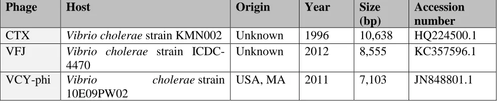
A list of Zot-encoding prophages in V. parahaemolyticus and V. cholerae is shown in Table 1.
Table 1. Prophages which harbor zot in V. parahaemolyticus and V. cholerae prophage strains
Phage Host Origin Year Size
(bp)
Accession number
CTX Vibrio cholerae strain KMN002 Unknown 1996 10,638 HQ224500.1
VFJ Vibrio cholerae strain ICDC-4470
Unknown 2012 8,555 KC357596.1
VCY-phi Vibrio cholerae strain
10E09PW02
VEJphi Vibrio cholerae strain MO45 India 2010 6,849 FJ904927
KSF1 Vibrio cholerae strain 55V71 Bangladesh 2005 7,107 AY714348.1
VSK Vibrio cholerae O139 India 2001 6,882 AF453500.3
VGJphi Vibrio cholerae strain SG25-1 India 2003 7,542 AY242528.1
fs1 Vibrio cholerae strain A1-4450 Unknown 1997 6,341 NC_004306.1
fs2 Vibrio cholerae strain MDO14 Unknown 1998 8,651 AB002632.1
Vf12 Vibrio parahaemolyticus strain Vp12
Unknown 1998 7,965 AB012574.1
Vf33 Vibrio parahaemolyticus strain Vp33
Unknown 1998 7,965 AB012573.1
VfO4K68 Vibrio parahaemolyticus strain O4:K68
Japan 2002 6,891 AB043679.1
VfO3K6 Vibrio parahaemolyticus strain O3:K6
Japan 2000 8,784 AB043678.1
We constructed a phylogenetic tree based on the amino acid sequences of Zots found in various Vibrio species, including the three Chilean non-toxigenic V. parahaemolyticus strains PMA2.15, PMA3.15 and PMC53.7 (Supplementary Table S1). As shown in Figure 1B, the Zot toxin present in PMA2.15 grouped with Zots of the pandemic strain and the V. parahaemolyticus phages O3:K6 and O3:K48. In contrast, the Zot toxin present in PMC53.7 was different in sequence and had greater similarity to sequences encoding Zots present in V. campbellii and other V. parahaemolyticus strains. The toxin of PMA 3.15 was the most different in sequence and shared an ancestor in common with the V. cholerae phage VFJ (Figure 1B). It should also be noted that the clade formed by the coding sequences for Zot present in V. cholerae does not include V. parahaemolyticus sequences (Figure 1B). Interestingly, the phylogenetic analysis of these Zot-like toxin proteins showed four different clusters, B1–B4 defined by Castillo et al., 2018b, where the B4 group included the Zot toxin encoded by prophage VfO3K6 identified in the pandemic V. parahaemolyticus clone. Our analysis showed that Zots found in Chilean non-toxigenic strains PMC53.7 and PMA2.15 belong to B4 while the Zot of PMA3.15 belongs to the B2 phylogenetic group (Figure 1B). Interestingly, all these zot-encoding prophages contained the Accessory Cholera Enterotoxin gene (ace), which has also been described in the CTX prophage.
Multiple sequence alignment (MSA) in different Zot proteins and their Walker A and Walker B motifs
of V. parahaemolyticus Chilean strains PMC53.7, PMA2.15 and PMA3.15. Both Walker motifs are marked with black squares.
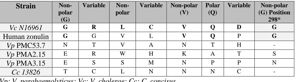
We noticed that three V. parahaemolyticus strains sequences have a glycine changed to a tyrosine in the Walker A motif (GXXXXYK[S/T]) as also was observed in V. cholerae. Both Walker motifs were located at the N-terminal side prior to the transmembrane domains (approximately 1–270, as defined for V. cholerae Zot). As these Walker A and Walker B motifs belong to the proteins of the p-loop containing the nucleoside triphosphate hydrolase (p-loop NTPase) superfamily, we used the entry identity IPR027417 in InterPro database [23]. As expected, we identified that the Zot proteins of V. parahaemolyticus had p-loop NTPase domains, the most prevalent domain of the several distinct nucleotide-binding protein folds (Figure 2, black squares). All sequences of the Zot proteins identified in V. parahaemolyticus aligned with 100% identity with the previously identified active domain of V. cholerae FCIGRL located in the C-terminal domain (Figure 3, black square) [19]. However, neither the other Zot sequences of C. concisus or N. meningitidis had the FCIGRL domain (Figure 3, black square). Di Pierro and coworkers showed that the eight amino acids shared by Zot and zonulin represent the putative receptor-binding site, characterized by the motif: non-polar (G)/variable/non-polar/variable/non-polar (V)/polar (Q)/variable/non-polar (G) [26]. They also showed that the glycine residue in position 298 has a key role in the activation of the intercellular TJ opening. We showed that PMC53.7, PMA2.15 and PMA3.15 do not have a glycine in position 298 (Table 2), but also C. concisus. Instead, PMA2.15 and PMA3.15 have a serine (S) and asparagine (N) residue, respectively, in this position (Table 2).
Table 2. Amino acid sequence of V. parahaemolyticus and C. concisus strains aligned with the octapeptide suggested as the Zot putative receptor-binding site of V. cholerae.
Strain
Non-polar (G)
Variable
Non-polar
Variable Non-polar (V)
Polar (Q)
Variable Non-polar (G) Position
298*
Vc N16961 G R L C V Q D G
Human zonulin G G V L V Q P G
Vp PMC53.7 N T V A N T H -
Vp PMA2.15 E R W H K A T S
Vp PMA3.15 E S S M N P P N
Cc 13826 T C L N N N C -
Vp: V. parahaemolyticus; Vc: V. cholerae; Cc: C. concisus
Secondary structure of the C-terminal region of V. parahaemolyticus Zot proteins.
As was described above, the active domain of V. cholerae FCIGRL was absent from Zot proteins of V. parahaemolyticus, C. concisus and N. meningitidis. However, the secondary structure of the
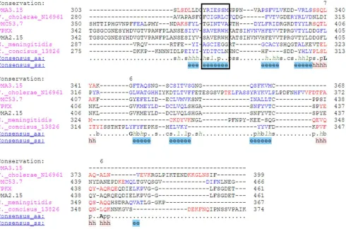
-sheet of sequences that aligned with this fragment using PROMALS3D alignment [28,29]was highly conserved among V. cholerae, N. meningitidis, C. concisus and all the V. parahaemolyticus strains (Figure 3).
Figure 3. Zot alignment considering the C-terminal secondary structure of PMC53.7, PMA2.15, PMA3.15, VpKX and N. meningitidis MC58, V. cholerae El Tor Inaba N16961 and C. concisus 13826. The C-terminal was calculated based on the information of domains reported for V. cholerae [26]. The conserved structure of the α-helix and -sheet can be observed under the alignment in red (hhhhhh) and blue (eeeee) respectively. The conserved structure of the FCIGRL fragment is marked with a black square. Complete alignment can be observed in Supplementary figure 1.
prediction using PSIPRED [31]. Zot proteins of V. parahaemolyticus, V. cholerae and C. concisus showed a conserved secondary structure in their C-terminal region (Supplementary Figure 2). Zots of PMC53.7, PMA2.15 and PMA3.15 contain 7, 8 and 9 α-helices, and 17, 19 and 17 β-strands respectively, compared to the 7 and 8 α-helices and 20 and 14 β-strands of V. cholerae and C. concisus. Interestingly, in all cases the region of the FCIGRL peptide of V. cholerae was part of conserved β-strand structure (see PMC53.7 in Supplementary Figure 2, PMA2.15 and PMA3.15 are not shown).
Prediction of the transmembrane domain in the Zots of V. parahaemolyticus
Figure 4. Prediction of transmembrane domains in Zot of V. parahaemolyticus strains using the Phobius server. (a) V. parahaemolyticus PMA3.15; (b) V. parahaemolyticus VpKX and PMA2.15; (c) V. parahaemolyticus PMC53.7; (d) V. cholerae used as control.
Structure prediction and 3D modeling
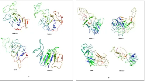
The Zot protein homology was determined using HHpred [33]. Amino acids aligned with a probability of 99.72% in the N-terminal region of the "Zonula Occludens Toxin" from N. meningitidis MC58 (template 2R2A from the PDB database), which has a crystallized structure of 199 amino acids from its N-terminal domain. The tridimensional structure was predicted with Phyre2 [34] and Swiss-Modell [35] using the template 2R2A, which had sequence identity with the target protein. Since to date there is no model for the C-terminal region of this protein, we could only align the first 250 amino acids of V. parahaemolyticus Zots using the crystallized Zot of N. meningitidis as template (partially available N-terminal structure). A total of 8 three-dimensional models were obtained (Figure 5).
Figure 5. Tertiary structure of V. parahaemolyticus Zot. Crystallized Zot of N. meningitidis 2R2A was used as template. (a) Swiss-Model, (b) Phyre2
Validity evaluation of the model
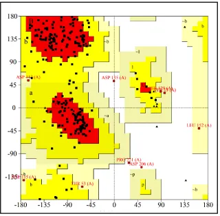
experimental structures deposited in PDB. Z-scores outside a range characteristic for native proteins indicate erroneous structures [36,37]. The results obtained are shown in Table 3. Comparing with calculated z-score, the model is shown to be valid according to the quality of the structure (Figure 6).
Figure 6. (a) ProSA analysis; Z-score plot (overall quality model) and (b) graphic plot (local model quality) of PMC53.7 Zot protein (Swiss-model 3D model).
Table 3. ProSA-web z-score results for the Zot 3D structure
Z-score PMC53.7 PMA2.15 PMA3.15 VpKX
Phyre2 -4.21 -2.91 -4.47 -3.75
Swiss-Model -5.28 -2.57 -3.64 -2.57
Figure 7. Ramachandran plot for PMC53.7 Zot (predicted 3D model) generated using PROCHECK server. The areas showing different colors i.e. red, yellow and light yellow represents most favored regions (76.7%), additional allowed regions (19.6%) and generously yellowed regions (2.7%) respectively. Residues in disallowed regions (0.9%).
3. Discussion
Our previous studies demonstrated that V. parahaemolyticus Zot proteins had low (about 24% identity) similarity to V. cholerae Zot, but they share some conserved regions located toward the N-terminal domain [11]. One of these domains is the Pfam PF05707 Zot domain, named for the homologue in the Vibrio CTX phage, which is essential for the assembly and export of phage virions. All Zot proteins of V. parahaemolyticus also contained Walker A and Walker B motifs, which are conserved motifs of the p-loop NTPase superfamily [27]. P-loop NTPase binds to NTP, typically ATP or GTP, through the Walker A and B motifs. Specifically, the N-terminal of Zot is predicted to act as an ATPase, powering the assembly and transport of phages through the envelope, as has been observed for Escherichia coli Ff-type phages [38].
The change of a glycine (non-polar aliphatic amino acid) to a tyrosine (aromatic amino acid) into the Walker A motif (GXXXXGK[S/T]) observed in most of V. parahaemolyticus strains was also observed in V. cholerae (GXXXXYK[S/T]) but non-observed in V. parahaemolyticus PMA3.15. This strain has a Glycine as also N. meningitidis and many Campylobacter species [23]. Despite this change, the Zot of V. cholerae maintains the functionality [15]. Also, a transmembrane domain was found in all V. parahaemolyticus Zot proteins, showing that Zots are transmembrane proteins in this species. We observed very low similarities (a, b, c values) among the C-terminal end of V. parahaemolyticus Zot proteins compared to Campylobacter, V. cholerae and N. meningitidis Zot proteins. No Zots of V. parahaemolyticus contained the active fragment described for V. cholerae in their sequences. However, since the Zot of C. concisus, lacking the FCIGRL fragment, also affects the paracellular pathway experimentally, it may be that the presence of this amino acid sequence would not be strictly necessary to perform the action of all Zot proteins. Similarly, a glycine in position 298 of V. cholerae Zot, with a crucial role in producing the opening of intracellular TJs, is also absent in C. concisus. These observations support that the structure and not the sequence would be responsible for the biological effects of Zot on the epithelial barrier. Indeed, all Zots of V. parahaemolyticus shared the secondary structure observed for the active fragment in V. cholerae and this -sheet structure is also observed in C. concisus. In summary, our results showed high variability in the amino acid sequences of Zot proteins between different bacterial species and between strains of V. parahaemolyticus, but all of them shared a general similarity in the secondary structure. However, it must be considered that the comparison of Zot amino acid sequences of V. parahaemolyticus strains against known toxin databases suggests that not all Zots would have a function, or at least not the same function. Interestingly, the sequence of PMA3.15, having not-matchwith any toxin, was the most different in sequence.
N-terminal region. These values indicate that the predicted structures are located in the range of native protein and are close to the database average, allowing the choice of the better model. Altogether, our results provide useful information for further examination of V. parahaemolyticus Zot proteins as potential virulence factors in non-toxigenic strains. Future studies will be conducted to determine the role of Zot in V. parahaemolyticus and the mechanism that could affect human cells.
4. Materials and Methods
Multiple sequence alignment (MSA) and phylogenetic analysis
The amino acid sequences of the Zot proteins from V. cholerae, C. concisus and N. meningitidis were obtained from UniprotKB. Multiple alignment was converted to PHYLIP format using Clustal Omega software [39]. It is shown in the InterPro database that the Zot family proteins (InterPro entry identity: IPR008900) belong to the p-loop NTPase superfamily. The proteins of the p-loop NTPase superfamily have Walker A and Walker B motifs [27]. Here we examined the presence of Walker A and Walker B motifs in V. parahaemolyticus Zot proteins by protein alignment using Clustal Omega software [39] and the PROMALS3D multiple sequence and structure alignment server [28,29] at http://prodata.swmed.edu/promals3d/promals3d.php. The Walker A motif has a sequence of GxxxxGK[S/T], where x is any residue, and the Walker B motif has a sequence of hhhh[D/E], where h is a hydrophobic residue [27]. The alignments for multiple protein sequences and secondary structure prediction were performed with PROMALS3D [28,29]. To reveal the phylogenetic relationship among genes encoding the identified Zot, amino acid sequences were aligned using ClustalW version 2.042 and phylogeny was inferred using Maximum Likelihood (1,000 bootstrap replicates) in Geneious version 10.1.338 [40].
Sequence-based analyses of the Zot protein
Sequence-based analysis of the Zot protein was performed using different web-based tools: the InterProScan https://www.ebi.ac.uk/interpro/; the PSIPRED server [31] at
http://bioinf.cs.ucl.ac.uk/psipred/; CD search tools [41,42]
https://www.ncbi.nlm.nih.gov/Structure/cdd/wrpsb.cgi and the NetPhos server [43] http://www.cbs.dtu.dk/services/NetPhos/ to detect domains and motifs, secondary structures, superfamily, and phosphorylation sites of the Zot protein, respectively. The prediction analysis of bacterial toxins was performed with the BTXpred program in SVM mode [45].
Prediction of transmembrane domains in V. parahaemolyticus Zot
The Phobius server [32] was used for prediction of transmembrane topology and signal peptides at https://www.ebi.ac.uk/Tools/pfa/phobius/.
Homology modeling of selected protein sequences was performed using Phyre2 [34] at http://www.sbg.bio.ic.ac.uk/phyre2/, the SWISS-MODEL automated comparative modeling server [35] at https://swissmodel.expasy.org/. Protein structure files were compiled from the protein data bank available at http://www.rcsb.org/pdb. Protein structures were viewed using CLC Sequence Viewer version 8.0.
Validation of the Zot protein model generated
The internal consistency and reliability of the model of the Zot protein (Swiss- Model and Phyre2) were evaluated using the ProSA-web server [36] at (https://prosa.services.came.sbg.ac.at/). The ProSA-web calculates the general quality score for a 3D structure. If the calculated scores are outside the native protein range, an error in the predicted structure is indicated. The overall quality of the targeted protein is validated with a graphical output map of local quality estimates. The z-score for homology modeling was calculated with a graphical plot, where the X-ray and NMR data from all the known protein sequences commencing to the PDB database are clearly depicted. Accordingly the residue score has been calculated using each amino acid sequence positions. Both 10 amino acid residue and 40 amino acid residue energy data are calculated for further consideration in experimental and theoretical structure validation [36]. Parallely the ProCheck server at http://servicesn.mbi.ucla.edu/PROCHECK/ has been applied for validation of the stereochemical quality of proteins structure using the Ramachandran plot [44].
Supplementary Materials
Figure 1S. Complete alignment of secondary structure of PMC53.7, PMA2.15, PMA3.15, VpKX and N. meningitidis MC58, V. cholerae El Tor Inaba N16961 and C. concisus 13826. The C-terminal was calculated based on the information of domains reported for V. cholerae [26]. The conserved structure of the α-helix and B-sheet can be observed under the alignment in red (hhhhhh) and blue (eeeee) respectively. The conserved structure of the FCIGRL fragment is marked with a black square.
Figure 2S. The secondary structure composition of the Zot protein of V. cholerae, C. concisus and V. parahaemolyticus PMC53.7 was predicted using the PSIPRED web server. The pink cylinders represent helices, the yellow arrows represent β-strands, and the black lines represent coiled structures. The height and color intensity of the blue bars indicate the confidence (conf) of the prediction. Pred: predicted secondary structure; AA: target sequence.
Table 1S. Amino acid sequences of Zots found in various Vibrio species.
Author contribution
performed the sequence alignment analysis and searching of conserved motifs, GC and RB performed the secondary structure analysis and determined the position of transmembrane domain, AP and CJB made the 3D structure prediction and validated the 3D models. All the authors read, discussed and approved the final version of this manuscript.
Conflict of Interest Statement
The authors declare that the research was conducted in the absence of any commercial or financial relationships that could be construed as a potential conflict of interest.
Funding
The authors acknowledge Fondecyt Iniciación 11140257 and 11160901, Fondecyt Regular 1190957, CONICYT, Chile; REDI170296 and Competitive Funds of Universidad de Las Américas PI2018026, Chile.
References
1. Letchumanan, V.; Chan, K.; Khan, T.M.; Bukhari, S.I.; Mutalib, N.A.; Goh, B.; Lee, L.; Lee, L. Bile Sensing : The Activation of Vibrio parahaemolyticus Virulence. 2017, 8, 1–6. 2. Raghunath, P. Roles of thermostable direct hemolysin (TDH) and TDH-related hemolysin
(TRH) in Vibrio parahaemolyticus. 2015, 5, 2010–2013.
3. Shinoda, S. Sixty Years from the Discovery of Vibrio parahaemolyticus and Some Recollections. Biocontrol Sci. 2011, 16, 129–137.
4. Nishibuchi, M.; Fasano, A.; Russell, R.G.; Kaper, J.B.; Pediatria, C.; Pugliese, O. Enterotoxigenicity of Vibrio parahaemolyticus with and without Genes Encoding Thermostable Direct Hemolysin. 1992, 60, 3539–3545.
5. Zhang, L.; Orth, K. Virulence determinants for Vibrio parahaemolyticus infection. Curr. Opin. Microbiol. 2013, 16, 70–77.
6. Broberg, C.A.; Calder, T.J.; Orth, K. Vibrio parahaemolyticus cell biology and pathogenicity determinants. Microbes Infect. 2011, 13, 992–1001.
7. Yu, Y.; Yang, H.; Li, J.; Zhang, P. Putative type VI secretion systems of Vibrio parahaemolyticus contribute to adhesion to cultured cell monolayers. 2012, 827–835. 8. Ceccarelli, D.; Hasan, N.A.; Huq, A.; Colwell, R.R. Distribution and dynamics of epidemic
and pandemic Vibrio parahaemolyticus virulence factors. Front. Cell. Infect. Microbiol.
2013, 3, 1–9.
9. Mahoney, J.C.; Gerding, M.J.; Jones, S.H.; Whistler, C.A. Comparison of the Pathogenic Potentials of Environmental and Clinical Vibrio parahaemolyticus Strains Indicates a Role for Temperature Regulation in Virulence. 2010, 76, 7459–7465.
10. Wagley, S.; Borne, R.; Harrison, J.; Baker-Austin, C.; Ottaviani, D.; Leoni, F. Galleria mellonella as an infection model to investigate virulence of Vibrio parahaemolyticus. 2018, 9, 197–207.
non-toxigenic Vibrio parahaemolyticus strains isolated in southern Chile. Front. Microbiol.
2018, 9, 1–15.
12. Pérez-Reytor, D.; García, K. Galleria mellonella : A model of infection to discern novel mechanisms of pathogenesis of non-toxigenic Vibrio parahaemolyticus strains. Virulence
2018, 9, 22–24.
13. Pérez-reytor, D.; Jaña, V.; Pavez, L.; Navarrete, P.; García, K. Accessory Toxins of Vibrio Pathogens and Their Role in Epithelial Disruption During Infection. 2018, 9, 1–11.
14. Fasano, A. Toxins and the gut: role in human disease. 2002, 9–14.
15. Schmidt, E.; Kelly, S.M.; Walle, C.F. Van Der Tight junction modulation and biochemical characterization of the zonula occludens toxin C-and N-termini. 2007, 581, 2974–2980. 16. Uzzau, S. Purification and preliminary characterization of the zonula occludens toxin
receptor from human (CaCo2) and murine (IEC6) intestinal cell lines. 2001, 194, 1–5. 17. Mahendran, V.; Liu, F.; Riordan, S.M.; Grimm, M.C.; Tanaka, M.M.; Zhang, L.
Examination of the effects of Campylobacter concisus zonula occludens toxin on intestinal epithelial cells and macrophages. Gut Pathog. 2016, 1–10.
18. Gopalakrishnan, S.; Pandey, N.; Tamiz, A.P.; Vere, J.; Carrasco, R.; Somerville, R.; Tripathi, A.; Ginski, M.; Paterson, B.M.; Alkan, S.S. Mechanism of action of ZOT-derived peptide AT-1002 , a tight junction regulator and absorption enhancer. 2009, 365, 121–130. 19. Goldblum, S.E.; Rai, U.; Tripathi, A.; Thakar, M.; Leo, L. De; Toro, N. Di; Not, T.; Ramachandran, R.; Puche, A.C.; Hollenberg, M.D.; et al. The active Zot domain (aa 288 – 293) increases ZO-1 and myosin 1C serine / threonine phosphorylation, alters interaction between ZO-1 and its binding partners, and induces tight junction disassembly through proteinase activated receptor 2 activation.
20. Vanuytsel, T.; Vermeire, S.; Cleynen, I. The role of Haptoglobin and its related protein, Zonulin, in inflammatory bowel disease. 2013, 1–9.
21. Zhang, L.; Lee, H.; Grimm, M.C.; Riordan, S.M.; Day, A.S.; Lemberg, D.A. Campylobacter concisus and inflammatory bowel disease. 2014, 20, 1259–1267.
22. Kaakoush, N.O.; Mitchell, H.M.; Man, S.M. Role of Emerging Campylobacter Species in Inflammatory Bowel Diseases. 2014, 20, 2189–2197.
23. Liu, F.; Lee, H.; Lan, R.; Zhang, L. Zonula occludens toxins and their prophages in Campylobacter species. Gut Pathog. 2016, 1–11.
24. Castillo, D.; Kauffman, K.; Hussain, F.; Kalatzis, P.; Rørbo, N.; Polz, M.F.; Middelboe, M. Widespread distribution of prophage-encoded virulence factors in marine Vibrio communities. Sci. Rep. 2018, 8, 2–10.
25. Fasano, A.; Fiorentini, C.; Donelli, G.; Uzzau, S.; Kaper, J.B.; Margaretten, K.; Ding, X.; Guandalini, S.; Comstock, L.; Goldblum, S.E. Zonula occludens toxin modulates tight junctions through protein kinase C-dependent actin reorganization, in vitro. J. Clin. Invest.
1995, 96, 710–720.
26. Di Pierro, M.; Lu, R.; Uzzau, S.; Wang, W.; Margaretten, K.; Pazzani, C.; Maimone, F.; Fasano, A. Zonula Occludens Toxin Structure-Function Analysis. J. Biol. Chem. 2002, 276, 19160–19165.
27. Hanson, P.I.; Whiteheart, S.W. Aaa+ proteins: have engine, will work. 2005, 6, 519–529. 28. Pei, J.; Kim, B.; Grishin, N. V PROMALS3D : a tool for multiple protein sequence and
structure alignments. 2008, 36, 2295–2300.
9781627036467.
30. Kaakoush, N.O.; Man, S.M.; Lamb, S.; Raftery, M.J.; Wilkins, M.R.; Mitchell, H. The secretome of Campylobacter concisus. 2010, 277, 1606–1617.
31. Mcguffin, L.J.; Bryson, K.; Jones, D.T. The PSIPRED protein structure prediction server.
2000, 16, 404–405.
32. Käll, L.; Krogh, A.; Sonnhammer, E.L. A Combined Transmembrane Topology and Signal Peptide Prediction Method. J. Mol. Biol. 2004, 338, 1027–1036.
33. Zimmermann, L.; Stephens, A.; Nam, S.; Rau, D.; Kübler, J.; Lozajic, M.; Gabler, F.; Söding, J.; Lupas, A.N.; Alva, V. A Completely Reimplemented MPI Bioinformatics Toolkit with a New HHpred Server at its Core. J. Mol. Biol. 2018, 430, 2237–2243.
34. Kelley, L.A.; Mezulis, S.; Yates, C.M.; Wass, M.N.; Sternberg, M.J.E. Europe PMC Funders Group The Phyre2 web portal for protein modelling , prediction and analysis. 2017, 10, 845–858.
35. Waterhouse, A.; Bertoni, M.; Bienert, S.; Studer, G.; Tauriello, G.; Gumienny, R.; Heer, F.T.; Beer, T.A.P. De; Rempfer, C.; Bordoli, L.; et al. SWISS-MODEL : homology modelling of protein structures and complexes. 2018, 46, 296–303
36. Wiederstein, M.; Sippl, M.J. ProSA-web : interactive web service for the recognition of errors in three-dimensional structures of proteins. 2007, 35, 407–410.
37. Droppa-Almeida, D.; Franceschi, E.; Padilha, F.F. Immune-informatic analysis and design of peptide vaccine from multi-epitopes against Corynebacterium pseudotuberculosis. Bioinform. Biol. Insights 2018, 12, 25–29.
38. Feng JN, Russel M, Model P A permeabilized cell system that assembles filamentous bacteriophage. 1997, 94, 4068–4073.
39. Sievers, F.; Wilm, A.; Dineen, D.; Gibson, T.J.; Karplus, K.; Li, W.; Lopez, R.; Thompson, J.D.; Higgins, D.G.; Mcwilliam, H.; et al. Fast , scalable generation of high-quality protein multiple sequence alignments using Clustal Omega. 2011.
40. Kearse, M.; Moir, R.; Wilson, A.; Stones-havas, S.; Sturrock, S.; Buxton, S.; Cooper, A.; Markowitz, S.; Duran, C.; Thierer, T.; et al. Geneious Basic : An integrated and extendable desktop software platform for the organization and analysis of sequence data. 2012, 28, 1647–1649.
41. Marchler-bauer, A.; Bryant, S.H. CD-Search : protein domain annotations on the fly. 2004, 32, 327–331.
42. Marchler-bauer, A.; Bo, Y.; Han, L.; He, J.; Lanczycki, C.J.; Lu, S.; Chitsaz, F.; Derbyshire, M.K.; Geer, R.C.; Gonzales, N.R.; et al. CDD / SPARCLE : functional classification of proteins via subfamily domain architectures. 2017, 45, 200–203.
43. Blom, N.; Sicheritz-Pontén, T.; Gupta, R.; Gammeltoft, S.; Brunak, S. Prediction of post-translational glycosylation and phosphorylation of proteins from the amino acid sequence. Proteomics 2004, 4, 1633–1649.
44. Laskowski, R.A.; Rullmann, J.A.C.; MacArthur, M.W.; Kaptein, R.; Thornton, J.M. AQUA and PROCHECK-NMR: Programs for checking the quality of protein structures solved by NMR. J. Biomol. NMR 1996, 8, 477–486.