Clinical/Scientific Notes
Diagnosis of neurolymphomatosis with
FDG PET
Sonja M. Rosso, MD, PhD; Hein G. de Bruin, MD, PhD; Ka Lung Wu, MD; and Martin J. van den Bent, MD, PhD
Neurolymphomatosis (NL) is a rare condition caused by intraneu-ral infiltration of malignant lymphocytes in peripheintraneu-ral nerves. Clinically, NL is characterized by a progressive and painful axonal polyneuropathy. It may develop in patients with widespread non-Hodgkin lymphoma (NHL) but may also be the first manifestation or sole relapse site of NHL. In 50% of cases, it is associated with leptomeningeal involvement. The diagnosis has to be confirmed by biopsy of an affected nerve showing lymphomatous infiltration; however, this may not be feasible or nondiagnostic. We present a case that shows the value of [18F]fluorodeoxyglucose (FDG) PET in such cases.1
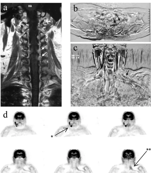
Case report. In March 2001, a 57-year-old woman presented with progressive paresis of the left and right arm, with radicular pain radiating to the left thumb. Her medical history revealed a swelling of the left orbit in 1998, diagnosed elsewhere as a possi-ble low-grade NHL, which had been treated with radiotherapy. At neurologic examination, there was atrophy of the left trapezius and deltoid muscles, with a paralysis and areflexia of the left arm and a mild paresis of the right deltoid muscle. Sensibility was diffusely impaired in the left arm. MRI showed enhancing roots C3 to C6 on the left and C4 and C5 on the right side (figure, A). CSF revealed a slightly increased monocytic cell count with 11% B-lymphocytes, 9% of which were of monoclonal origin, and the diagnosis of B-cell NHL was made. Further staging, including immunologic examination of peripheral blood and bone marrow and CT of neck, thorax, and abdomen did not reveal other localiza-tions of the NHL. Electromyography (EMG) showed widespread denervation signs in the left arm but intact sensory nerve poten-tials, consistent with a polyradiculopathy and without evidence of peripheral nerve involvement. At this stage, the patient developed progressive motor disturbances of the tongue, right arm, and left leg. She was treated with radiotherapy of the neuraxis followed by intrathecal Ara-C. Initially, the symptoms improved dramatically, and strength improved to a paresis of Medical Research Council (MRC) grade 3 to 4. However, in July 2001, the pain in the left arm returned, and the function of the left arm deteriorated to a paresis MRC grade 2 to 3 despite ongoing intrathecal treatment. T1-weighted subtraction MR images showed gadolinium-enhanced signal of root C8, continuing outside the dural sac (figure, B). Also, a dubious gadolinium-enhanced signal of the cervicobrachial plexus on the left side was seen (figure, C). Repeat EMG showed a decreased amplitude of sensory potentials of the left arm. CSF showed a persistent mild monocytic pleiocytosis, without a mono-clonal cell proliferation. Renewed staging showed no evidence of systemic NHL. An FDG PET was performed, which showed two regions of increased metabolism: one in the left cervicobrachial region consistent with plexus involvement and another in the right submandibular region (figure, D). Ultrasound identified an enlarged submandibular lymph node, which was biopsied; micros-copy showed a large B-cell NHL. The patient was treated with six cycles of CHOP (cyclophosphamide, doxorubicin, vincristine, and prednisone) chemotherapy with intrathecal methotrexate and Ara-C. The function of the left arm improved during the treatment up to a global MRC scale score of 4, and the pain decreased. Currently, 4 years after treatment, there is no evidence of tumor activity.
Discussion. Our patient with leptomeningeal lymphoma with-out evidence of systemic disease was initially treated with radio-therapy of the neuraxis. After an initial dramatic improvement, clinical signs and symptoms quickly recurred. Based on the clinical findings and suggestive but uncertain MRI findings, we suspected intraneural lymphoma localization within the cervico-brachial plexus. This suspicion was substantiated by the FDG PET scan, which also suggested asystemic lymphoma localization in a submandibular gland.
and monitoring treatment responses in patients with cancer, in-cluding lymphoma.4,5As shown in the current case, FDG PET may have clinical value when the diagnosis of NL is considered, espe-cially when lymphomatous infiltration in large peripheral nerves or plexus is suspected and histologic findings of the biopsy is negative or not possible.6,7
The treatment of choice for NL is systemic chemotherapy. Al-though in some patients, the outcome is poor, the durable re-sponse in our patient shows that intraneural lymphoma localizations can be susceptible to chemotherapy with agents that do not readily penetrate an intact blood– brain barrier, although penetration in peripheral nerves may be different.
From the Departments of Neurology/Neuro-Oncology (S.M.R., M.J.v.d.B.), Radiology (H.G.d.B.), and Hematology (K.L.W.), Daniel den Hoed Cancer Center/Erasmus University Hospital, Rotterdam, the Netherlands. Disclosure: The authors report no conflicts of interest.
Received January 31, 2006. Accepted in final form April 19, 2006.
Figure. (A) Gadolinium-enhanced T1-weighted images of the cervical spine made at the time of first presentation, showing enhancing cervical roots on the left more pro-nounced than on the right (arrows). (B) Transverse MR subtraction image of the thoracic outlet region made after neuraxis radiotherapy, made by subtracting the T1-weighted image made after gadolinium administration from the same image before gadolinium administration (showing contrast enhancement as dark areas), showing enhancement of the proximal plexus region (arrows). (C) Anteroposterior MR subtraction images showing question-able enhancement of the left cervical plexus region (arrow). (D) [18F]Fluorodeoxyglucose PET images, showing clear
Copyright © 2006 by AAN Enterprises, Inc.
References
1. van den Bent MJ, de Bruin HG, Bos GJM, Brutel de la Riviere G, Sillevis Smit PAE. Negative sural nerve biopsy in neurolymphomatosis. J Neurol 1999;246:1159–1163.
2. van den Bent MJ, de Bruin HG, Beun GDM, Vecht ChJ. Neurolympho-matosis of the median nerve. Neurology 1995;45:1403–1405.
3. Quinones-Hinojosa A, Friedlander RM, Boyer PJ, Batchelor TT, Chiocca EA. Solitary sciatic nerve lymphoma as an initial manifestation of dif-fuse neurolymphomatosis. Case report and review of the literature. J Neurosurg 2000;92:165–169.
4. Kasamon YL, Wahl RL, Swinnen LJ. FDG PET and high-dose therapy for aggressive lymphomas: towards a risk-adapted strategy. Curr Opin Oncol 2004;16:100–105.
5. Schot B, van Imhoff G, Pruim J. Predictive value of 18F-fluorodeoxyglucose positron emission tomography in chemotherapy sen-sitive relapsed lymphoma. Br J Haematol 2003;123:282–287.
6. Kanter P, Zeidman A, Streifler J. PET-CT imaging of combined bra-chial and lumbosacral neurolymphomatosis. Eur J Haematol 2006;74: 66–69.
7. Bokstein F, Goor O, Shihman B. Assessment of neurolymphomatosis by brachial nerve biopsy and PET/CT. J Neuro-Oncol 2005;72:163–167.
Reversible dementia with parkinsonian
features associated with budesonide use
Calin I. Prodan, MD; Marilee Monnot, PhD; Elliot D. Ross, MD; and Anton E. Coleman, MDA 51-year-old man was referred for neurologic consultation be-cause of increasing forgetfulness, difficulty walking, frequent falls, apathy, and decreased motivation to perform activities of daily living such as bathing, cooking, and driving, over a 6-month pe-riod. He had a history of Crohn disease (CD), diabetes, and coro-nary artery disease. Current medications included amlodipine, rosiglitazone, hydrochlorothiazide, and budesonide. He was treated with oral prednisone for CD in the past with excellent response. Two years before evaluation, he developed multiple side-effects including weight gain, edema, and hypertension that pre-cluded the use of prednisone. Nine months prior to his presentation, he was started on oral budesonide at 9 mg/day. He tolerated the new medication well and had none of the side-effects previously noted. However, he experienced a relapse of his CD 6 weeks after an attempt was made to discontinue budesonide, and the medication was restarted.
On initial examination, he had a Mini-Mental State Examina-tion (MMSE) score of 23 of 30 (missing points on orientaExamina-tion, naming, attention, and recall), mild hypophonia, decreased speech output with mild anomia, loss of facial expression, bradykinesia, mild nuchal and limb rigidity, postural instability, and mild rest-ing more than action tremor of the upper extremities. Additional neuropsychological testing showed evidence of long-term more than short-term memory deficits that improved with cuing, mark-edly impaired phonemic and categorical naming, decreased perfor-mance on block designs and clock drawing, and a forward digit span of four. A variety of laboratory tests including blood count, serum chemistry, liver function tests, HIV, syphilis and Lyme disease serologies, folic acid, serum vitamin B12 levels, megalo-blastic anemia profile, thyroid studies, serum immunoelectro-phoresis, antinuclear antibody test, and serum and urinary levels of arsenic, lead, and mercury were all negative or within normal limits. CSF studies showed only an elevated protein level at 94 mg/dL (normal 15 to 45 mg/dL) with cell count, protein electro-phoresis, myelin basic protein, immunoglobulin G (IgG) index, IgG synthesis rate, oligoclonal band determination, VDRL, and cul-tures all normal or negative. MRI of the brain showed very mild frontal and anterior temporal atrophy, which could well have been normal for age. EEG and PET scan were normal. Because of the pattern of behavioral and cognitive deficits, a diagnosis of fronto-temporal dementia with parkinsonian features was entertained,1 and a trial of donepezil was initiated, with no obvious effects over the next 6 months.
Over the next 14 months, his condition remained stable. Dur-ing this time, multiple attempts were made to discontinue budes-onide, but he experienced CD relapses that required restarting the medication. On average, he was on budesonide for 9 months/year during this period. The longest period off medication was 6 weeks without reported changes in cognitive symptoms. The patient and his family declined immunosuppressive therapy for CD. Repeat examination and brain MRI were unchanged 2 years after the initial evaluation.
Twenty-four months after his initial evaluation, budesonide was decreased gradually from 9 to 1.5 mg/day over 4 months. He
reported gradual cognitive improvement 2 months after initiating the medication taper. Repeat examination performed 30 months after the initial visit, on 1.5 mg/day of budesonide, demonstrated an MMSE score of 29 of 30, mild resting tremor with normal speech, language, facial expression, muscle tone, and postural bal-ance. Repeat neuropsychological testing showed a performance within the expected range for age. Three years after the initial visit, budesonide was discontinued, and the patient is currently attending vocational training.
Steroid dementia2 is a reversible cause of cognitive decline even in the absence of psychosis. Sustained exposure to steroids has been shown to affect the hippocampal formation.3However, recent data suggest that the cognitive deficits are due to neuro-toxic effects on both the hippocampal and the prefrontal areas.4 Both short-term and long-term use of steroids has been associated with cognitive deficits.5A recently reported case presented with reversible cognitive symptoms suggestive of Alzheimer disease af-ter a 3-month course of prednisone.6Due to side-effects associated with steroid use in chronic diseases, newer preparations are being used currently. Budesonide is a glucocorticoid with low systemic availability due to extensive first-pass liver metabolism that has fewer side-effects than traditional steroids.7The oral delayed re-lease formulation is approved by the Food and Drug Administra-tion for the treatment of CD at 9 mg/day, and recent clinical practice follow-up data have confirmed its effectiveness.7The case presented here illustrates that long-term use of this medication at recommended doses may cause a clinical picture of cognitive de-cline with parkinsonian features that is reversible with reduction in dosage.
From the Department of Neurology (C.I.P., M.M., E.D.R.), University of Oklahoma Health Sciences Center, Oklahoma City, and ISJ Clinic–Mayo Health System (A.E.C.), Mankato, MN.
Disclosure: The authors report no conflicts of interest.
Received January 10, 2006. Accepted in final form May 2, 2006.
Address correspondence and reprint requests to Dr. C.I. Prodan, 711 S.L. Young Blvd., PPOB, Suite 215, Oklahoma City, OK 73104; e-mail: [email protected]
Copyright © 2006 by AAN Enterprises, Inc.
References
1. Neary D, Snowden JS, Gustafson L, et al. Frontotemporal lobar degener-ation: a consensus on clinical diagnostic criteria. Neurology 1998;51: 1546–1554.
2. Varney NR, Alexander B, MacIndoe JH. Reversible steroid dementia in patients without steroid psychosis. Am J Psychiatry 1984;141:369–372. 3. Starkman MN, Giordani B, Gebarski SS, Berent S, Schork MA,
Schtein-gart DE. Decrease in cortisol reverses human hippocampal atrophy fol-lowing treatment of Cushing’s disease. Biol Psychiatry 1999;46:1595– 1602.
4. Wolkowitz OM, Lupien SJ, Bigler E, et al. The “steroid dementia syn-drome”: an unrecognized complication of glucocorticoid treatment. Ann NY Acad Sci 2004;1032:191–194.
5. Keenan PA, Jacobson MW, Soleymani RM, et al. The effect on memory of chronic prednisone treatment in patients with systemic disease. Neurol-ogy 1996;47:1396–1402.
6. Sacks O, Shulman M. Steroid dementia: an overlooked diagnosis? Neu-rology 2005;64:707–709.
Resolution of SUNCT after removal of a
pituitary adenoma in mild acromegaly
Todd D. Rozen, MD
The syndrome of short-lasting unilateral neuralgiform headache attacks with conjunctival injection and tearing (SUNCT) is a tri-geminal autonomic cephalalgia. It is marked by multiple daily episodes of headache each lasting less than 250 seconds and ac-companied by autonomic features. SUNCT is recognized as a treatment refractory headache, but there has been some success with lamotrigine,1gabapentin,2and topiramate.3 The etiology of SUNCT is unknown but neuroimaging has indicated a hypotha-lamic influence.4SUNCT also occurs in individuals with pituitary tumors suggesting involvement of the hypothalamic-pituitary axis. A recent study suggested that SUNCT arises only in patients with acromegaly or with prolactinomas.5Thus, growth hormone or dopamine or both may play a role in SUNCT pathogenesis. There are no reports of the course of SUNCT after pituitary tumor re-moval and normalization of pituitary hormones.
Case report. A 37-year-old man presented with a 4-year his-tory of daily left-sided retro-orbital headaches. Each headache would last 60 to 120 seconds, would occur on average 30 times per day, and were associated with conjunctival injection, lacrimation, and ptosis. The pain was described as sharp and as if the eye was being pulled into the head. In between attacks he would be pain-free. The headaches were disabling and caused him to miss a considerable amount of work. A diagnosis of SUNCT was made. He was treatment refractory, failing lamotrigine (500 mg), a com-bination of topiramate (400 mg) and gabapentin (3,600 mg), verapamil (480 mg), indomethacin (150 mg), oxcarbazepine (1,200 mg), and valproic acid (1,500 mg). He had a positive but short-lasting response to clomiphene citrate. He failed inpa-tient hospitalization with multiple parenteral medications including chlorpromazine, droperidol, magnesium, diphenhy-dramine, ketorolac, valproate sodium, and hydrocortisone. He had no persistent response to interventional procedures includ-ing greater occipital nerve blockade and a C2-C3 selective nerve root block with pulsed radiofrequency. Brain MRI and MRA of the intracranial and extracranial circulation, looking for any secondary cause, were read as normal. Neurologic evaluation in between attacks was normal. During attacks he had evidence of a left-sided ptosis and injected conjunctiva. One year into treat-ment a repeat brain MRI was completed because of the refrac-tory nature of the pain. A 6 mm mass in the left anterior pituitary was noted, suggesting a microadenoma (figure). When his old scans were reviewed, the mass had been present but missed by radiology. The patient was sent for endocrinology evaluation and was found to have acromegaly based on elevated growth hormone (GH) (1.93 ng/mL; normal 0.01 to 1.00 ng/mL) and serum insulin-like growth factor (IGF) 1 levels (372 ng/mL; normal 109 to 284 ng/mL). He had questionable acromegalic features on examination. His prolactin level was normal (9.3 ng/mL; normal 2.6 to 13.1 ng/mL). The endocrinologist did not feel that the tumor had to be surgically removed as it was causing no mass effect and the patient showed only minimal clinical signs of pituitary dysfunction. Because his headaches were treatment resistant and as a result he had a poor quality of life, a transsphenoidal tumor resection was performed. Post procedure his headaches immediately ceased and he has been pain free for 8 months even after stopping all headache preven-tives. His GH and IGF-1 levels normalized after tumor resection.
Conclusion. Pituitary tumor associated SUNCT may occur in patients with acromegaly. Removal of an acromegalic mi-croadenoma led to complete cessation of SUNCT attacks, sug-gesting a possible role for growth hormone in SUNCT pathogenesis. Interestingly, octreotide, a somatostatin ana-logue and growth hormone inhibitor, has shown success as an abortive agent in cluster headache, another hypothalamus-influenced headache syndrome.6One patient with SUNCT has also been reported who had a decrease in headache frequency and severity after treatment with octreotide.5 Octreotide was
not given to the present patient. As the tumor was ipsilateral to the side of the SUNCT attacks a possible mass effect or me-chanical action may have played a role in SUNCT onset. How-ever, the relatively small size of the lesion argues against this and more for a hormonally mediated syndrome.
SUNCT may be the initial presentation of pituitary disease. Cases of SUNCT should be investigated for underlying pituitary adenomas, both with imaging and neurohormonal analysis (at least prolactin, GH, and IGF-1 levels). Removal of functionally active pituitary tumors, regardless of size, should be considered in treatment-refractory SUNCT. As pituitary surgery is not without substantial risks more data are needed before pituitary tumor removal can be recommended for cases of treatment resistant SUNCT.
From Michigan Head-Pain and Neurological Institute, Ann Arbor. Disclosure: The author reports no conflicts of interest.
Selected as a poster presentation for the 58th American Academy of Neurol-ogy annual meeting, San Diego, CA, 2006.
Received February 7, 2006. Accepted in final form May 4, 2006.
Address correspondence and reprint requests to Dr. Todd Rozen, MHNI, 3120 Professional Drive, Ann Arbor, MI 48104; e-mail: [email protected]
Copyright © 2006 by AAN Enterprises, Inc.
References
1. D’Andrea G, Granella F, Ghiotto N, Nappi G. Lamotrigine in the treat-ment of SUNCT syndrome. Neurology 2001;57:1723–1725.
2. Graff-Radford SB. SUNCT syndrome responsive to gabapentin (Neuron-tin). Cephalalgia 2000;20:515–517.
3. Rossi P, Cesarino F, Faroni, et al. SUNCT syndrome successfully treated with topiramate: case reports. Cephalalgia 2003;23:998–1000.
4. May A, Bahra A, Buchel C, Turner R, Goadsby PJ. Functional magnetic resonance imaging in spontaneous attacks of SUNCT. Short-lasting neu-ralgiform headache with conjunctival injection and tearing. Ann Neurol 1999;46:791–794.
5. Levy MJ, Matharu MS, Meeran K, Powell M, Goadsby PJ. The clinical characteristics of headache in patients with pituitary tumors. Brain 2005;128:1921–1930.
6. Matharu MS, Levy MJ, Meeran K, Goadsby PJ. Subcutaneous octreotide in cluster headache: randomized placebo-controlled double-blind cross-over study. Ann Neurol 2004;56:488–494.
Alexia without agraphia in a child with acute
disseminated encephalomyelitis
Robert D. Little, MD; and Joshua L. Goldstein, MD
Alexia without agraphia is a neurologic syndrome when a patient cannot read but has normal writing and verbal language skills. We present alexia without agraphia in an 8-year-old boy with acute disseminated encephalomyelitis.
Case report. A previously healthy normally developing 8-year-old boy presented to an emergency department with the chief complaint of the inability to read. On questioning, the pa-tient was noted to have fallen several times while getting ready for school that day but otherwise seemed fine to his parents. At school he reported to his teachers that he was unable to read. He and several family members had recently been ill with fever, vomiting, and diarrhea several days prior to this admission, but all had recovered completely.
On examination his formal mental status examination was normal aside from an isolated marked difficulty in reading. When trying to read, he could identify individual letters and some short words but could not sound out larger words. At baseline he was in regular classes in the third grade, reading at age appropriate levels. He could identify objects, had fluent spontaneous and re-petitive speech, and could write well. The remainder of his de-tailed neurologic examination was normal aside from difficulty in identifying colors in his right visual field, mild truncal ataxia, and a very mild right hemiparesis involving his arm and leg equally. He was, however, able to identify fingers and movements in all visual fields.
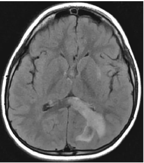
MRI was performed on the first day of admission and demon-strated multiple foci of T1 hypointense, T2 hyperintense abnor-malities throughout the white matter. One prominent lesion was located in the left occipital lobe extending to the splenium of the corpus callosum (figure).
A lumbar puncture was performed. The CSF had 0 leukocytes, 0 red blood cells, glucose 67 mg/dL, and protein 17 mg/dL. Cytol-ogy was sent on the spinal fluid and was normal. Myelin basic protein was also normal.
The patient was given IV methylprednisolone for 5 days at 30 mg/kg/day and experienced improvements in his ataxia and hemi-paresis after approximately 48 hours of treatment. His reading also slowly improved over the course of the hospitalization. He was seen in clinic 6 weeks later, and his neurologic examination was normal. A repeat MRI at that time showed significant im-provement in the previously noted lesions; no new lesions were noted.
Discussion. Alexia without agraphia was first described by Dejerine in 1892.1Clinically, the patient described was unable to read but had intact writing, naming, and recall. The patient also had a right hemiachromatopsia with intact visual fields.
Our patient showed many of the classic findings for the syn-drome of alexia without agraphia. He had intact writing skills, naming, and recall but could not read words. He had a right hemiachromatopsia. He had additional findings on examination, such as ataxia and a right hemiparesis, which were likely due to multifocal lesions seen on MRI. The clinical findings of pure alexia and hemiachromatopsia combined with the lesion in the left occip-ital lobe and splenium of the corpus callosum correlate well with previous descriptions of alexia without agraphia. Although a right hemianopsia is often part of this syndrome, there have been many reported cases without a visual field deficit.2
The most common etiology of alexia without agraphia is an infarction in the territory of the left posterior cerebral artery. However, this syndrome has been reported in association with a number of other causes.3-5Alexia without agraphia has rarely been reported in children.2,5
Acute disseminated encephalomyelitis (ADEM) is a monopha-sic, inflammatory demyelinating disease of the CNS. It most often follows an acute illness, either viral or bacterial. The presenting signs and symptoms are variable; the most common are long tract signs, hemiparesis, and altered mental status.6 MRI typically shows multifocal demyelinating lesions. CSF is often abnormal, with elevated protein and sometimes a mild lymphocytic
pleocyto-sis. However, the CSF may be normal in up to 30% of cases of ADEM.7
A lesion in the left occipital lobe extending to the splenium of the corpus callosum can cause alexia without agraphia regardless of the etiology. Although this syndrome has not been reported in association with ADEM, it is reasonable to assume that a demyeli-nating lesion in the appropriate location could produce these find-ings. Furthermore, alexia without agraphia has been reported in association with multiple sclerosis,3 a condition with a similar pathophysiology.
From Children’s Memorial Hospital, Northwestern University, Chicago, IL. Disclosure: The authors report no conflicts of interest.
Received January 30, 2006. Accepted in final form May 10, 2006. Address correspondence and reprint requests to Dr. Joshua Goldstein, Chil-dren’s Memorial Hospital, 2300 ChilChil-dren’s Plaza, Box 51, Chicago, IL 60614; e-mail: [email protected]
Copyright © 2006 by AAN Enterprises, Inc.
References
1. Dejerine J. Contribution a l’etude anatomoclinique et clinique des differ-entes varietes de cecite verbal. Mem Soc Biol 1892;4:61–90.
2. Damasio A, Damasio H. The anatomic basis of pure alexia. Neurology 1983;33:1573–1583.
3. Mao-Draayer Y, Panitch H. Alexia without agraphia in multiple sclero-sis: case report with magnetic resonance imaging localization. Mult Scler 2004;10:705–707.
4. Sabet HY, Blake P, Nguyen D. Alexia without agraphia in a postpartum eclamptic patient with factor V Leiden deficiency. AJNR: Am J Neurora-diol 2004 ;25:419–420.
5. Tamhankar MA, Coslett HB, Fisher MJ, Sutton LN, Liu GT. Alexia without agraphia following biopsy of a left thalamic tumor. Pediatr Neu-rol 2004;30:140–142.
6. Tenembaum S, Chamoles N, Fejerman N. Acute disseminated encepha-lomyelitis: a long-term follow-up study of 84 pediatric patients. Neurol-ogy 2002;59:1224–1231.
7. Murthy SN, Faden HS, Cohen ME, et al. Acute disseminated encephalo-myelitis in children. Pediatrics 2002;110 (2 pt 1):e21.
Multiple system atrophy presenting with
language impairment
L.G. Apostolova, MD; I. Klement, MD; Y. Bronstein, MD; H.V. Vinters, MD; and J.L. Cummings, MD
Multiple system atrophy (MSA) typically presents with parkinson-ism, cerebellar ataxia, corticospinal dysfunction, and autonomic failure.1We describe a case of pathologically confirmed MSA with semantic language impairment.
Case report. A 55-year-old right-handed psychiatrist pre-sented with language difficulties since age 50. He had shrink-ing vocabulary, spellshrink-ing, and readshrink-ing difficulties, loss of foreign language skills, and difficulty with comprehension of complex conversations. His speech was fluent, grammatically correct, with occasional anomic pauses and substitution of low-frequency words with general words (e.g., “thing” and “it”). He had hyperreflexia in the right arm and leg, right-sided Babin-ski, writer’s cramp, and mildly impaired fine motor skills in the right hand. Neuropsychological evaluation revealed anomia (Boston Naming Test [BNT] score 35/60, ⬍1%), intact verbal fluency (FAS 88%), surface dysgraphia (“curteus” instead of “courteous,” “medeval” instead of “medieval”), surface dyslexia (could not read “subtlety”), and moderate retrieval-type verbal memory deficit. His receptive vocabulary skills were below ex-pectation given his advanced education (Peabody Picture Vo-cabulary Test, 48%). His performance on tests of attention, visual memory, visuospatial, frontal/executive, and parietal functions was superior. His full-scale IQ was 141 with discor-dance between his performance IQ score of 150 and verbal IQ score of 123. CT and nuclear MR brain scans, EEG, and carotid duplex did not reveal abnormalities.
Five months later, he was unable to name high-frequency words (e.g., “horse”) and common medical terms (e.g., “cornea,” “stethoscope”) or interpret low-frequency words such as “nauti-cal,” “mercantile,” and “constellation.” His BNT performance declined from 35/60 to 18/60. He showed impaired word catego-rization and conceptual tracking. Reading, comprehension, rep-etition, spelling, and verbal abstract reasoning were within normal range but below expectation given his advanced educa-tion. Visuospatial and arithmetic abilities, social behavior, and judgment were preserved. He manifested frontal/executive impairment (Wisconsin Card Sorting Test 4/6 categories [7%]; perseverations ⬍1%), profound anomia, and impaired single-word comprehension due to semantic knowledge loss. His speech, albeit fluent, became progressively empty. Seven years after symptom onset, he quit his general psychiatry practice.
MRI performed 9 years into the illness revealed mild generalized atrophy with slight left perisylvian predominance. SPECT revealed diffuse left-predominant frontotemporal hypometabolism.
Sixteen years later (21 years into his illness), he was mute and dependent for all activities of daily living. He lost motor skills in his right arm and could not ambulate. He never developed parkin-sonian or cerebellar signs and dysautonomia. He died 22 years after dementia onset.
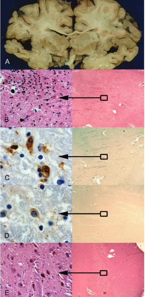
The postmortem examination was limited to the brain. It showed relatively circumscribed atrophy of the inferior and middle temporal gyri and planum temporale more severe on the left than the right (figure, A). Atrophy of the parietal, occipital, and frontal lobes (including Broca area) was minimal. The cer-ebellum and brainstem appeared normal. Moderate atrophy and brownish discoloration with corresponding severe neuron loss, reactive gliosis, and iron deposition were noted bilaterally in the putamen (figure, B). Similar focal but less severe degen-eration was seen in the thalamus. Changes within the substan-tia nigra were subtle with only mild overall reduction in numbers of pigmented neurons (figure, E). Immunohistochemis-try demonstrated only rare Alzheimer-type neurofibrillary tan-gles, no Pick bodies or other neuronal inclusions, but many glial cytoplasmic inclusions (GCIs) within the putamen (figure, C), lateral temporal cortex and white matter, and focally within the substantia nigra. Occasional GCIs were immunoreactive for
␣-synuclein (figure, D) and ubiquitin. Neuronal ubiquitin-positive inclusions were absent.
GCIs are a common feature of MSA, but the tau variant involved appears to be different from tau associated with Alzheimer disease.3
Our case is an unusual neurodegenerative disorder presenting with frontal executive and semantic language deficits and cortico-spinal system involvement, pathologically most consistent with MSA. Although nonfluent aphasia,4,5executive disturbance,6and “fluent, circumlocutory speech”7 have been described, our case shows semantic language deficits with underlying MSA pathology.
From the Departments of Neurology (L.G.A., J.L.C.), Pathology (I.K., H.V.V.), and Psychiatry and Biobehavioral Neuroscience (J.L.C.), University of California Los Angeles, and Kaiser Permanente Medical Group (Y.B.), Woodland Hills, CA.
Disclosure: The authors report no conflicts of interest. Received June 27, 2005. Accepted in final form May 11, 2006.
Address correspondence and reprint requests to Dr. L.G. Apostolova, Reed Neurological Research Center, Suite 2-238, 710 Westwood Blvd., Box 951769, Los Angeles, CA 90095-1769; e-mail: [email protected]
Copyright © 2006 by AAN Enterprises, Inc.
References
1. Gilman S, Low PA, Quinn N, Albanese A, Ben-Shlomo Y, Fowler CJ, et al. Consensus statement on the diagnosis of multiple system atrophy. J Neurol Sci 1999;163:94–98.
2. Wenning GK, Tison F, Shlomo B, Daniel SE, Quinn NP. Multiple system atrophy: a review of 203 pathologically proven cases. Mov Disord 1997; 12:133–147.
3. Cairns NJ, Atkinson PF, Hanger DP, Anderton BH, Daniel SE, Lantos PL. Tau protein in the glial cytoplasmic inclusions of multiple system atrophy can be distinguished from abnormal tau in Alzheimer’s disease. Neurosci Lett 1997;230:49–52.
4. Konagaya M, Konagaya Y, Miwa S, Matsuoka Y. [Clinico-MRI study of hemispheric disorder in long-term follow-up cases of multiple system atrophy]. Rinsho Shinkeigaku 1998;38:1031–1036.
5. Konagaya M, Sakai M, Matsuoka Y, Konagaya Y, Hashizume Y. Multi-ple system atrophy with remarkable frontal lobe atrophy. Acta Neuro-pathol (Berl) 1999;97:423–428.
6. Robbins TW, James M, Lange KW, Owen AM, Quinn NP, Marsden CD. Cognitive performance in multiple system atrophy. Brain 1992;115:271– 291.
7. Schlossmacher MG, Hamman C, Cole AG, Gonzalez RG, Frosch MP. Case 27-2004: a 79-year-old woman with disturbances in gait, cognition, and autonomic function. N Engl J Med 2004;351:912–922.
Correction
The G526R glycyl-tRNA synthetase gene mutation in distal hereditary motor neuropathy type V
In the article “The G526R glycyl-tRNA synthetase gene mutation in distal hereditary motor neuropathy type V” by O. Dubourg et al. (Neurology2006;66:1721–1726), on page 1722 the authors describe the mutation identified in three of the dHMN-V families as a “c.2094 A3G” nucleotide change leading to the G526R mutation in theGARSgene. The correct mutation is c.2094 G3C leading to the same amino acid change.