TISSUE SPECIFIC VARIATIONS IN THE UREA SENSITIVITY OF THE E, ESTERASE IN MAIZE1
TORU END02 AND DREW SCHWARTZ
Department of Botany, Indiana University, Bloomington
Received February 2.8, 1966
N
a previous publication (SCHWARTZ 1964a) evidence was presented to theI
effect that enzymes specified by the same allele functioning in two different parts of the maize plant are not identical. The NN p H 7.5 esterases synthesized under the control of the E: allele in the seedling and endosperm show identical electrophoretic migration rates in starch gel electrophoresis; however, they differ in their inactivation by urea. This finding was interpreted as reflecting a differ- ence in the total structure of the enzymes. The enzymes could vary in the com- pactness of folding around the active site, their conjugation with uncharged co- factors which could influence enzyme stability, or the like. These results were based on comparison of the relative intensities of the pH 7.5 esterase bands in zymograms developed after electrophoresis of control and urea-treated homog- enates of the two plant parts. This is not a very quantitative method for inacti- vation studies, but the method was employed in order to compare the urea sensi- tivity of the enzymes in the crude preparations minimizing possible alterations of the structure of the enzyme by purification procedures. Comparison of total esterase activity in treated and untreated crude homogenates is meaningless since both plumule and endosperm material contain a number of esterases specified by other genes.In this paper, we report the results of our studies on the comparison of the NN
pH 7.5 esterase of the two plant parts utilizing partially purified enzyme prepa- rations. Since this enzyme preparation is free of all the other esterases specified by nonallelic genes, more quantitative procedures could be employed for assay of enzymatic activity. The results confirm the previously reported tissue specific differences in urea sensitivity of the NN esterase. These differences are eliminated when the enzyme is converted to an acidic form by treatment with glyceralde- hyde.
MATERIALS A N D METHODS
The inveshgations were conduced on the FF and N N isozymes of the pH 7.5 esterase in maize which are specified by the E,' and E l N alleles respectively (SCHWARTZ 1960). The comparisons were made on the enzymes synthesized in 16 day immature endosperm and plumules of 3 to 7 day-old seedlings. Inbred lines homozygous for each allele were sibcrossed. Some of the ears were harvested 16 days after pollination for source of immature endosperm material. The kernels were cut off the cobs and frozen in plastic bags at -20°C. The remainder of the ears were harvested a t
This work was supported by National Sclence Foundation grants GB 2823 and GB 4178 Present address National Institute of Genetxs, Misnna, Japan
234 T. E N D 0 A N D D. SCHWARTZ
maturity. The kernels from the matured ears were germinated in Petri dishes on moistened filter paper at 30°C. The plumules were cut at the point of attachment to the seed and stored at -20°C for a few days prior to use.
The pericarp and embryo were removed from the immature kernels and the remaining endo- sperm washed a few times in distilled water. Thirty to 60 grams of endosperm or plumule mate- rial was macerated in a mortar and centrifuged. All centrifugations in the purification procedure were a t 27,000 x g for 15 minutes. The p H of the supernatant was adjusted to 4.5 with N HC1, centrifuged and then readjusted to pH 7.5 with N NaOH. The 50 to 80% ammonium sulfate precipitate was dissolved in 0.005 p (ionic strength) Triscitrate (TC) buffer, pH 6.4, and dialyzed overnight against the same buffer. After centrifugation to remove the small amount of precipitate which formed, the dialysate was passed through a carboxymethylcellulose column (1 X 7 cm) equilibrated at pH 6.4 with the TC buffer. The column was washed with about 300 ml of 0.005 p TC buffer, p H 6.4, and the pH 7.5 esterase eluted from the column with 0.1 p TC buffer at the same pH. The fractions which contained esterase activity were dialyzed overnight against distilled water, refrigerated and used within 10 days. The enzyme was stable under these stored conditions. Starch gel electrophoretic analysis showed the eluted fractions to contain only the NN or FF esterase iozymes. Undoubtedly other proteins were present but the fractions were free from the other esterases specified by nonallelic genes which are found in the crude preparation.
Glyceraldehyde treatment consisted of adding m-glyceraldehyde to the partially purified enzyme solution to a final molarity of 0.2 M. The mixture was incubated a t 37°C for 24 hours and the precipitate which formed was removed by centrifugation.
For the urea sensitivity tests a series of 8 M urea solutions, buffered with 0.04 p TC at p H s ranging from 5.0 to 9.0 at 0.5 pH unit intervals, were used. Urea treatment was carried out at room temperature by mixing one part enzyme solution with three parts buffered urea solution resulting in a final concentration of 6 M urea and 0.03 p buffer. Controls were treated identically except that the urea was omitted.
Enzymatic activity was measured by determining the rate of hydrolysis of l-naphthylacetate following a modification of the method of GOMORI (1953). The reaction mixture was composed of 2.4 ml water, 1 ml 0.3 M sodium phosphate buffer, pH 7.7, 1 ml 0.5% solution of polyvinylpyr- rolidone, 0.1 ml of enzyme or enzyme plus urea solution at the various pH values and 0.5 ml of a solution of 1 miv l-naphthylacetate in 10% acetone. The final p H of the reaction mixtures which contained the enzyme-urea solution buffered at p H 8.5 and 9.0 was adjusted to 7.7 by the addition of 0.01 and 0.02 ml N HC1 respectively. After incubation at 30°C for 20 minutes, 1 ml of reagent containing 0.15% diazonium salt (Fast Blue RR) and 6% sodium laurylsulfate was added to the reaction tubes. The detergent serves to stop enzyme activity and solubilize the diazonium-naphthol complex. The tubes were reincubated at 30°C for 25 minutes and absorbancy read in a Klett colorimeter at 520 mp. For the urea time-course experiments the buffered urea and enzyme solutions were mixed and 0.1 ml aliquots of the mixture removed at intervals and diluted 45-fold by addition to tubes containing 4.4 ml of the reaction mixture complete except for the 1-naphthylacetate substrate. After the last sample was taken, the substrate was added and the reaction allowed to proceed at 30°C.
RESULTS A N D DISCUSSION
T I S S U E S P E C I F I C ESTERASE VARIATIONS 235
5.0 5.5 6.0 6.5 7.0 7.5 0.0 0.5 9.0
PH
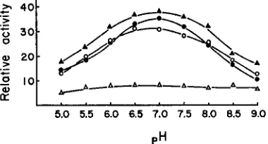
FIGURE 1 .-Sensitivity of FF and N N pH 7.5 esterases of endosperm and plumule to 15 minute treatment with 6 M urea, expressed as percent of activity in the controls.
(A)=
FF endosperm,( 0 ) = F F plumule. ( A ) = N N endosperm, (0) = N N plumule.
enzyme is most resistant around pH 7.0 and becomes increasingly sensitive as the pH is reduced or raised. The inactivation is increased almost threefold at pH 5 and 9. The pH dependence was observed for FF of the endosperm and plumule and NN of the plumule. The small amount of the activity remaining after 15 minutes treatment of NN endosperm with 6 M urea did not show pH dependence. The residual activity of NN endosperm after the urea treatment may be due to slight contamination of the pH 7.5 esterase preparation by another urea insensitive esterase. A striking pH dependence similar to that observed for the other enzyme preparations is found when NN endosperm enzyme is exposed to urea for only 1 minute (Figure 2 ) .
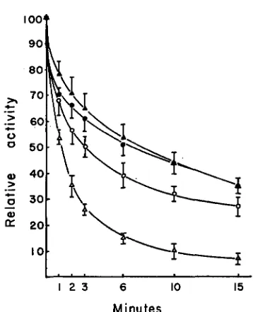
The differential urea sensitivity of the NN enzyme of plumule and endosperm is also clearly seen when the relative activity is plotted as a function of length of exposure of the enzyme preparation to urea (Figure 3 ) .
The inactivation of the pH 7.5 esterases by urea is not dependent on enzyme
I . . . o . . . . .
5.0 5.5 6.0 6.5 7.0 7.5 0.0 0.5 9.0
PH
FIGURE 2.-Sensitivity of FF and N N pH 7.5 esterases of endosperm and plumule to 1 minute treatment with 6 M urea, expressed as percent of activity in the controls.
(A)=
FF endosperm,236 T. END0 A N D D. SCHWARTZ
I O O t
1 2 3 6 10 15 Minutes
FIGURE 3.-Sensitivity of F F and NN pH 7.5 esterases of endosperm and plumule with length of exposure to 6 M urea at pH 7.0, expressed as percent of activity in the untreated controls. The confidence limits represent standard errors.
(A)
= FF endmperm, ( 0 ) = FF plumule, ( A ) = NN endosperm, (0) = NN plumule.concentration (four-fold dilution of the enzyme preparations does not affect the urea sensitivity). The sensitivity of the enzymes from either tissue to 6 M urea
is not altered by treatment of the N N enzyme with 1 mM EDTA at p H 7.0 for 24 hours at 5°C.
The results presented in this paper on the quantitative comparison of urea inactivation of the N N pH 7.5 esterase of plumule and endosperm origin confirm the previously published results ( SCHWARTZ 1964a). The NN enzyme of the endosperm is much more sensitive to urea than the corresponding enzyme of the plumule even though both are specified by the same allele. The suggestion was advanced in the earlier paper that this differential urea inactivation might reflect a difference in the tertiary or quarternary structure of the enzyme synthesized in the different tissues. The N N endosperm enzyme may be combined with some substance which renders it urea sensitive. Ribonuclease is sensitized to urea in the presence of polyvalent anions such as pyrophosphate and 2’-cytidylic acid (NELSON et al. 1962) and chymotrypsin is insensitive in the presence of calcium anions (MARTIN and FRAZIR 1963). If a similar situation is to account for the difference in urea sensitivity of the pH 7.5 esterase, the factor which confers sensitivity or resistance must remain strongly bound to the enzyme during the course of the fractionation procedure used in purification. It must also be un- charged and of small enough size as not to perceptibly alter the migration rate of the enzymes in starch gel electrophoresis.
TISSUE SPECIFIC ESTERASE VARIATIONS 23 7
dynamic hypothesis for the determination of tertiary structure of proteins. This hypothesis proposes that the particular configuration a protein assumes under any specific set of conditions is the one that is thermodynamically the most stable. Since the intracellular environment of endosperm and plumule material is un- doubtedly different, it would not be surprising to find that the NN enzyme in the two tissues would have different folded configurations. The thermodynami- cally most stable configuration of the enzyme in the endosperm might not be the most stable form in the plumule even though the enzymes in the two tissues have identical primary structures.
Bacillus cereus produces two forms of penicillinase, a cell-bound and a n exo- enzyme. Both forms are inducible by penicillin and are present in the constitutive line, 569H, suggesting that they may be specified by the same structural gene. The two enzyme forms show identical substrate specificity, Michaelis constants and sedimentation rates but differ immunologically as well as in their sensitivity to iodine (POLLOCK 1956). Furthermore, treatment of the exoenzyme with urea or guanidine hydrochloride results in the conversion of the enzyme from the iodine resistant form to an iodine sensitive form characteristic of the cell-bound penicillinase (CITRI, GARBER and SELA 1960). BERSON and YALLOW (1961) were able to distinguish immunologically between pork and sperm whale insulin even though these have identical amino acid sequences.
238 T . E N D 0 A N D D. SCHWARTZ
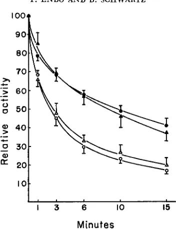
I oo*
1 3 6 10 15
Minutes
FIGURE 4.-Sensitivity of glyceraldehyde-converted FF and NN pH 7.5 esterases of endosperm snd plumule with length of exposure to 6 M urea at pH 7.0, expressed as percent of activity in the
controls. The confidence limits represent standard errors.
(A)
= FT endosperm, ( 0 ) = FF plumule, (A) = NN endosperm, (0) = NN plumule.polypeptides is still detectable, even a€ter the conversion, by the difference in urea sensitivity.
S U M MARY
The urea sensitivities of the pH 7.5 esterases specified by the same
E,
allele functioning in different parts of the maize plant were compared. PurifiedE;'
enzyme preparations from the plumule and endosperm differed significantly in inactivation by urea. Treatment with glyceraldehyde converts theEl
esterases into an acidic form eliminating the charge differences between the allelic iso- zymes. The treatment eliminates the tissue specific but not the allele specific differences in urea sensitivity.LITERATURE CITED
BERSON, S. A., and R. S. YALLOW, 1961
CITRI, N., N. GARBER, and M. SELA, 1960
EPSTEIN, C. J., R. F. GOLDBERGER, and C. B. ANFINSEN, 1963
Immunochem'cal distinction between insulins with identical amino-acid s-quznces. Nature 191 : 1392-1393.
The effect of urza and guanidine hydrochloride on activity and optical rotation of penicillinase. J. Biol. Chem. 235: 364-3459.
The genetic control of tertiary protein structure: Studies with model systems. Cold Spring H a r b x Symp. Quant. Biol. 28: 439-4.49.
GOMORI, G., 1953
MARTIN, C. J., and A. R. FRAZIR, 1963
Human esterases. J. Lab. Clin. Med. 42: 44.5-453.
T I S S U E SPECIFIC ESTERASE VARIATIONS 239
Stabilization of pan- creatic ribonuclease against urea denaturation by anion binding. J. Biol. Chem. 237: 1575- 1580.
NELSON, C. A., J. P. HUMMEL, C. A. SWANSON, and L. FRIEDMAN, 1962
POLLOCK, M. R., 1956
SCHWARTZ, D., 1960
The cell-bound penicillinase of Bacillus cereus. J. Gen. Microbiol. 15:
Genetic studies on mutant enzymes in maize: Synthesis of hybrid enzymes by heterozygotes. Proc. Natl. Acad. Sci. U.S. 46: 1210-1215.
-
1964a Genetic studies on mutant enzymes in maize. IV. Comparison of pH 7.5 esterase synthesized in seedling and endosperm. Genetics 49: 373-377. ~ 1964.b Genetic studies on mutant enzymes inmaize. V. In vitro interconversion of allelic isozymes. Proc. Natl. Acad. Sci. U.S. 52 : 222- 226.