_____________________________________________________________________________________________________
*Corresponding author: E-mail: [email protected];
13(3): 1-7, 2016, Article no.BJMMR.22602 ISSN: 2231-0614, NLM ID: 101570965
SCIENCEDOMAIN international www.sciencedomain.org
A Review of the Crown Lengthening Surgery; The
Basic Concepts
Mohammad Reza Talebi Ardakani
1, Fatemeh Khalilian
1*, Zahra Nateghi
1,
Azade Esmaeilnejad
1and Noushin Janbakhsh
11
Department of Periodontics, Shahid Beheshti University of Medical Sciences, Tehran, Iran.
Authors’ contributions
This work was carried out in collaboration between all authors. Author MT designed the study. Author FK wrote the protocol. Author ZN wrote the first draft of the manuscript. Author AE managed the literature searches. Author NJ wrote the final manuscript. All authors read and approved the final manuscript.
Article Information
DOI: 10.9734/BJMMR/2016/22602
Editor(s):
(1) Emad Tawfik Mahmoud Daif, Professor of Oral & Maxillofacial Surgery, Cairo University, Egypt.
Reviewers:
(1) S. Bizzarro, Academic Center for Dentistry, Amsterdam, Netherlands. (2)Cherif Adel Mohsen, Minia University, Egypt. (3)Murat Tozlu, Yeditepe University, Turkey. (4)Anna Carolina Ratto Tempestini Horliana, Nove de Julho University, Brazil. (5)Ketij Mehulic, University of Croatia, Croatia. Complete Peer review History:http://sciencedomain.org/review-history/12904
Received 14th October 2015 Accepted 22nd December 2015 Published 5th January 2016
ABSTRACT
Clinical crown of the tooth is the distance from gingival margin to incisal edge or occlusal surface of the tooth. A short clinical crown is defined as any tooth with less than 2 mm of sound, opposing parallel walls remaining after occlusal and axial reduction. Although implants have reasonably high success rate, recent literature showed that keeping patient’s tooth has numerous benefits. Crown lengthening is a surgical procedure designed to increase the extent of the supragingival tooth structure, so that the clinician can restore the tooth. The aim of the current study is to review the implications of CL in routine dental practice. To reach this aim, diagnosis requirements, restorative procedures after crown lengthening, stability of crown lengthening and esthetic crown lengthening are discussed in different sessions.
Keywords: Crown lengthening; periodontology; diagnosis; surgery; restorative dentistry; treatment; review.
1. INTRODUCTION
Clinical crown of the tooth is the distance from gingival margin to incisal edge or occlusal surface of the tooth [1]. A short clinical crown is defined as any tooth with less than 2 mm of sound, opposing parallel walls remaining after occlusal and axial reduction [2,3] which might be because of subgingival caries, subgingival crown fractures, too short tooth crown for restoration retention, excess of gingiva and partially opened anatomical tooth crown [4,5].
In such occasions the dentist should weight the clinical findings and patients’ concerns in the balance to determine if the tooth or teeth should be extracted (and replaced by dental implants) or be restored. Although implants have reasonably high success rates, implant failures must be considered before establishing treatment plan [6,7].
It was demonstrated that retaining a tooth over time is the most economical option compared to replacing it with implant prosthesis. Therefore, the importance of preserving the natural dentition cannot be ignored [7-9]. If clinician decides to keep the tooth, increasing the size of crown is required to regain ferrule effect, which is identified as a 360 degree metal crown collar surrounding parallel walls of dentin and extending coronal to the shoulder of the preparation [10].
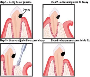
One of the processes to obtain appropriate size of clinical crown is crown lengthening (CL). According to the definition of the American Academy of Periodontology, CL is “a surgical procedure designed to increase the extent of the supragingival tooth structure for restorative or esthetic purposes by apically positioning the gingival margin, removing supporting bone or both” [8,11] (Fig. 1).
Despite periodontists are most likely specialists to perform CL surgery, there is no reason for general dentists not to perform the procedure if the procedure lies inside their comfort zone. An alternative to this treatment plan would be orthodontic extrusion which maintains the bone level better [12].
The aim of the current study is to review the implications of CL in routine dental practice. To reach this aim, diagnosis requirements, restorative procedures after crown lengthening, stability of crown lengthening and esthetic crown lengthening are discussed in different sessions.
2. DIAGNOSIS REQUIREMENTS
A short clinical crown cannot be evaluated by visual inspection alone. Hence, a comprehensive examination that includes clinical examination, radiographic examination and diagnostic cast analysis is essential for successful rehabilitation [2,3,13]. When performing clinical examination, it is necessary to evaluate if the periodontal biological width has been infringed. The biologic width is defined as the dimension of the soft tissue, which is attached to the portion of the tooth coronal to the crest of the alveolar bone. This term was based on the work of Gargiulo et al. who described the dimensions and relationship of the dentogingival junction in humans. Measurements were made from the dentogingival components of 287 individual teeth from 30 autopsy specimens established that there is a definite proportional relationship between the alveolar crest, the connective tissue attachment, the epithelial attachment, and the sulcus depth. Following mean dimensions were obtained: A sulcus depth of 0.69 mm, an epithelial attachment of 0.97 mm, and a connective tissue attachment of 1.07 mm. Based on this work, the biologic width is routinely stated to be 2.04 mm, which represents the sum of the epithelial and connective tissue measurements [14,15]. In addition, periodontal probing helps the clinician to better understand the supporting tissues [12,15-17].
During radiographic analysis the clinician should consider crown to root ratio. This phenomenon plays an important role on final decision of performing CL surgery or extracting the tooth [18].
An accurate clinical and paraclinical examination is an essential factor for a successful treatment. The dentist should keep it in mind that
inadequate diagnosis and improper treatment plan may not ensure satisfactory results.
3. CONTRAINDICATIONS AND CLINICAL OUTCOMES OF CROWN LENGTHENING
Despite the fact that surgical CL is a commonly performed treatment, little is known about the specific surgical endpoints of the procedure or the stability of the newly attained crown height over time [19,20].
With restorable teeth, crown lengthening is contraindicated when there is an unfavorable crown/ root ratio because of short roots or reduced bone support. Without sufficient periodontal support, it seems unreasonable to achieve appropriate results. Another related factor to the failure of the procedure deemed to be the presence of furcation in a multi-rooted tooth. Furcation exposure introduces potential periodontal breakdown and puts prognosis of the tooth in question [3,21-23]. Patients with a high smile line can also be a contraindication if the total esthetic outcome is not considered. Single anterior tooth CL causes uneven gingival contour, which is esthetically unpleasing, especially on patients with a high smile line. Moreover, CL is contraindicated on anterior teeth with long clinical crowns since it causes already long crowns to be even longer and results in an inappropriate esthetic view [24-26].
Although not an absolute contraindication for periodontal surgery, cigarette smoking can impair wound healing and is detrimental to the success of the surgery. Hence, patients who smoke may experience unpredictable surgical outcomes. Other factors such as patient compliance, oral hygiene and history of periodontal disease can also influence surgical outcome [21,27,28].
The results of a study aiming at investigating biologic width revealed that during surgical CL, the bone level was lowered for the placement of the prosthetic margin and reestablishment of the biological width. The biological width at the treated sites was reestablished to its original vertical dimension by 6 months. In addition a consistent 3 mm gain of coronal tooth structure was observed at the 3 and 6 month examination [29].
surgery. Suturing the flap ≤ 3 mm from the osseous crest and thick-flat biotype were associated with greater tissue rebound [30].
The results of another clinical investigation demonstrated that during 1 year healing period following apically positioned flap surgery and osseous resection the marginal periodontal tissue showed a distinct tendency to grow in a coronal direction from the level defined at surgery. At the end of the study, the gingival margin was 3.2 mm (interproximal) and 2.9 mm (buccal/lingual) coronally from where the osseous crest was located immediately following procedure [31].
In a study performed on 15 subjects preoperative and postoperative measurements of length of the clinical crown, width of attached gingival, gingival zenith and interdental papilla height were taken comparing three different surgical techniques of crown lengthening procedures of gingivectomy, apically repositioned flap and Surgical extrusion using periotome. The study presented that clinical crown lengthening by surgical extrusion using periotome offers several advantages rather than other surgical approaches since there was no change in the width of attached gingiva, interdental papilla height and gingival zenith level in pre- and post-operative measurements [32].
The positional changes of the periodontal tissues, particularly the biologic width, following surgical crown-lengthening in 15 human subjects were evaluated in another investigation. The results showed a significant apical displacement in the free gingival margin at the treated sites, which provided adequate exposure of the crown tooth structure to be restored without impinging on the biological width. There was no statistically significant difference in biologic width and the biologic width was reestablished to the original vertical dimension at all sites [33].
As a conclusion, CL surgery has a high success rate if proper patient’s selection is applied. Nonetheless, as any procedure, the patient needs to be informed of any potential complications such as possible poor aesthetics after surgery, root resorption and transient mobility of the teeth.
4. RESTORATIVE PROCEDURES AFTER CROWN LENGTHENING
Predictable long-term restorative success requires a combination of restorative principles
with the correct management of the periodontal tissues. Improper management of the periodontal tissues during restorative procedures is a common cause of failure. When a restoration is placed, the preservation of an intact, healthy periodontium is necessary to maintain the tooth or teeth being restored [20,34,35].
When planning restorative treatments, one of the most important questions is how long a clinician should wait to begin the procedures to ensure stable results? In fact the answer to the question is still controversial. However, many authors
quote range of 1 month or 3 months or up to 6 months [34,36-38]. More clinical research is needed to come to a conclusion on this
question.
Good communication between the restoring dentist and the periodontist is important to achieve optimal results with CL surgery, particularly in esthetically demanding cases (see session esthetic crown lengthening) [39,40]. In addition to establishing the smile line, the dentist should evaluate the anterior and posterior occlusal planes for harmony and balance, as well as the anterior and posterior gingival contours [32,41]. This information allows the dentist to determine the ideal incisogingival length and mesiodistal width of the anterior maxillary teeth. On the basis of these projections, the periodontist recontours and relocates the gingival margin and the alveolar crest to achieve both an esthetically pleasing appearance and periodontal health [27,32,40].
As a conclusion, an accurate restorative
treatment planning prior to surgical CL would be beneficial to the dentist to achieve
appropriate results. Commencing restorative
treatments after proper healing of surgical CL site would also help in more guaranteed
results.
5. ESTHETIC CROWN LENGTHENING
Gingival contour and tooth abnormalities play an important role in the social life of the patients. For example the results of a recent study performed in the US showed that excessive gingival display did negatively affect how attractive a person’s smile is judged to be. In addition, how friendly, trustworthy, intelligent, and self-confident a person was perceived to be was inversely related to the amount of gingival display. Surprisingly, untrained laypeople were aesjust as sensitive to
After a proper diagnosis, the first step in esthetic CL must include an understanding of the patient's concerns. Esthetic perceptions between dentists and laypeople can vary [45,46]. The use of a well-made diagnostic wax-up cast can provide valuable information to the dentist, laboratory, and patient which can be otherwise difficult to communicate [13].
The appearance of the gingival tissues surrounding the teeth plays an important role in the esthetics of the anterior maxillary region of the mouth. Abnormalities in symmetry and contour can significantly affect the harmonious appearance of the natural or prosthetic dentition [27,47]. Surgical CL can be a viable option for facilitating restorative therapy or improving esthetic appearance [7].
During the treatment, the dentist might use a single discipline, such as restorative treatments, periodontics, endodontics, orthodontics or oral and maxillofacial surgery. However, usually conditions will be related to a combination of two or more treatments. A healthy periodontium and incisal wear with adequate tooth structure for restorations may require only restorative treatment. A sound, intact dentition with gingival hyperplasia may require only periodontal treatment. A tooth that has been damaged by caries or trauma to the extent that there is less than 3 mm of sound tooth structure coronal to the alveolar crest will require periodontal and perhaps orthodontic treatment prior to fabrication of a definitive restoration [28,38,48,49].
6. CONCLUSION
CL is a common periodontal surgery in routine dental practice. A comprehensive examination that includes clinical examination, radiographic examination and diagnostic cast analysis is essential for successful rehabilitation. It is safe to conclude that the success rate of the treatment is high if appropriate case selection is considered. The clinician must consider patient’s concerns and expectations also. Studies showed that clinician should wait 1 to 3 months to begin the restorative procedures to ensure stable results.
CONSENT
It is not applicable.
ETHICAL APPROVAL
It is not applicable.
COMPETING INTERESTS
Authors have declared that no competing interests exist.
REFERENCES
1. Planciunas L, Puriene A, Mackeviciene G. Surgical lengthening of the clinical tooth crown. Stomatologija / issued by public institution "Odontologijos studija", et al. 2006;8(3):88-95.
2. Seol HW, Koak JY, Kim SK, Heo SJ. Full mouth rehabilitation of partially and fully edentulous patient with crown lengthening procedure: A case report. The Journal of Advanced Prosthodontics. 2010;2(2):50-3. 3. Sharma A, Rahul GR, Poduval ST, Shetty
K. Short Clinical Crowns (SCC) - Treatment considerations and techniques. Journal of Clinical and Experimental Dentistry. 2012;4(4):e230-e6.
4. Park JB. Restoration of the severely decayed tooth using crown lengthening with simultaneous tooth-preparation. European Journal of Dentistry. 2010; 4(2):197-201.
5. Bateman GJ, Karir N, Saha S. Principles of crown lengthening surgery. Dental Update. 2009;36(3):181-2,4-5.
6. Hempton TJ, Esrason F. Crown lengthening to facilitate restorative treatment in the presence of incomplete passive eruption. Journal of the California Dental Association. 2000;28(4):290-1,4-6,8.
7. Hempton TJ, Dominici JT. Contemporary crown-lengthening therapy: A review. Journal of the American Dental Association (1939). 2010;141(6):647-55. 8. Tseng SC, Fu JH, Wang HL. Immediate
temporization crown lengthening. Compendium of continuing education in dentistry (Jamesburg, NJ : 1995). 2011;32(3):38-43.
9. da Cruz MK, Martos J, Silveira LF, Duarte PM, Neto JB. Odontoplasty associated with clinical crown lengthening in management of extensive crown destruction. Journal of Conservative Dentistry : JCD. 2012;15(1):56-60.
11. Commonly Used Terms. American Academy of Periodontology’s glossary of definitions for common periodontal terms. American Academy of Periodontology Web site; 2001.
Available:http://www.perio.org/consumer/gl ossary.htm(Accessed December 18, 2009) 12. Cunliffe J, Grey N. Crown lengthening surgery-indications and techniques. Dental Update. 2008;35(1):29-30,2,4-5.
13. Malik K, Tabiat-Pour S. The use of a diagnostic wax set-up in aesthetic cases involving crown lengthening-A case report. Dental Update. 2010;37(5):303-4, 6-7. 14. Khuller N, Sharma N. Biologic width:
Evaluation and correction of its violation. J Oral Health Comm Dent. 2009;3(1):20-5. 15. Nugala B, Kumar BS, Sahitya S, Krishna
PM. Biologic width and its importance in periodontal and restorative dentistry. Journal of Conservative Dentistry: JCD. 2012;15(1):12-7.
16. Zanatta FB, Giacomelli BR, Dotto PP, Fontanella VR, Rosing CK. Comparison of different methods involved in the planning of clinical crown lengthening surgery. Brazilian Oral Research. 2010;24(4):443-8. 17. Oh SL. Biologic width and crown lengthening: Case reports and review. General Dentistry. 2010;58(5):e200-5. 18. Becker W, Ochsenbein C, Becker BE.
Crown lengthening: The periodontal-restorative connection. Compendium of continuing education in dentistry (Jamesburg, NJ : 1995). 1998;19(3):239-40,42,44-6 passim; quiz 56.
19. Deas DE, Moritz AJ, McDonnell HT, Powell CA, Mealey BL. Osseous surgery for crown lengthening: A 6-month clinical study. Journal of Periodontology. 2004;75(9):1288-94.
20. Levine DF, Handelsman M, Ravon NA. Crown lengthening surgery: A restorative-driven periodontal procedure. Journal of the California Dental Association. 1999;27(2):143-51.
21. Hildebrand CN. Crown lengthening for optimum restorative success. Compendium of continuing education in dentistry (Jamesburg, NJ:1995). 2003;24(8):620-2,4-9.
22. Ducar JP, Tsutsui F, Merin RL. Therapeutic choices in the molar region. Journal of the California Dental Association. 2002;30(5):355-61.
23. Dibart S, Capri D, Kachouh I, Van Dyke T, Nunn ME. Crown lengthening in
mandibular molars: A 5-year retrospective radiographic analysis. Journal of Periodontology. 2003;74(6):815-21. 24. Yeh S, Andreana S. Crown lengthening:
basic principles, indications, techniques and clinical case reports. The New York State Dental Journal. 2004;70(8):30-6. 25. Pitman DP. Surgical crown lengthening for
enhanced clinical success. Dentistry Today. 2002;21(11):112-5.
26. Nemcovsky CE, Artzi Z, Moses O. Preprosthetic clinical crown lengthening procedures in the anterior maxilla. Practical Procedures & Aesthetic Dentistry : PPAD. 2001;13(7):581-8;quiz 9.
27. Lai JY, Silvestri L, Girard B. Anterior esthetic crown-lengthening surgery: A case report. Journal (Canadian Dental Association). 2001;67(10):600-3.
28. Lee EA. Aesthetic crown lengthening: classification, biologic rationale, and treatment planning considerations. Practical Procedures & Aesthetic Dentistry: PPAD. 2004;16(10):769-78;quiz 80. 29. Lanning SK, Waldrop TC, Gunsolley JC,
Maynard JG. Surgical crown lengthening: Evaluation of the biological width. Journal of Periodontology. 2003;74(4):468-74. 30. Kolhatkar S, Mason SA, Janic A, Bhola M,
Haque S, Winkler JR. Surgical crown lengthening in a population with human immunodeficiency virus: A retrospective analysis). Journal of Periodontology. 2012;83(3):344-53.
31. Pontoriero R, Carnevale G. Surgical crown lengthening: A 12-month clinical wound healing study. Journal of Periodontology. 2001;72(7):841-8.
32. Nethravathy R, Vinoth SK, Thomas AV. Three different surgical techniques of crown lengthening: A comparative study. Journal of Pharmacy & Bioallied Sciences. 2013;5(Suppl 1):S14-6.
33. Shobha KS, Mahantesha, Seshan H, Mani R, Kranti K. Clinical evaluation of the biological width following surgical crown-lengthening procedure: A prospective study. Journal of Indian Society of Periodontology. 2010;14(3):160-7.
34. Ganji KK, Patil VA, John J. A comparative evaluation for biologic width following surgical crown lengthening using gingivectomy and ostectomy procedure. International Journal of Dentistry. 2012;2012:479241.
case report. Quintessence international (Berlin, Germany : 1985). 2004;35(7):514-8.
36. Arora R, Narula SC, Sharma RK, Tewari S. Evaluation of supracrestal gingival tissue after surgical crown lengthening: A 6-month clinical study. Journal of Periodontology. 2013;84(7):934-40. 37. Diniz DE, Okuda KM, Fonseca CR,
Gonzalez MK, Greghi SL, do Valle AL, et al. Surgical crown lengthening: A 12-month study - Radiographic results. Journal of Applied Oral Science: Revista FOB. 2007;15(4):280-4.
38. Ribeiro FV, Hirata DY, Reis AF, Santos VR, Miranda TS, Faveri M, et al. Open-flap versus flapless esthetic crown lengthening: 12-Month clinical outcomes of a randomized-controlled clinical trial. Journal of Periodontology; 2013.
39. Lack JD. Aesthetic crown lengthening: a step by step surgical guide and biologic considerations. The Alpha Omegan. 2009; 102(4):133-41.
40. Lowe RA. Minimally invasive dentistry combined with laser gingival plastic surgery: maximize your aesthetic results. Dentistry Today. 2008;27(8):102,4-5. 41. Borges I, Jr, Ribas TR, Duarte PM. Guided
esthetic crown lengthening: case reports. General Dentistry. 2009;57(6):666-71.
42. Malkinson S, Waldrop TC, Gunsolley JC, Lanning SK, Sabatini R. The effect of esthetic crown lengthening on perceptions of a patient's attractiveness, friendliness, trustworthiness, intelligence, and self-confidence. Journal of Periodontology. 2013;84(8):1126-33.
43. Blue AH. Periodontal plastic procedures in esthetic dentistry. Texas Dental Journal. 2001;118(10):972-6.
44. Oringer RJ, Iacono VJ. Periodontal cosmetic surgery. Journal of the International Academy of Periodontology. 1999;1(3):83-90.
45. Hartwig AC. Gingival esthetics. Journal of the American Dental Association (1939). 2003;134(6):678. 80; author reply 80. 46. Mahn DH. Crown lengthening in the
aesthetic zone. Dentistry Today. 2011; 30(1):158, 60.
47. Kao RT, Dault S, Frangadakis K, Salehieh JJ. Esthetic crown lengthening: appropriate diagnosis for achieving gingival balance. Journal of the California Dental Association. 2008;36(3):187-91. 48. Jorgensen MG, Nowzari H. Aesthetic
crown lengthening. Periodontology 2000. 2001;27:45-58.
49. Fitzgibbon D. Crown lengthening surgery-the relevance of biological width. Journal of the New Zealand Society of Periodontology. 2007;90:12-6.
_________________________________________________________________________________ © 2016 Talebi et al.; This is an Open Access article distributed under the terms of the Creative Commons Attribution License (http://creativecommons.org/licenses/by/4.0), which permits unrestricted use, distribution, and reproduction in any medium, provided the original work is properly cited.
Peer-review history: