283
Task Decomposition Strategy for Four Class Classification of
Skin Lesions
Srinivas Bachu1, Sri Harsha Davuluri2
1, 2
Associate Professor, Department of ECE
1, 2
Guru Nanak Institutions Technical Campus (Autonomous)
1, 2
Hyderabad, Telangana, INDIA
1
[email protected], [email protected]
Abstract— This paper proposes another PC
helped strategy for the skin injury arrangement
appropriate to both melanocytic skin injuries
(MSLs) and nonmelanocytic skin sores
(NoMSLs). The PC supported skin sore
grouping has drawn consideration as a guide for
recognition of skin malignancies. A few
specialists have created techniques to recognize
melanoma and nevus, which are both arranged
as MSL. Be that as it may, the vast majority of
these studies did not concentrate on NoMSLs,
for example, basal cell carcinoma (BCC), the
most widely recognized skin disease and
seborrheic keratosis (SK) regardless of their high
rate rates. It is desirable over manage these
NoMSLs and in addition MSLs particularly for
the potential clients who are insufficient
equipped for diagnosing pigmented skin sores all
alone, for example, dermatologists in preparing
and doctors with various aptitude. We assessed
the models with 964 dermoscopy pictures and
demonstrated that the layered model beat the
two level models. The layered model with 25
highlights accomplished a recognition rate of
90% for melanomas and more than 80% for each
of the three different sorts of skin sores.
Keywords – Task Decomposition, Four class classification, Skin lesions, HSV.
1. INTRODUCTION
Incidence of skin cancer has been
increasing over the decades and early treatment
is becoming more and more important. The five
year survival rate of melanoma, the most fatal
skin cancer is only 9–15% at stage IV, while this
rate increases to 85–99% if detected early at
stage II. Basal cell carcinoma (BCC), the most
common skin cancer is rarely fatal, but it
destroys surrounding tissue if left untreated
[1-3].
Early detection and appropriate treatment
are essential. Identification of skin growths is
troublesome because of the befuddling
appearance of wide assortment of skin injuries.
Melanomas and nevi are particularly hard to
separate. Indeed, even with dermoscopy, which
utilizes an amplifying glass with a polarization
channel and a uniform light source, the precision
of melanoma conclusion by master
dermatologists stays at 75–84% [2,4]. Biopsy
gives a conclusive finding; be that as it may, it
can bring about metastasis, and thusly, is just
permitted in view of the reason of taking after
surgical operation inside a month. Moreover,
these are intrusive operations and make
repulsive encounters to the patient. To keep
284
researched noninvasive PC helped techniques to
recognize melanomas from nevi utilizing
dermoscopy pictures. These methods usually
consist of three steps: 1) border detection of skin
tumour; 2) feature extraction; and 3)
classification. The border detection process finds
the border of the tumour in the dermoscopy
image, which is essential for an accurate skin
lesion classification. Several methods have been
proposed such as the dermatologist like method,
SRM, hybrid thresholding, threshold fusion, and
so on. The component extraction process gets
separating picture highlights that encourage
order, for example, general shading
measurements, form shape, and surface data [3].
In this paper, we concentrate on the main
issue, i.e., the confinement of pertinent skin sore
sorts. That is, the vast majority of the ordinary
works took care of just melanocytic skin injuries
(MSLs, for example, melanomas and nevi,
which start from melanocytes,whereas
nonmelanocytic skin lesions, (NoMSLs)
indicating all the other pigmented skin lesions
except MSLs such as BCCs and seborrheic
keratosis (SKs) have been relatively neglected
[4].
2. RELATED WORK
Face recognition: A literature overview by W.
Zhao, R. Chellappa, P. J. Phillips and A.
Rosenfeld
This paper gives an up and coming basic
review of still-and video-based face
acknowledgment research. There are two
fundamental inspirations for us to think of this
study paper: the first is to give a cutting-edge
survey of the current writing, and the second is
to offer a few bits of knowledge into the
investigations of machine acknowledgment of
appearances. To give a thorough review, we
order existing acknowledgment strategies as
well as present nitty gritty depictions of agent
techniques inside every class. Also, applicable
subjects, for example, psychophysical
concentrates on, framework assessment, and
issues of enlightenment and stance variety are
secured [5].
Expelling Camera Shake from a Solitary Photo
by Loot Fergus, Barun Singh, Aaron Hertzmann,
Sam T. Roweis and William T. Freeman
Camera shakes amid introduction
prompts shocking picture obscure and ruins
numerous photos. Ordinary visually impaired
de-convolution techniques commonly accept
recurrence area limitations on pictures, or
excessively simple parametric structures for the
movement way amid camera shake. Genuine
camera movements can take after convoluted
ways, and a spatial area earlier can better keep
up outwardly striking picture qualities. We
acquaint a strategy with expel the impacts of
camera shake from genuinely obscured pictures.
The strategy expects a uniform camera obscure
over the picture and unimportant in-plane
camera pivot. Keeping in mind the end goal to
gauge the obscure from the camera shake, the
client must indicate a picture district without
285
an assortment of advanced photos taken from
individual photograph accumulations [6].
High-quality Movement De-blurring from a
Solitary Picture, by Qi Shan, Jiaya Jia and
Aseem Agarwala
We introduce another calculation for
expelling movement obscure from a solitary
picture. Our strategy processes a deblurred
picture utilizing a uni_ed probabilistic model of
both obscure portion estimation and unblurred
picture rebuilding. We show an examination of
the reasons for regular ancient rarities found in
current deblurring techniques, and afterward
present a few novel terms inside this
probabilistic model that are motivated by our
investigation. These terms incorporate a model
of the spatial arbitrariness of commotion in the
obscured picture, too another nearby smoothness
earlier that diminishes ringing antiques by
compelling difference in the unblurred picture
wherever the obscured picture displays low
differentiation. At long last, we portray an
ef_cient improvement conspire that interchanges
between obscure bit estimation and unblurred
picture reclamation until meeting. As an
aftereffect of these strides, we can deliver great
deblurred results in low calculation time.We are
even ready to create consequences of similar
quality to systems that require extra data pictures
past a solitary hazy photo, and to techniques that
require extra equipment [7].
Expelling Non-Uniform Movement Obscure from
Pictures, by Sunghyun Cho, Yasuyuki
Matsushita and Seungyong Lee
We propose a technique for expelling
non-uniform movement obscure from various
foggy pictures. Conventional strategies
concentrate on assessing a solitary movement
obscure portion for the whole picture.
Conversely, we mean to restore pictures
obscured by obscure, spatially fluctuating
movement obscure bits brought on by various
relative movements between the camera and the
scene. Our calculation at the same time appraises
various movements, movement obscure bits, and
the related picture portions. We plan the issue as
a regularized vitality work and explain it
utilizing a rotating streamlining strategy. Real
world tests exhibit the viability of the proposed
technique [8].
Richardson-Lucy Deblurring for Scenes under
Paperive Movement Way, by Yu-Wing Tai, Ping
Tan, Long Gao and Michael S. Chestnut
This paper addresses the issue of
displaying and revising picture obscure created
by camera movement that takes after a paperive
movement way. We present another Paperive
Movement Obscure Model that regards the
obscured picture as a joining of a reasonable
scene under a grouping of paperive changes that
portray the camera's way. The advantages of this
movement obscure model is that it minimally
speaks to spatially changing movement obscure
without the requirement for express hazy spots
286
neighborhood districts with the same spatially
invariant obscure. We demonstrate to alter the
Richardson-Lucy (RL) calculation to fuse our
paperive Movement Obscure Model to appraise
the first clear picture [9].
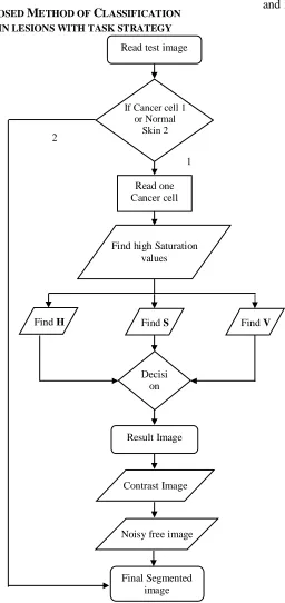
3. PROPOSED METHOD OF CLASSIFICATION OF SKIN LESIONS WITH TASK STRATEGY
Flow Chart of Classification of skin
lesions with task strategy is shown in figure 1
Skin lesions are classified into three modules.
They are
i.Border detection, ii. Feature extraction
and iii. Classification
Fig. 1 Flow Chart of Classification of skin lesions with task strategy Read test image
If Cancer cell 1 or Normal
Skin 2
Read one Cancer cell
1
Find high Saturation values
Find H Find S Find V
Result Image Decisi
on
Contrast Image
Noisy free image
287 i. Border detection
From each skin lesion image, we
extracted the border between the tumour and the
surrounding normal skin area. Accurate border
detection usually results in better classification
performance. Conventional automated methods
of border detection mostly focused on only
melanocytic skin lesions (MSLs). In our
previous study, we developed a general border
detection algorithm for both MSLs and
NoMSLs. The core of the algorithm is color
thresholding, removal of artifacts such as
microscope border and hair, and inclusion of
bright area seen specifically in NoMSLs [10,
12].
ii. Feature extraction
After determining the border of the
tumour, we segmented the skin lesion image into
four regions as normal skin, peripheral, central
tumour, and whole tumour. The whole tumour
consists of all pixels within the extracted border.
In contrast, the normal skin is all pixels on the
outside of the border. The peripheral is the first
30% of the whole tumour area, obtained by
going inward from the border as in our previous
studies. Finally, the central tumour is obtained
by removing the peripheral from the whole
tumour [11, 12].
iii.Classification
We used linear classifiers over nonlinear
ones in order to gain a clear understanding of the
relationship between the inputs and the outputs
of the models and to facilitate a comparison of
the classification performance.
Layered model (proposed):
The first-step classifier “MN-BS”
identifies the input skin lesion as MSL if the
output value is greater than the classifier’s
threshold value or as NoMSL otherwise. If the
result is an MSL, the second-step classifier
“M-N” distinguishes melanoma from nevus in the
same manner by comparing its output value with
the threshold value.
Flat models (performance baseline):
We introduce two types of flat models,
namely the ‘flat model I” and the ‘flat model II”
as the performance baseline. Each of the flat
models has four linear classifiers: “M,” “N,”
“B,” and “S” whose output values estimate the
presence/absence of the corresponding classes:
melanoma, nevus, BCC, and SK, respectively.
This kind of classification model is typically
288
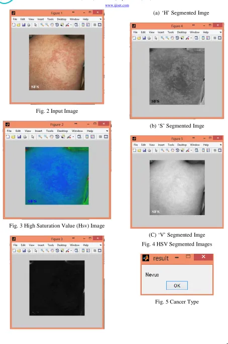
4. RESULTS AND DISCUSSIONS Input Image
In this image a person having cancer, is shown
figure 2, and this image is considered as an input
image.
HSV Image
HSV is the most commonly used cylindrical
representation of points in a RGB color model is
shown figure 3. SL, HSV, and related models
can be derived via geometric strategies, or can
be thought of as specific instances. In this the
HSV is again further divided into 3 images to
know the exact perception of the type of cancer
cell. These three images are depict more clearer
value of data needed for further investigation of
the cell is as shown figure 4.
Detected Cancer Type
Here the type of cancer among the four types of
cancer cells is detected while running the
MATLAB software. The result is shown figure
5, appears in the form of a name and the cancer
cell is identified.
Contrast Adjustment
By performing scaling operation here the
contrast is adjusted and also as this program
consists of multiple inputs and multi svm
technique is used for clearer display of the image
as shown in figure 6.
Cleared Image
Here imborder and erosion are used to remove
borders and also for the more elegant look of the
image without borders which makes it further
easy for the specialist to understand and analyses
the cancer cell keenly as shown the cleared
image in figure 7.
Final Segmented Image
Final segmented image is the image consisting
of the image after performing the segmentation
process as shown in figure 8. If any holes occur
in this image they can be filled by the dilation
process. The dilation process fills up the hole
with pixels and finally a final segmented image
is appeared which is helpful for the physicians
289
Fig. 2 Input Image
Fig. 3 High Saturation Value (Hsv) Image
(a) ‘H’ Segmented Imge
(b) ‘S’ Segmented Imge
(C) ‘V’ Segmented Imge
Fig. 4 HSV Segmented Images
290
Fig. 6 Contrast Adjustment Image
Fig. 7 Cleared Images
Fig. 8 Final Segmented Image
5. CONCLUSION
In our paper, we proposed a method to
distinguish among melanomas, nevi, BCCs,
and SKs. For the classification model, we
introduced a layered model for task
decomposition and two flat models to serve
as the baseline. We evaluated the models
with 964 dermoscopy images and showed
that the layered model outperformed the two
flat models. The layered model with 25
features achieved a detection rate of 90% for
melanomas and over 80% for each of the
three other types of skin lesions. The result
of this study shows promise for broadening
the range of users for classification and
enhancing the capability of the
computer-aided skin lesion classification.
REFERENCES
1. W. Zhao, R. Chellappa, P. J. Phillips, and
A. Rosenfeld, “Face recognition: A
literature survey,” ACM Comput. Surv.,
vol. 35, no. 4,pp. 399–458, Dec. 2003.
2. R. Fergus, B. Singh, A. Hertzmann, S. T.
Roweis, and W. T. Freeman, “Removing
camera shake from a single photograph,”
ACM Trans. Graph., vol. 25, no. 3, pp.
787–794, Jul. 2006.
3. Q. Shan, J. Jia, and A. Agarwala,
“High-quality motion deblurring from a single
image,” ACM Trans. Graph., vol. 27, no.
3, pp. 73:1–73:10,Aug. 2008.
291
5. A. Levin, Y. Weiss, F. Durand, and W.
T. Freeman, “Understanding blind
deconvolution algorithms,” IEEE Trans.
Pattern Anal. Mach. Intell., vol. 33, no.
12, pp. 2354–2367, Dec. 2011.
6. M. Šorel and F. Šroubek, “Space-variant
deblurring using one blurred and one
underexposed image,” in Proc. 16th
IEEE Int. Conf. Image Process., Nov.
2009, pp. 157–160.
7. H. Ji and K. Wang, “A two-stage
approach to blind spatially-varying
motion deblurring,” in Proc. IEEE Conf.
Comput. Vis. Pattern Recognit., Jun.
2012, pp. 73–80.
8. S. Cho, Y. Matsushita, and S. Lee,
“Removing non-uniform motion blur
from images,” in Proc. Int. Conf.
Comput. Vis., Oct. 2007, pp. 1–8.
9. Y.-W. Tai, P. Tan, and M. S. Brown,
“Richardson-Lucy deblurring for scenes
under a paperive motion path,” IEEE
Trans. Pattern Anal. Mach. Intell., vol.
33, no. 8, pp. 1603–1618, Aug. 2011.
10.O.Whyte, J. Sivic, A. Zisserman, and J.
Ponce, “Non-uniform deblurring for
shaken images,” Int. J. Comput. Vis.,
vol. 98, no. 2, pp. 168–186, 2012.
11.A. Gupta, N. Joshi, L. Zitnick, M.
Cohen, and B. Curless, “Single image
deblurring using motion density
functions,” in Proc. Eur. Conf. Comput.
Vis., 2010, pp. 171–184.
12. Z. Hu and M.-H. Yang, “Fast
non-uniform deblurring using constrained
camera pose subspace,” in Proc. Brit.
Mach. Vis. Conf., 2012, pp. 1–11.
13. C. Paramanand and A. N. Rajagopalan,
“Non-uniform motion de-blurring for
bilayer scenes,” in Proc. IEEE Conf.
Comput. Vis. Pattern Recognit., Jun.
2013, pp. 1115–1122.
CONTRIBUTORS
Sri Harsha Davuluri received his Master of Technology in VLSI System Design & Bachelor of Technology in ECE Stream from JNT University, Hyderabad. He has more than 10 years of Teaching Experience at various engineering colleges. At present he is working as an Associate
Professor, Department of ECE at Guru Nanak Institutions Technical Campus (Autonomous). He
contributed several more than 10 research papers in peer - reviewed International Journals & Conferences. He is one of the Reviewer Board Member in 5 International Journals. His areas of interest are VLSI & Embedded Systems & much interested towards learning new tools.
Srinivas Bachu, Research Scholar from GITAM University Hyderabad, and received M.Tech
degree from JNT University, Hyderabad, has 10 years of teaching experience. At present Srinivas Bachu working as an Associate Professor, Department of ECE at Guru Nanak Institutions
Technical Campus (Autonomous), Telangana. He is the Life Member of ISTE, AMIE, and