binds αvβ3 integrin with antibody-like affinity
Thesis by Gene Kym
In Partial Fulfillment of the Requirements for the degree of
Doctor of Philosophy
CALIFORNIA INSTITUTE OF TECHNOLOGY Pasadena, California
2014
ACKNOWLEDGEMENTS
ABSTRACT
TABLE OF CONTENTS
Acknowledgements ... iii
Abstract ... iv
Table of contents ... v
Figures and tables ... vi
Abbreviations ... viii
Chapter I: Introduction to alternative scaffolds ... 1
Chapter II: Discobody engineering and characterization ... 25
Appendix A: Detailed discobody data ... 61
FIGURES AND TABLES
Figure 1-1. Antibody domain schematic ... 19
Figure 1-2. Early antibody alternatives ... 21
Figure 1-3. Alternative scaffold structures ... 23
Figure 2-1. Topology diagrams of discobody and traditional scaffolds ... 40
Figure 2-2. Flow cytometry plots of discobody loop grafts ... 41
Figure 2-3. FACS plots of sequential library sorts and Eng-Db sequence ... 42
Figure 2-4. Binding characterization of Eng-Db ... 43
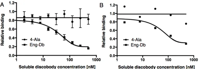
Figure 2-5. Soluble Eng-Db competition binding ... 43
Figure 2-6. Crystal structure of Eng-Db ... 44
Figure 2S-1. Potential linker orientations of bispecific Dbs ... 45
Figure 2S-2. On rate of Eng-Db using multiple antigen concentrations ... 46
Figure 2S-3. Off rate of Eng-Db using single antigen concentration ... 48
Figure 2S-4. On rate of Eng-Db using single antigen concentration ... 50
Figure 2S-5. Equilibrium binding constant of Eng-Db ... 52
Figure 2S-6. Melting temperature of Eng-Db ... 54
Figure 2S-7. Gel filtration elution profiles of Eng-Db ... 54
Figure 2S-8. Displayed Eng-Db competition with Dbs ... 55
Figure 2S-9. Displayed fibronectin competition with Dbs ... 56
Figure 2S-10. Eng-Db structure compared to RGD peptide ... 57
Table 2-1. Eng-Db characterization summary ... 58
Table 2S-1. Crystal structure data collection and refinement statistics ... 59
Figure A-1. Discoidin family structural information ... 61
Figure A-2. Factor VIII and the C2 domain ... 62
Figure A-3. Hydrophobic feet and the 4-Ala mutation of the C2 domain ... 62
Figure A-4. αvβ3 integrin and the RGD motif ... 63
Figure A-5. Yeast surface display schematics ... 63
Figure A-6. Yeast display expression of 4-Ala by flow cytometry ... 64
Figure A-7. Soluble expression of 4-Ala ... 64
Figure A-8. AgRP binding control by flow cytometry ... 65
Figure A-10. AgRP equilibrium titration curve fit ... 66
Figure A-11. Measurements for loop grafting ... 67
Figure A-12. Close look at loop graft measurements ... 67
Figure A-13. SGRGDNDLV loop graft equilibrium titration ... 68
Figure A-14. SGRGDNDLV loop graft curve fit ... 68
Figure A-15. AVTGRGDSPASS loop graft equilibrium titration ... 69
Figure A-16. AVTGRGDSPASS loop graft curve fit ... 69
Figure A-17. Normalized binding curves for control and loop grafts ... 70
Figure A-18. Library construction schematics ... 71
Figure A-19. FACS library population lift per sorting round ... 71
Figure A-20. Sequences from sort round I-III ... 72
Figure A-21. Converged sequences from sort round V ... 72
Figure A-22. RPRGDIE equilibrium titration by flow cytometry ... 73
Figure A-23. RPRGDIE curve fit ... 73
Figure A-24. ACRGDTC equilibrium titration by flow cytometry ... 74
Figure A-25. ACRGDTC curve fit ... 74
Figure A-26. Db constructs for bacterial expression ... 75
Figure A-27. Db bacterial expression gel ... 75
Figure A-28. TEV cleavage of Eng-Db ... 76
Figure A-29. Db constructs for yeast secretion expression ... 76
Figure A-30. Db yeast secretion expression gel filtration and gel ... 77
Figure A-31. Db yeast display levels compared to traditional scaffolds ... 77
Figure A-32. Db yeast display levels quantified ... 78
Figure A-33. Db soluble protein thermal melts ... 78
Figure A-34. Crystals collected for X-ray diffraction ... 79
Figure A-35. Diffraction during pre-data collection ... 79
ABBREVIATIONS
C h a p t e r 1 change how mAbs are engineered [5]. Additionally, antibody engineering innovations [6] have lead to novel mAb formats, including bispecifics [7-9], small domains [10-12], and fusions to cytotoxic payloads [13] or nanoparticles [14]. Small domain alternative scaffolds such as fibronectin or DARPins are driving many of these later generation technologies [11,12,15]. While alternative scaffolds have demonstrated value in protein engineering and as reagents, their clinical utility is just beginning to be explored [10,16].
Antibodies
The Fab region of the mAb is evolved through a series of VDJ recombination gene rearrangements and somatic hypermutation evolution that introduces point mutations for greater stability of the mAb as well as affinity and specificity of antigen binding. Clones are filtered through a negative selection step that removes self-antigens during B-cell development in the immune system. Traditionally, an antigen of interest that has been tethered to or formulated with immune-stimulating haptens is injected into a mouse. The murine immune system recognizes the antigen as foreign, and activates individual B-cell clones harboring a mAb sequence that recognize the foreign target. This clone is affinity matured and expanded, producing large amounts of antigen-specific antibodies. These B-cells can be isolated from the mouse spleen and fused with an immortalized myeloma cell line to produce hybridomas, which are then screened for the mAb of interest. These hybridomas can be multiplied and utilized to make clonal mAbs [17].
Fully human mAbs have been engineered using display technologies or genetically engineered mice with human antibody repertoires [20]. Phage display is the most prominent display method, and utilizes the M13 bacteriophage with antibody libraries fused to the viral p3 coat protein [21]. Yeast display is another useful technology that expresses antibody libraries on the yeast surface as Aga1p-Aga2p yeast mating protein fusions [22]. While larger libraries can be accessed using phage display (109-1011) than yeast display (107-109), the yeast eukaryotic protein processing system may allow for greater library expression [23]. Because display libraries are made from synthetic DNA, including cloned human B-cell sequence repertoires, it is possible to isolate fully human mAbs. Adalimumab (Humira, AbbVie) is an example of a fully human mAb from phage display. A number of genetically engineered mice with completely human B-cell repertoires have been created, including the XenoMouse, which lead to the discovery and commercialization of panitumumab (Vectibix, Amgen) [20,21].
four isotyopes of human IgG bind each of these receptors with differing affinities, and therefore have varying effector functions. Fc regions can be engineered to modulate half-life and effector function [24-27].
Several early protein engineering advances demonstrated that the mAb IgG protein scaffold was not the only option for targeted protein therapy (Fig. 1-2). Etanercept (Enbrel, Amgen/Pfizer) is a chimeric tumor necrosis factor (TNF) inhibitor made by fusing the extracellular domain of TNF receptor 2 to an IgG1 Fc, essentially replacing the Fab portion of the mAb with a natural receptor (Fig. 1-2A) [28]. Domain antibodies (dAbs, GlaxoSmithKline) are VH domains engineered for binding and resistance to aggregation via phage display (Fig. 1-2B). It was hoped that dAbs would show better tissue and tumor penetration due to their small size, and that they could easily be tethered together to make bispecific molecules [29,30]. The 10th repeat of fibronectin III (fibronectin or Adnectin, Bristol-Myers Squibb) was the first alternative scaffold to be engineered. Fibronectin is a β-sandwich with three loops clustered on one end that resembles a dAb (Fig. 1-2C). Fibronectin exhibits thermal stability above 80°C and its binder derivatives have been extensively studied [31-38]. Etanercept, dAbs, and fibronectin have influenced subsequent mAb variants and alternative scaffolds for use as therapeutics.
engineering to ensure a heterodimeric mAb pair. Single-chain variable fragment (scFv) formats, which string together the VH and VL domains via a long glycine and serine rich (Gly-Ser) linker, are not stable [40]. Additionally, the use of Gly-Ser linkers can potentially be immunogenic [8]. While the 150 kDa size of mAbs is good for a long serum half-life, the large molecular weight can prevent deep tissue penetration and tumor uptake when compared with small domains [11,12,15,39]. Additionally, the complex patent situation around mAbs can make freedom to operate analyses difficult, since many alternative scaffolds have clear-cut intellectual property associated with them [41].
Since Shohei Koide’s group demonstrated that fibronectin could be engineered to bind new targets [31], other non-antibody proteins have been explored as alternative scaffolds and have enjoyed varying degrees of success. Besides fibronectin, the most prominent examples include DARPins, Avimers, Anticalins, Affibodies, and peptide-based scaffolds [10,15,16]. Here we review the different scaffolds with a focus on protein engineering and clinical applications. Structures of their respective folds are shown in Figure 3, with engineered regions highlighted in red [16,42-49].
Fibronectin
ribosome display, are in vitro technologies which are most useful for small domains and peptides [51].
Using loop length variability and recursive mutagenesis in yeast display, Hackel et al. extended the affinity limit of the fibronectin scaffold to 1 picomolar against hen egg lysozyme [34]. This is the tightest known engineered protein-protein interaction, though some scFvs can bind hydrophobic molecules such as fluorescein with femtomolar affinities [52]. The bottom AB loop of fibronectin and the β-strands have also been engineered to bind targets, demonstrating the further utility of this scaffold [38,53].
CT-322 (Angiocept, Bristol-Myers Squibb) is a polyethylene glycol (PEG)ylated fibronectin engineered against vascular endothelial growth factor receptor 2 (VEGFR-2). It binds both human and mouse receptors with high affinities, allowing for use of mouse xenograft models for preclinical testing. 322 blocks the interaction of VEGFR-2 with VEGF-A, VEGF-C, and VEGF-D. CT-322 had a serum half-life of 100 hours, and in clinical trials demonstrated small decreases in tumor volume in 4/34 patients. 24 patients showed stable disease. Additionally, a PEGylated bispecific fibronectin against epidermal growth factor receptor (EGFR) and insulin-like growth factor 1 receptor (IGF1R) showed greater anti-proliferative effects against various human cancer cell lines than either single domain, demonstrating that the scaffold works in bispecific formats [16,54].
to current mAb therapeutics [55]. Novel mAb formats such as Ab-Fn3 highlight the potential of therapies based on alternative scaffolds and mAbs. Folds similar to fibronectin have been engineered for use as therapeutics, including a domain from human tenascin C (Tn3, Medimmune) and a fibronectin consensus domain (Centyrin, Janssen Pharmaceuticals) [56,57].
DARPins
DARPins have been utilized in creative applications for therapeutics [16,58,59]. Of particular interest is viral retargeting to tumors for gene therapy. One study used a tetrameric DARPin expressed separately from the virus, with three of the DARPins binding an adenovirus surface protein. The remaining DARPin was used to target HER2 positive cells. Upon infection with the adenovirus-DARPin assembly, the cells showed a significant increase in luciferase transduction. Because the DARPin construct was expressed independently of the virus, complications associated with expressing virus fused to extraneous constructs were avoided [62]. DARPins have also been used to redirect adeno-associated viruses (AAVs) as direct coat-protein fusions [63].
Because DARPins lack cysteines, they express well in bacterial hosts and can be site-specifically conjugated by the addition of individual cysteine residues, which are amenable to various bioconjugate chemistries. DARPins have been conjugated to protamine polymers complexed with small interfering RNAs (siRNAs), and demonstrated delivery of anti-bcl-2 siRNA to tumor cells via epithelial cell adhesion molecule (EpCAM) binding DARPins. EpCAM binding DARPins have also been attached to protein toxins such as Pseudomonas exotoxin A, and this fusion demonstrated potency against EpCAM-positive tumor cells. As with the Ab-Fn3 fibronectin example mentioned before, DARPins have been fused to form multiepitopic binders against EGFR. These molecules showed activity equivalent to, and in some cases better than, cetuximab, an EGFR targeting mAb. Anti-VEGF DARPins are undergoing phase II clinical trials for age-related macular degeneration (AGN 150998, Allergan) [16,58,59].
Avimers
(Fig. 1-3C). Over 200 different A-domains are found in human proteins, including low-density lipoprotein receptors. 28 positions per subunit are used for diversity generation, similar to the 25-30 residues involved in mAb binding. Avimer libraries were created using exon shuffling. Unlike other domains, where affinity maturation may be required, the first binding A-domain is subsequently tethered to a new library, where a second randomized A-domain separated by a five amino acid linker is generated. In this fashion, up to eight A-domains were engineered into a single Avimer. Avimers bind with subnanomolar affinities and can be expressed in high yields, even with eight domains linked together [41,64].
Avimers have been tested clinically, and were found to have some advantages over their mAb counterparts. mAbs against c-met or CD28 cause adverse effects, possibly because each mAb has two Fab regions and can bind two antigens at once, leading to receptor clustering and activation. Avimers against these targets did not demonstrate negative effects because each Avimer only binds a single antigen, although on multiple epitopes. An anti-IL-6 triepitopic Avimer inhibited IL-6-induced cell proliferation and showed activity in mice. Since Avimers are small proteins, they are rapidly cleared from the serum through the kidneys. This problem was solved by tethering an anti-IgG domain as part of the Avimer to take advantage of the natural serum half-life extension of the IgG-FcRn interaction [16]. Amgen has since acquired Avimer technology for further development.
Anticalins
interface that can accommodate small molecule binding in a cavity made by the binding loops and β-barrel structure. In nature, this cavity is used to bind small molecule vitamins and hormones for transport, storage, and sequestration. Anticalin libraries can be made with 16-24 random residues in the loops as well as select residues in the small molecule binding pocket, allowing for great diversity generation. Due to their unique structure, they can be engineered to bind proteins or small molecules [16,65,66].
Anticalins have been conjugated to fluorescent molecules via site-specific cysteine insertions, or PEGylated for extended half-life. PEGylated Anticalins against VEGF showed anti-angiogenic and anti-tumor activity in mouse xenograft models. PRS-050, a site-specifically PEGylated Anticalin that binds to all splice forms of human VEGF-A as well as rodent orthologues, was tested in a phase I dose escalation study. PRS-050 showed no immunogenicity, with only a single patient (out of 26) showing ADAs after 17 doses. Half-life was approximately six days, and no free VEGF-A was detected after drug treatment, while PRS-050-VEGF-A complexes were detected up to three weeks after dosing [67]. Bispecific Anticalins, called duocalins, have been made to target two antigens at once [16]. A89Zn-labeled anti-MET Anticalin was used to visualize MET-positive tumor expression in mouse models via positron-emission tomography (PET) imaging. Tumor uptake was shown to be dose-dependent [68]. Pieris AG is the company developing Anticalins.
Affibodies
three α-helices (Fig. 1-3E). Anti-HER2 Affibodies have been generated with affinities down to 22 picomolar [16,69].
Radiolabelled variants were used to image HER2-positive xenograft tumors with high contrast in mouse models. Anti-HER2 Affibodies fused to albumin binding domains lead to molecules with increased serum half-life due to the long circulation times of albumin, and had a concomitant increase in tumor uptake. Anti-HER2 Affibodies were fused to Pseudomonas exotoxin A and killed BT-474 breast cancer cells in tumor xenograft models. EGFR binding Affibodies were also generated for radiolabelled imaging and showed accumulation in tumors. Anti-EGFR Affibodies were tethered via a Gly-Ser linker to anti-HER2 Affibodies to make bivalent molecules, since both HER2 and EGFR are co-expressed on some tumor types [10,16,69].
Affibodies have also been developed as Fc fusions and in large complexes for drug delivery via viruses, nanoparticles, and liposomes. Affibodies are of particular interest in imaging due to their small size and rapid plasma clearance, which can increase contrast ratios. They have been conjugated to fluorescent proteins, dyes, and radiolabels for such applications [69]. Affibody AB is developing the Affibody technology.
Peptides
selectivity profiles, since many of their parental molecules demonstrate promiscuity in binding partners [10,16,39]. The universal applicability of these small scaffolds to create de novo binders has yet to be demonstrated.
Knottins are 30 amino acid (3 kDa) cystine knot proteins with three internal disulfide bonds that confer stability onto the protein, preventing denaturation even in extreme acid or base conditions. Several knottins, including the human Agouti-related protein (Fig. 1-3F) and the Ecballium elaterium trypsin inhibitor were engineered with RGD-based libraries to bind to integrin. Variants with different subtype selectivity profiles against αvβ3, αvβ5, and αvβ1 integrin were engineered with affinities down to 10 nanomolar. These peptides were selected for in yeast display, and binders were synthesized chemically for testing [70,71]. The molecules have been labeled for PET imaging in early cancer detection [72,73]. ω-conotoxins are another class of disulfide-rich peptides, in this case derived from the cone snail Conus magus [74]. SNX-111 (Ziconotide, Prialt) is a Food and Drug Administration (FDA) approved therapeutic for chronic pain that blocks N-type voltage-gated calcium channels [75].
Νext generation alternative scaffolds
Alternative scaffolds have been engineered to bind a variety of targets, and have been linked together to make multi-specific molecules. Exciting next generation applications of alternative scaffolds are beginning to emerge, including mAb fusions targeting up to five ligands at once, small bicyclic peptides with chemically conjugated hydrophobic cores, and engineered super ligands that are tailor-made to modulate specific biological pathways.
The aforementioned triepitopic Ab-Fn3 against EGFR demonstrated receptor clustering and efficacy against monotherapy resistant cell lines. This concept has also been demonstrated against five different antigens at once using mAbs fused to small alternative scaffolds. These molecules are known as zybodies (Zyngenia). By tethering Affibodies, knottins, or peptides (referred to as “modular recognition domains,” or “MRDs” in this study) with varying degrees of structure to the N- and C-termini of HER2 or EGFR targeting mAbs, zybodies showed better efficacy than trastuzumab (anti-HER2) in an angiogenesis-dependent xenograft tumor model [80].
1.5 nanomolar. The bicyclic peptides can be synthesized chemically [81], and Bicycle Therapeutics is developing the technology for clinical use.
Interleukins (IL) are immune signaling molecules that bind to receptor complexes consisting of different subunits. Affinity towards the receptor depends on which subunits interact with the bound IL. IL-2 is one such molecule that exerts immune stimulating effects. The IL-2 receptor consists of α, β, and γ subunits that all form interactions with the IL-2 ligand. IL-2 can bind to the αβγ complex with high affinity, or the βγ complex alone with a lower affinity. IL-2 variants were made to bind either the αβγ or βγ complexes with increased affinity, allowing selective IL-2 activation on different receptor assemblies. Certain cell types, such as leukocytes, that are targets of IL-2 activation can be specifically modulated by the βγ binding IL-2 variant. Using this strategy, background cells which express the αβγ complexes, such as lung endothelial cells, can be avoided [82].
Factor VIII
fVIIIa binding to membranes catalyzes the interaction of factors IXa and X (fIXa, fX), which leads to further downstream processing that ultimately results in coagulation [49,83-88].
The C2 domain of fVIII is of critical importance. Hemophilia A can also be acquired through autoimmune antibodies against the C2 domain, and ADAs that form against the C2 domain of fVIII drugs can lead to decreased efficacy [89]. The HAMSTeRS database (Hemophilia A Mutation, Structure, Test, and Resource Site) is a compilation of fVIII genetic mutations [96]. The C2 domain has been co-crystallized with a variety of inhibitory mAbs. Additionally, alanine scanning mutagenesis, which tests the importance of certain residues for binding, has been performed on the C2 domain. These mutants were tested for binding to a panel of inhibitory mAbs, helping identify which residues can be engineered to avoid B-cell epitopes for future drug development [97].
Discoidin Domain Family
The C2 domain is part of the discoidin domain family, a 150 amino acid (18 kDa) domain that mediates many biological processes, including cellular adhesion, vasculogenesis, angiogenesis, migration, and development. DDR1 and DDR2 are receptor tyrosine kinases that contain the discoidin domain and are involved in extracellular matrix remodeling, cell adhesion, and proliferation. Neurexins and neuropilins also contain discoidin domains, and are important in nervous system development. Discoidin domains bind to a variety of cognate ligands such as galactose, collagen, growth factors, phospholipids, and other non-charged lipids. Discoidin domains are found as subunits of larger proteins, and can be arrayed in a repeated fashion or found as single units within the protein. The domain was first identified in the amoeba Dictyostelium discoideum, and was described as a lectin that bound galactose with high affinity (10-6 to 10-8 M Kd). Since then, genome sequencing has identified over 100 eukaryotic and 300 prokaryotic proteins that contain the discoidin domain [98-101].
β-strands support three conformationally flexible spikes on one end that make up the binding interface (Fig. 1-3H). While the core β-barrel structure is fairly conserved within the discoidin domain family, the three binding spikes demonstrate diversity as result of the range of ligands that the family can bind to. However, each individual discoidin domain is only known to bind to a single cognate ligand [101]. The fVIII discoidin domain contains hydrophobic residues at the ends of spikes one and three. These intercalate into phospholipid membranes, and have positively charged residues at the base of the spikes that interact with the negatively charged phospholipid head groups [83,86]. In the case of the Dictyostelium discoideum discoidin domain, the spikes specifically coordinate binding to galactose residues to provide specific binding. The N- and C-termini of the discoidin domain are adjacent to one another, and bound by a disulfide in eukaryotic discoidin domains. The discoidin domain is believed to have originated from a single gene. Its modular architecture facilitates evolution into new proteins via domain shuffling [98-101].
Discobody Alternative Scaffold
Fig. 1-1. Domain schematic of a mAb. (A) Upper left: Fv in light blue, with VL and VH domains
Fig. 1-2. Early mAb alternatives. (A) Etanercept, or Enbrel, a chimeric protein consisting of the
Fig. 1-3. Alternative scaffold structures in grey with engineerable binding interfaces highlighted
C h a p t e r 2
DISCOBODY ENGINEERING AND CHARACTERIZATION
Monoclonal antibodies (mAbs) are an important part of the biotechnology industry and have demonstrated value as therapeutics, as research reagents, and in diagnostic assays [1-3]. mAbs can be engineered to bind specific molecular targets with high affinity. However, this ability is not limited to mAbs, as in recent years alternative scaffolds based on fibronectin, protein A, ankyrin repeats, lipocalins, and other proteins have been engineered to bind various antigens. Alternative scaffolds can be engineered to have binding affinities and specificities characteristic of mAbs. Many of these scaffolds have also demonstrated superior biochemical and biophysical properties, such as high bacterial expression levels and thermal stability [15]. These attributes are important for reagents and diagnostic tools, where low material cost and high thermal stability (to facilitate storage and use in real-world conditions) are priorities [1,104,105].
The therapeutic utility of alternative scaffolds is assessed with different criteria [11,106]. Immunogenicity is a critical issue for any protein therapeutic. Because immunogenicity remains difficult to predict a priori [106], it is important to mitigate this risk by starting with a human protein [11]. The first mAbs were of non-human origin and, despite successes in laboratory settings, were not suitable as human drugs. They were too immunogenic for repeated dosing and required humanization to make them useful as a therapeutic [107]. In contrast, scaffolds derived from human proteins, such as fibronectin [31], have elicited less immunogenic responses in patients. A Phase I trial of CT-322, a fibronectin engineered to bind VEGFR-2, only showed anti-drug antibodies (ADAs) against the engineered binding loops and not the scaffold itself.
not associated with any adverse events [54]. It remains to be seen whether alternative scaffolds built from consensus designs [combinations of homologous sequences that were fully [57] or partially [108] derived from human proteins] will form ADAs upon repeated intravenous injections [106]. Other alternative scaffolds from non-human sources, such as camelid nanobodies, have been humanized prior to clinical use [109].
Alternative scaffolds have been touted for their small size, which could lead to enhanced tumor penetration [11,15,105,110]. Additionally, monomeric scaffolds can be strung in tandem to form bispecifics that target two different antigens, or recognize two different sites on the same antigen and thus enhance binding. Bispecifics have been created using fibronectin [37], camelid
nanobodies [111], DARPins [112,113], and more [15]. Another use of alternative scaffolds is to tether them to mAbs to form multi-specific molecules. Various proteins have been appended to mAbs including fibronectin [55], peptides, knottins, and affibodies [80]. The majority of these domains have N- and C-termini on opposing ends of the molecule (Fig. 2-1A, C, D, and F). This architecture requires a long and potentially immunogenic linker [8] when connecting tandem domains (Supplementary Fig. 2-S1A).
fVIII is a blood coagulation protein encoded by the F8 gene, which is involved in the formation of blood clots after injury. Mutation to the F8 gene results in the medical condition hemophilia A, requiring hemophiliacs to receive injections of functional fVIII to aid blood clotting [85,114]. The C2 domain of fVIII is an 18 kDa distorted β-barrel that contains three loops, or “spikes,” on its binding interface. Spikes 1 and 3 extend the farthest and contain hydrophobic residues at their ends. The native function of these spikes is to insert into phosphatidylserine (PS)-rich platelet membranes to initiate the coagulation cascade [83,87]. An important feature of our chosen scaffold is that it is based on the β-barrel topology. β-barrel folds are similar to the β-sandwich structures of the canonical mAb immunoglobulin and fibronectin scaffolds. Much like the mAb immunoglobulin and fibronectin folds, the discoidin β-barrel has loops clustered on one end [101] that may be engineered to bind specific antigens (Fig. 2-1).
We chose to validate the fVIII C2 scaffold by engineering a binder against αvβ3 integrin, a cell surface protein and a target of interest in oncology [102]. Additionally, αvβ3 is a well-studied protein and is known to interact with the linear Gly-Arg-Gly-Asp-Ser (GRGDS) peptide, exhibiting a 740 nM IC50 in extracellular matrix (ECM) competition assays [115]. The Arg-Gly-Asp (RGD) motif is found in ECM proteins such as fibronectin and is used by αvβ3 integrin-expressing cells as footholds to navigate the ECM for cellular motility [116]. αvβ3 integrin is overexpressed in a number of cancers [103], and the RGD-αvβ3 interaction contributes to tumor growth, angiogenesis, and metastasis. Blocking this interaction could provide therapeutic value [102]. We sought to build an RGD-based library of discobody variants and select for binders against αvβ3 integrin.
Materials and Methods
Reagents and strains
αvβ3 integrin was purchased from R&D Systems. Yeast display Saccharomyces cerevisea strain EBY100, yeast display plasmid pPNL6, yeast secretion strain YVH10, and yeast secretion plasmid pPNL9 were obtained from Pacific Northwest National Laboratory. All yeast display protocols, buffers, and reagents were used as previously described [22,70]. Oligonucleotides were obtained from Integrated DNA Technologies, and PCR assembly primers were designed using DNAWorks from NIH’s Helix Systems [117]. KOD Hot Start Polymerase from Novagen was used to PCR assemble gene inserts. The inserts had flanking 20-40 bp overlaps with the desired cloning site in their respective, linearized plasmids. These were assembled into the appropriate plasmids using either yeast homologous recombination or Gibson cloning into TOP10
Strep-Tag II cloned onto the C-terminus. Purification resins included HIS-select HF Nickel Affinity Gel from Sigma and StrepTactin Sepharose High Performance from GE Healthcare Life Sciences. Unless otherwise stated, all chemicals were from Sigma, and all E. coli strains from Life Technologies. Soluble protein expression was performed in 2.5 L Ultra Yield Flasks from Thomson. Protein was concentrated in Amicon 10,000 Da MWCO centrifugal filters from EMD Millipore. DNA extraction was performed with kits from Qiagen.
Loop grafts, library construction, flow cytometry, and sorting
RGD-based integrin binding loops from six different engineered knottin peptides [70,71] and from wild-type fibronectin [43] were grafted into the spike 1 region of the discobody starting template (4-Ala C2 discoidin domain of fVIII). Yeast displayed protein was expressed at 20ºC and 250 RPM for 16 hours. Displayed protein was incubated with αvβ3. Flow cytometry was performed on a BD Biosciences FACSCalibur, and data was analyzed with FlowJo from Tree Star. 50,000 cell counts were collected for loop graft binding experiments. All yeast display experiments were performed in Integrin Binding Buffer (IBB) as previously described [70]. Libraries were constructed by high efficiency yeast electroporation [22]. FACS was performed on the MoFlo XDP instrument from Beckman Coulter using polypropylene BD Falcon FACS tubes. Sorted sequences were represented using WebLogo [119].
Expression and characterization of soluble discobodies
Discobodies were expressed both in YVH10 yeast secretion culture and BL21(DE3) E. coli.
from Life Technologies. This culture was expressed at 20ºC, 200 RPM, for 48 hours. Pellets were spun down and discarded twice, and the supernatant was ammonium sulfate (VWR) precipitated at 80% salt. Precipitate was resuspended in YVH10 binding/wash buffer (300 mM NaCl, 20 mM sodium phosphate, pH 7.8, 0.05% tween 20, 2.5% glycerol, 10 mM imidazole) and loaded onto HIS-select resin. After washing with the same buffer, samples were eluted in YVH10 elution buffer (300 mM NaCl, 20 mM sodium phosphate, pH 7.8, 0.05% tween 20, 2.5%
glycerol, 200 mM imidazole), concentrated, and run over an analytical Superdex-75 column from Amersham Pharmacia on the ÄKTA FPLC system. Sample volume was 0.5 mL, run at 0.5 mL/min in discobody storage buffer (200 mM NaCl, 20 mM Tris, pH 7.4). Typical yields were 1 mg/L from yeast secretion culture.
For E. coli expression, single colonies of BL21(DE3) containing the discobody construct in pET11a with a TEV-cleavable Strep-Tag II were picked and inoculated in 5 mL LB cultures with 100 ug/mL amplicillin (LB-amp) and grown overnight at 37ºC, 250 RPM. The next day, we inoculated 4 L of LB-amp per clone, at 1 L per flask, with a 1:333 dilution of overnight culture and grew it at 37ºC until it reached an OD600 of 0.5. Cultures were inoculated with 1 mM IPTG and grown at 16ºC, 200 RPM, overnight to express protein. The following day, 4 L of the
discobody clone were spun down and pooled, after which the pellet was split between two 50 mL conical Falcon tubes. The concentrated pellets were frozen at -20ºC then thawed at room
temperature. Each 50 mL tube corresponded to 2 L worth of pellet and weighed 6.5 g. We added 35 mL of BugBuster Master Mix from EMD Millipore to each 50 mL tube with heavy vortexing, then nutated the mix at room temperature for 30 minutes. This was then spun for 45 minutes at 15,000 rcf, 4ºC on an angled rotor to separate the pellet from the discobody-containing
Tris pH 8). This mix was run through equilibrated StrepTactin resin in a gravity column, washed once, then eluted in StrepTactin elution buffer (150 mM NaCl, 1 mM EDTA, 2.5 mM desthiobiotin, 100 mM Tris pH 8). Eluate was concentrated and purified by gel filtration using discobody storage buffer as described above. Yields were 1 mg/L.
Binding kinetics in display
Kinetics were performed as described [24]. All incubations were performed while nutating at room temperature. Quenched samples were placed on ice in a 4ºC cold room. All work with cold samples was performed at 4ºC with chilled tips and a dedicated cold room centrifuge. Samples were handled in 1.5 mL Eppendorf tubes, then transferred to a Nunc polypropylene 96-well V-bottom plate prior to addition of secondary antibody for ease of handling and standardization between samples. Competition experiments were performed entirely in 96-well plates. Prior to flow cytometry analysis, samples were spun down and kept as pellets on ice, covered from light. Immediately prior to analysis, individual samples were resuspended in 0.5 mL of IBB and transferred to a polystyrene BD Falcon FACS tube for loading. 20,000 cell counts were collected for each sample in kinetic assays. Analysis was performed in GraphPad Prism version 6.0d for OSX 10.9 from GraphPad Software. Kd was fit to the Michaelis-Menten model, kon calculated association kinetics of two or more concentrations of hot ligand, and koff was determined via one phase exponential decay dissociation kinetics. kon values using one ligand concentration
Crystallization
Initial crystal screening was performed using an Index screen (Hampton Research). Crystallization was conducted in 96-well plate format using a MRC sitting-drop crystallization plate (Hampton Research) at 20ºC; protein drops at 10 mg/mL were mixed with precipitant in a 0.5 µL x 0.5 µL ratio and were equilibrated with 50 µL of precipitant. Four initial hits were observed in 25% PEG3350 in different buffers. Upon crystal optimization, the best diffracting crystal was obtained in 0.1 M Bis-Tris pH 6.5, 10% PEG750 methyl ether (Sigma). Crystals started to appear after four days and grew to maturation in a week with an approximate length of 300 µm.
X-ray data collection and structure determination
Crystals were harvested and soaked in a cryoprotectant solution containing Fomblin Y 16/6 (Sigma). Crystals were frozen and sent to the Stanford Synchrotron Radiation Laboratory (SSRL). The diffraction data were collected at SSRL beamline 12-2 equipped with a PILATUS 6M PAD detector. The diffraction data were indexed, integrated, and scaled using XDS [120]. Initial phase was obtained by molecular replacement using PhaserMR [121] of the scaled experimental data using the previously solved C2 domain of human factor VIII [87] (PDB ID: 1D7P). Model building was done using Coot [122], and the structure was refined using Phenix.Refine [123]. Composite omit map was calculated using CCP4 suite [124].
Results
The search process
adopt many of the lessons learned from engineering loop-based binders and apply them to our scaffold, possibly also allowing loop grafting from known mAbs or other binding proteins. In addition to these biochemical search terms, we searched for “blood” or “serum” as keywords, and limited the search to human proteins. We expected that this constrained search would find a scaffold that behaved well as an intravenously injectable human therapeutic. An initial hit of the C2 discoidin domain of factor V, another blood coagulation factor, stood out to us. Upon further review of the discoidin domains involved in coagulation, we settled on the C2 domain of factor VIII, a very similar protein that has been more extensively characterized [83,84,86].
Display of template discoidin scaffold, loop grafting, and binding to integrin
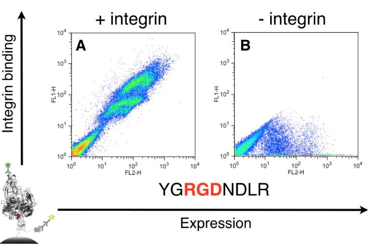
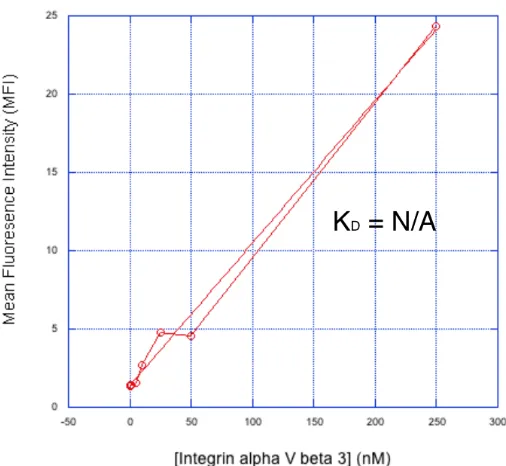
The C2 domain of fVIII contains hydrophobic residues on the most solvent-exposed tips of spikes 1 and 3. Previous studies [83] have shown that mutating these residues (Met-Phe of spike 1 and Leu-Leu of spike 3) to Ala-Ala on each spike to produce a four-point mutant (called 4-Ala) results in a 35-fold reduction in binding to PS membranes and a 91% reduction in specific activity in the activated partial thromboplastin time assay, one way of measuring fVIII activity. The 4-Ala mutant C2 domain expresses in yeast surface display [22] format, so we used this approach to determine binding to our target protein, αvβ3 integrin, as measured by flow cytometry. As shown in Fig. 2-2A, 4-Ala shows no inherent binding to αvβ3 integrin and was therefore chosen as our starting scaffold for engineering.
To assess whether our discobody scaffold could bind a new target, we grafted a number of loops from αvβ3 binding proteins into the spike 1 region of the 4-Ala template, replacing the
FTNAAAT spike 1 sequence. These sequences were taken from engineered integrin binding peptides (Fig. 2-2B–G) as well as fibronectin (Fig. 2-2H), which naturally binds αvβ3
these data, we proceeded to build an RGD-containing degenerate codon library into spike 1 of 4-Ala.
Library generation and FACS selection
We mutated the three most solvent-exposed amino acids at the tip of spike 1 to RGD, without changing its length, to ensure that the RGD motif was available to guide binding. Because both the position of RGD in the loop and its flanking amino acids significantly affect binding affinity [70,115,127], we randomly mutated two residues on each side of the motif using NNS degenerate codons. The FTNAAAT sequence of spike 1 was replaced with XXRGDXX, where “X”
represents the NNS codon. NNS encodes all twenty amino acids as well as a single stop codon [128]. The theoretical library size was 1.6 x 10⁵, and we were able to obtain ~1000-fold coverage, as we attained a transformation efficiency of 108 into the EBY100 yeast display strain.
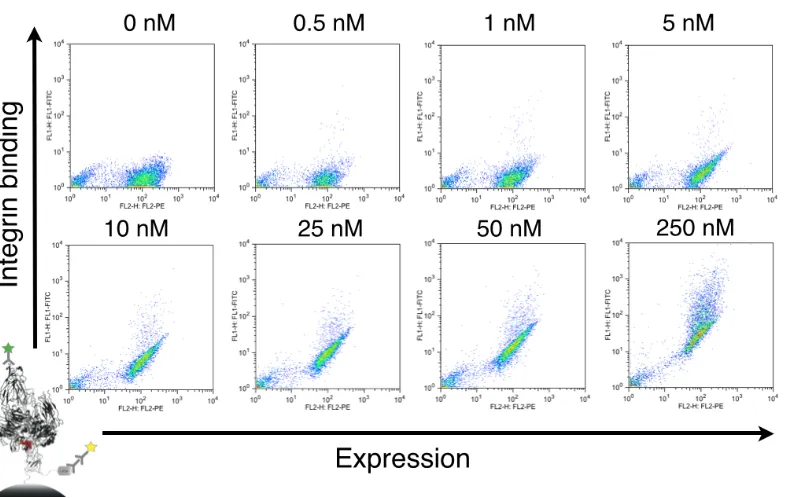
We expressed a library size of 109 and performed five sequential fluorescence-activated cell sorting (FACS) sorts with decreasing concentrations of αvβ3 integrin. Sorting against 200 nM for the first two sorts, 100 nM for the third and fourth sorts, and 10 nM for the final sort, we sorted the top 1-5% of each population for both antigen binding and surface display levels. We
monitored each round by sequencing 10 clones each time. By round five, the XXRGDXX library converged upon a dominant clone with the loop sequence ACRGDTC (Fig. 2-3). This engineered variant (Eng-Db) was selected for further characterization and analysis.
Binding and characterization of engineered discobody
incubating aliquots of the displayed discobody with 20, 50, and 100 nM αvβ3 integrin and quenching on ice at time points up to four hours. Quenching was performed by washing the sample in cold buffer and keeping it on ice until analysis. The kon was fit to 1.3 ± 0.2 (103 M-1s-1) (Fig. 2-4A, Supplementary Fig. 2-S2). This slow association may be due to the conformational change that αvβ3 integrin undergoes when binding to RGD-containing partners [129-131]. We measured koff by incubating yeast displaying Eng-Db with 200 nM antigen for four hours,
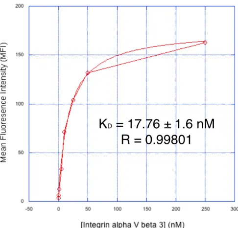
washing aliquots at different time points, and leaving them with 1 uM soluble Eng-Db competitor at room temperature for intervals up to 26 hours to allow antigen dissociation. We calculated a koff of 2.0 ± 0.1 (10-5 s-1) and a t1/2 of 9.6 hours (Fig. 2-4B, Supplementary Fig. 2-S3). Using this koff value, we measured the kon again using a single concentration of 200 nM αvβ3 integrin and obtained a kon of 2.1 ± 0.4 (103 M-1s-1), which is similar to the value obtained using different antigen concentrations (Supplementary Fig. 2-S4). We also performed an equilibrium titration to determine Kd at equilibrium. Yeast displaying Eng-Db were incubated with varying
concentrations of αvβ3 integrin (0–250 nM) for 24 hours. We measured a binding affinity of 16.2 ± 1.7 nM (Fig. 2-4C, Supplementary Fig. 2-S5). Binding measurements are summarized in Table I.
in a dose-dependent manner, but soluble 4-Ala did not, indicating that Eng-Db functions outside of display (Fig. 2-5A, Supplementary Fig. 2-S8).
Integrin αvβ3 naturally binds to the ECM protein fibronectin [116]. To see if our protein competes for the same binding site, we displayed fibronectin in yeast surface display and incubated it with αvβ3 integrin in the presence of soluble 4-Ala or Eng-Db. As before, Eng-Db blocked binding in a dose-dependent manner and 4-Ala did not, indicating that Eng-Db competes with the fibronectin binding site (Fig. 2-5B, Supplementary Fig. 2-S9).
X-ray crystal structure of engineered discobody
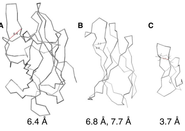
To gain further insight into Eng-Db, we obtained its crystal structure at 2.1 Å resolution (Fig. 2-6A, Supplementary Table I). The engineered variant looks very similar to previously solved wild-type fVIII C2 domain structures [87,127]. The C2 domain’s membrane-binding spikes exhibit some conformational heterogeneity, as illustrated in a structural overlay (Fig. 2-6C). Eng-Db’s loops fall within the observed range. The RGD tripeptide motif is positioned at the most solvent exposed tip, as we had planned for during library design. Note the presence of an additional disulfide bond within spike 1, which may account for the 6ºC increase in Tm (Fig. 2-6A).
Discussion
Discoidin domain as an alternative scaffold
allows for library construction [101]. Because the binding interface is made up of loops,
similar to those of antibodies and engineered fibronectins [31,126], it is possible to transfer loops from preexisting binders onto the discobody scaffold, as has been demonstrated in our loop graft experiments (Fig. 2-2).
Discobodies begin fully human, but loop mutations still need to be chosen with care to avoid introducing immunogenic sequences [106]. Certain hemophiliac patients who receive fVIII injections develop ADAs [89], but it seems unlikely that this would happen in an individual with endogenous fVIII due to negative selection during immune system development (personal communication, Gary Gilbert).
αvβ3 integrin and RGD-containing peptides
To minimize the number of variables while validating a new scaffold, we chose the well understood system of αvβ3 integrin and the RGD motif. Additionally, αvβ3 integrin is over-expressed in a number of cancers and is a drug target currently being pursued by the
pharmaceutical industry [102].
αvβ3bound to a cyclic Arg-Gly-Asp-{D-Phe}- {N-methyl-Val-} pentapeptide has been
crystalized [130] (PDB ID: 1L5G). The cyclic pentapeptide from this structure can be overlaid with spike 1 of Eng-Db (Supplementary Fig. 2-S10A), and the backbone RMSD of the respective Arg-Gly-Asp sequences is only 0.3 Å (Supplementary Fig. 2-S10B), demonstrating that the two binding sites may be very similar. In Eng-Db, the Arg and Asp sidechains are directed away from each other, much like in the target-bound peptide. Although the side chains differ slightly starting from their respective Cβs (Supplementary Fig. 2-S10A), their conformation may change upon binding to αvβ3integrin.
Future work for a universal discobody scaffold
Conclusions
Figures and Tables
Fig. 2-1. Topology diagrams comparing (A) the antibody immunoglobulin fold, (B) the discoidin
Fig. 2-2. Flow cytometry plots showing binding of 100 nM integrin to (A) 4-Ala and various
Fig. 2-3. FACS plots for sequential sorts (A-E) of the library against decreasing concentrations of
Fig. 2-4. Binding characterization of Eng-Db. (A) On rate measured with different concentrations
of integrin. (B) Off rate measured with 200 nM integrin. (C) Equilibrium titration of displayed Eng-Db against integrin.
Fig. 2-5. (A) Binding of displayed Eng-Db to 125 nM integrin with competing soluble 4-Ala or
Supplementary Fig. 2-S1. Orientations of N- and C-termini of discobodies allow for shorter
Supplementary Fig. 2-S2. (A) Flow cytometry diagrams of displayed Eng-Db binding to 20,
Supplementary Fig. 2-S3. (A) Flow cytometry diagrams of displayed Eng-Db dissociating
Supplementary Fig. 2-S4. (A) Flow cytometry diagrams of displayed Eng-Db binding to 200
Supplementary Fig. 2-S5. (A) Flow cytometry diagrams of displayed Eng-Db binding to
Supplementary Fig. 2-S6. Melting temperature (Tm) as assessed by the Thermofluor
denaturation assay shows that Eng-Db is 6ºC more thermostable than 4-Ala.
Supplementary Fig. 2-S7. Gel filtration profiles of soluble (A) 4-Ala and (B) Eng-Db. (C)
Supplementary Fig. 2-S8. (A) Flow cytometry diagrams of displayed Eng-Db binding to 125
Supplementary Fig. 2-S9. (A) Flow cytometry diagrams of displayed fibronectin binding to 125
Supplementary Fig. 2-S10. (A) Line representations of spike 1 of Eng-Db (magenta) overlaid
with that of the Arg-Gly-Asp-{D-Phe}- {N-methyl-Val-} integrin-binding cylic pentapeptide from the 1L5G αvβ3-RGD-Mn structure (cyan). RGD from spike 1 of Eng-Db is shown in red and the disulfide is in yellow; RGD from the pentapeptide is shown in blue. (B) Line
Table 2-I. Eng-Db binding and thermal stability characterization
A p p e n d i x A
DETAILED DISCOBODY DATA
Here we present extensive data related to the Discobody engineering project that supports the main thesis body. All methods were detailed in the preceding chapters, and will not be covered here. Due to the supplementary nature of the section, most information will be presented in figure format with corresponding legends.
Figures and Tables
Fig. A-1. Discoidin domain structural information. (A) The C2 domain of factor V (PDB ID:
Fig. A-2. Factor VIII. (A) The complete fVIII structure (PDB ID 2R7E) with the C2 domain
highlighted in dark grey. (B) The isolated C2 domain from the same structure.
Fig. A-3. Hydrophobic residues mediate lipid binding. (A) The wild type fVIII C2 discoidin domain
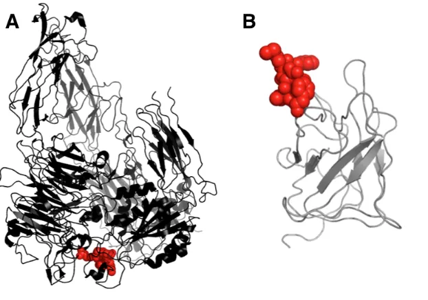
Fig. A-4. αvβ3 integrin and the RGD motif. (A) αvβ3 integrin in black bound to an RGD peptide in red. (B) The discobody with the position of an inserted RGD noted in red.
Fig. A-5. Yeast surface display schematic. (A) Diagram of yeast surface display constructs with
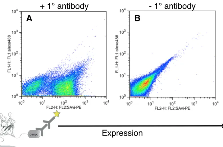
Fig. A-6. Flow cytometry plots demonstrate surface expression of 4-Ala discobody. (A) Display
constructs with primary anti-cmyc antibody and secondary with fluorophore. (B) Secondary antibody only.
Fig. A-7. Coomassie stain of soluble expression and purification from yeast of 4-Ala demonstrates
Fig. A-8. Flow cytometry plots of integrin binding AgRP control peptides from Silverman, et al, in
Fig. A-9. Affinity titration of control AgRP peptide against αvβ3 integrin, data are flow cytometry plots for displayed peptide. Incubation was performed for 1 hour at room temperature.
Fig. A-11. Ribbon diagrams measuring backbone distances for loop grafting from onto (A) the
discoidin domain. Loops containing RGD from (B) Fibronectin and (C) AgRP peptide were grafted onto the 4-Ala scaffold. Measured distances indicated below in angstroms. PDB IDs (A) 2R7E, (B) 1FNA, and (C) 1HYK.
Fig. A-13. Affinity titration of 4-Ala with the SGRGDNDLV loop graft against αvβ3 integrin via flow cytometry of displayed protein.
Fig. A-15. Affinity titration of 4-Ala with the AVTGRGDSPASS loop graft against αvβ3 integrin via flow cytometry of displayed protein.
Fig. A-17. Binding curves normalized by expression for AgRP (Figure A-10), JRC-5
Fig. A-18. Library construction schematics. (A) Overlap PCR oligos for constructing the degenerate
codon insert in green, RGD and flanking diverse regions shown in red. (B) Insert for cloning into blue pPNL6 yeast display vector via yeast homologous recombination.
Fig. A-19. Percent lift of library population per FACS sorting round. Library “lift” is defined as the
Fig. A-20. Sequence diversity of each FACS sorting round of the library. Round I-III demonstrate
no convergence.
Fig. A-21. Sequences converge in sort round V. (A) VL, a minor population, converges upon
Fig. A-22. Overlaid flow cytometry plots for affinity titration of RPRGDIE in display format. αvβ3 integrin was incubated with displayed RPRGDIE discobody for 30 minutes at room temperature and the geometric mean of the center of each population was used to calculate lift.
Fig. A-24. Overlaid flow cytometry plots for affinity titration of ACRGDTC in display format.
αvβ3 integrin was incubated with displayed ACRGDTC discobody for 30 minutes at room temperature and the geometric mean of the center of each population was used to calculate lift. Later titrations were performed for much longer equilibrium times due to the slow on-rate.
Fig. A-25. Curve fit for ACRGDTC data from Figure A-24. Later titrations with much longer
Fig. A-26. Constructs for bacterial expression. Discobody constructs are separated by a TEV
cleavable linker to the Strep-II tag.
Fig. A-27. UV image of protein gel for soluble discobody clones. “S” is supernatant and “P” is
Fig. A-28. TEV cleavage of ACRGDTC discobody (Eng-Db). (A) Cut and uncut schematics. (B)
Protein gel of cut and uncut proteins show clear difference in molecular weight, “C” and “UC,” respectively. (C) Gel filtration of cut and uncut shows different elution times, the cut peak represents several milligrams of protein, while the uncut peak is 100 micrograms for comparison.
Fig. A-29. Constructs for yeast secretion expression. Discobody constructs are separated by a TEV
Fig. A-30. Gel filtration and reducing SDS-PAGE gel of soluble discobodies from yeast. (A) Gel
filtration data shows monomeric elution profiles for all three constructs, though RPRGDIE is slightly wider at its base. (B) SDS-PAGE gel of three constructs shows expected molecular weight.
Fig. A-31. Yeast display levels of different discobody clones compared to scFv and fibronectin
Fig. A-32. Relative expression levels from Figure A-31 plotted. Expression levels correlate with
known stabilities of each protein.
Fig. A-33. Thermal melts of soluble discobody constructs via thermofluor denaturation. “RP” is
Fig. A-34. Crystal collected for diffraction.
Table A-1. Sequences of discobody constructs
Wild type sequence of fVIII C2 domain : hydrophobic feet in bold, spikes 1-3 underlined in order. Sequence is residues L2171-Y2332 from 2R7E structure.
LNSCSMPLGMESKAISDAQITASSYFTNMFATWSPSKARLHLQGRSNAWRPQVNNPKEW LQVDFQKTMKVTGVTTQGVKSLLTSMYVKEFLISSSQDGHQWTLFFQNGKVKVFQGNQ DSFTPVVNSLDPPLLTRYLRIHPQSWVHQIALRMEVLGCEAQDLY
4-Ala mutant : with MF/LL ! AA/AA mutations in bold, same underline scheme as above.
LNSCSMPLGMESKAISDAQITASSYFTNAAATWSPSKARLHLQGRSNAWRPQVNNPKEW LQVDFQKTMKVTGVTTQGVKSAATSMYVKEFLISSSQDGHQWTLFFQNGKVKVFQGNQ DSFTPVVNSLDPPLLTRYLRIHPQSWVHQIALRMEVLGCEAQDLY
DNA sequence of 4-Ala mutant : XhoI cut site in bold, spike 1 underlined. This construct was used to clone degenerate codon libraries into pPNL6 yeast display vector.
RGD library schematic : NNS codons denoted with “X” and spikes 1-3 underlined in order.
LNSCSMPLGMESKAISDAQITASSYXXRGDXXWSPSKARLHLQGRSNAWRPQVNNPKEW LQVDFQKTMKVTGVTTQGVKSAATSMYVKEFLISSSQDGHQWTLFFQNGKVKVFQGNQ DSFTPVVNSLDPPLLTRYLRIHPQSWVHQIALRMEVLGCEAQDLY
Eng-Db sequence : spikes 1-3 underlined in order.
BIBLIOGRAPHY
1. Borrebaeck CA: Antibodies in diagnostics - from immunoassays to protein chips. Immunol Today (2000) 21(8):379-382.
2. Nissim A, Chernajovsky Y: Historical development of monoclonal antibody therapeutics. Handb Exp Pharmacol (2008) 181):3-18.
3. Reichert JM: Monoclonal antibodies as innovative therapeutics. Curr Pharm Biotechnol (2008) 9(6):423-430.
4. Eccles SA: Monoclonal antibodies targeting cancer: 'Magic bullets' or just the trigger? Breast cancer research : BCR (2001) 3(2):86-90.
5. Page DB, Postow MA, Callahan MK, Allison JP, Wolchok JD: Immune modulation in cancer with antibodies. Annual review of medicine (2014) 65(185-202.
6. Boder ET, Jiang W: Engineering antibodies for cancer therapy. Annual review of chemical and biomolecular engineering (2011) 2(53-75.
7. Stork R, Campigna E, Robert B, Muller D, Kontermann RE: Biodistribution of a bispecific single-chain diabody and its half-life extended derivatives. J Biol Chem (2009) 284(38):25612-25619.
8. Spiess C, Merchant M, Huang A, Zheng Z, Yang NY, Peng J, Ellerman D, Shatz W, Reilly D, Yansura DG, Scheer JM: Bispecific antibodies with natural architecture produced by co-culture of bacteria expressing two distinct half-antibodies. Nature biotechnology (2013) 31(8):753-758.
9. Baeuerle PA, Reinhardt C: Bispecific t-cell engaging antibodies for cancer therapy. Cancer Res (2009) 69(12):4941-4944.
10. Stern LA, Case BA, Hackel BJ: Alternative non-antibody protein scaffolds for molecular imaging of cancer. Curr Opin Chem Eng (2013) 2(4).
11. Skerra A: Alternative non-antibody scaffolds for molecular recognition. Curr Opin Biotechnol (2007) 18(4):295-304.
12. Gebauer M, Skerra A: Engineered protein scaffolds as next-generation antibody therapeutics. Current Opinion in Chemical Biology (2009) 13(3):245-255.
14. Cardoso MM, Peca IN, Roque AC: Antibody-conjugated nanoparticles for therapeutic applications. Current medicinal chemistry (2012) 19(19):3103-3127.
15. Lofblom J, Frejd FY, Stahl S: Non-immunoglobulin based protein scaffolds.
Curr Opin Biotechnol (2011) 22(6):843-848.
16. Weidle UH, Auer J, Brinkmann U, Georges G, Tiefenthaler G: The emerging role of new protein scaffold-based agents for treatment of cancer. Cancer genomics & proteomics (2013) 10(4):155-168.
17. Murphy K, Travers P, Walport M, Janeway C: Janeway's immunobiology.
Garland Science, New York (2012).
18. Hwang WY, Foote J: Immunogenicity of engineered antibodies. Methods
(2005) 36(1):3-10.
19. Stern M, Herrmann R: Overview of monoclonal antibodies in cancer therapy: Present and promise. Critical reviews in oncology/hematology (2005) 54(1):11-29.
20. Lonberg N: Fully human antibodies from transgenic mouse and phage display platforms. Current opinion in immunology (2008) 20(4):450-459. 21. Barbas CF: Phage display : A laboratory manual. Cold Spring Harbor
Laboratory Press, Cold Spring Harbor, NY (2001).
22. Chao G, Lau WL, Hackel BJ, Sazinsky SL, Lippow SM, Wittrup KD: Isolating and engineering human antibodies using yeast surface display. Nat Protoc
(2006) 1(2):755-768.
23. Bowley DR, Labrijn AF, Zwick MB, Burton DR: Antigen selection from an hiv-1 immune antibody library displayed on yeast yields many novel
antibodies compared to selection from the same library displayed on phage.
Protein engineering, design & selection : PEDS (2007) 20(2):81-90.
24. Kontermann R, Dübel S, SpringerLink (Online service): Antibody engineering. In:Springer-Verlag Berlin Heidelberg,, Berlin, Heidelberg (2010).
25. Chames P: Antibody engineering : Methods and protocols. Humana Press, New York (2012).
26. Lazar GA, Dang W, Karki S, Vafa O, Peng JS, Hyun L, Chan C, Chung HS, Eivazi A, Yoder SC, Vielmetter J et al: Engineered antibody fc variants with enhanced effector function. Proceedings of the National Academy of Sciences of the United States of America (2006) 103(11):4005-4010.
27. Vaccaro C, Zhou J, Ober RJ, Ward ES: Engineering the fc region of
immunoglobulin g to modulate in vivo antibody levels. Nature biotechnology
28. Maini R, St Clair EW, Breedveld F, Furst D, Kalden J, Weisman M, Smolen J, Emery P, Harriman G, Feldmann M, Lipsky P: Infliximab (chimeric anti-tumour necrosis factor alpha monoclonal antibody) versus placebo in rheumatoid arthritis patients receiving concomitant methotrexate: A randomised phase iii trial. Attract study group. Lancet (1999)
354(9194):1932-1939.
29. Holt LJ, Herring C, Jespers LS, Woolven BP, Tomlinson IM: Domain antibodies: Proteins for therapy. Trends Biotechnol (2003) 21(11):484-490. 30. Jespers L, Schon O, Famm K, Winter G: Aggregation-resistant domain
antibodies selected on phage by heat denaturation. Nature biotechnology
(2004) 22(9):1161-1165.
31. Koide A, Bailey CW, Huang X, Koide S: The fibronectin type iii domain as a scaffold for novel binding proteins. Journal of molecular biology (1998) 284(4):1141-1151.
32. Batori V, Koide A, Koide S: Exploring the potential of the monobody scaffold: Effects of loop elongation on the stability of a fibronectin type iii domain. Protein Eng (2002) 15(12):1015-1020.
33. Lipovsek D, Lippow SM, Hackel BJ, Gregson MW, Cheng P, Kapila A, Wittrup KD: Evolution of an interloop disulfide bond in high-affinity antibody mimics based on fibronectin type iii domain and selected by yeast surface display: Molecular convergence with single-domain camelid and shark antibodies. Journal of molecular biology (2007) 368(4):1024-1041.
34. Hackel BJ, Kapila A, Wittrup KD: Picomolar affinity fibronectin domains engineered utilizing loop length diversity, recursive mutagenesis, and loop shuffling. Journal of molecular biology (2008) 381(5):1238-1252.
35. Hackel BJ, Ackerman ME, Howland SW, Wittrup KD: Stability and cdr composition biases enrich binder functionality landscapes. Journal of molecular biology (2010) 401(1):84-96.
36. Hackel BJ, Wittrup KD: The full amino acid repertoire is superior to
serine/tyrosine for selection of high affinity immunoglobulin g binders from the fibronectin scaffold. Protein engineering, design & selection : PEDS (2010) 23(4):211-219.
37. Emanuel SL, Engle LJ, Chao G, Zhu RR, Cao C, Lin Z, Yamniuk AP, Hosbach J, Brown J, Fitzpatrick E, Gokemeijer J et al: A fibronectin scaffold approach to bispecific inhibitors of epidermal growth factor receptor and insulin-like growth factor-i receptor. MAbs (2011) 3(1):38-48.
39. Banta S, Dooley K, Shur O: Replacing antibodies: Engineering new binding proteins. Annu Rev Biomed Eng (2013) 15(93-113.
40. Welschof M, Krauss Jr: Recombinant antibodies for cancer therapy : Methods and protocols. Humana Press, Totowa, N.J. (2003).
41. Jeong KJ, Mabry R, Georgiou G: Avimers hold their own. Nature biotechnology (2005) 23(12):1493-1494.
42. Arnoux B, Ducruix A, Prange T: Anisotropic behaviour of the c-terminal kunitz-type domain of the alpha3 chain of human type vi collagen at atomic resolution (0.9 a). Acta crystallographica Section D, Biological crystallography
(2002) 58(Pt 7):1252-1254.
43. Dickinson CD, Veerapandian B, Dai XP, Hamlin RC, Xuong NH, Ruoslahti E, Ely KR: Crystal structure of the tenth type iii cell adhesion module of human fibronectin. Journal of molecular biology (1994) 236(4):1079-1092.
44. Schonfeld D, Matschiner G, Chatwell L, Trentmann S, Gille H, Hulsmeyer M, Brown N, Kaye PM, Schlehuber S, Hohlbaum AM, Skerra A: An engineered lipocalin specific for ctla-4 reveals a combining site with structural and conformational features similar to antibodies. Proceedings of the National Academy of Sciences of the United States of America (2009) 106(20):8198-8203. 45. Fass D, Blacklow S, Kim PS, Berger JM: Molecular basis of familial
hypercholesterolaemia from structure of ldl receptor module. Nature (1997) 388(6643):691-693.
46. Bolin KA, Anderson DJ, Trulson JA, Thompson DA, Wilken J, Kent SB, Gantz I, Millhauser GL: Nmr structure of a minimized human agouti related protein prepared by total chemical synthesis. FEBS letters (1999) 451(2):125-131.
47. Eigenbrot C, Ultsch M, Dubnovitsky A, Abrahmsen L, Hard T: Structural basis for high-affinity her2 receptor binding by an engineered protein.
Proceedings of the National Academy of Sciences of the United States of America
(2010) 107(34):15039-15044.
48. Bandeiras TM, Hillig RC, Matias PM, Eberspaecher U, Fanghanel J, Thomaz M, Miranda S, Crusius K, Putter V, Amstutz P, Gulotti-Georgieva M et al:
Structure of wild-type plk-1 kinase domain in complex with a selective darpin. Acta crystallographica Section D, Biological crystallography (2008) 64(Pt 4):339-353.
50. Xu L, Aha P, Gu K, Kuimelis RG, Kurz M, Lam T, Lim AC, Liu H, Lohse PA, Sun L, Weng S et al: Directed evolution of high-affinity antibody mimics using mrna display. Chemistry & biology (2002) 9(8):933-942.
51. Lipovsek D, Pluckthun A: In-vitro protein evolution by ribosome display and mrna display. Journal of immunological methods (2004) 290(1-2):51-67. 52. Boder ET, Midelfort KS, Wittrup KD: Directed evolution of antibody
fragments with monovalent femtomolar antigen-binding affinity.
Proceedings of the National Academy of Sciences of the United States of America
(2000) 97(20):10701-10705.
53. Koide A, Abbatiello S, Rothgery L, Koide S: Probing protein conformational changes in living cells by using designer binding proteins: Application to the estrogen receptor. Proceedings of the National Academy of Sciences of the United States of America (2002) 99(3):1253-1258.
54. Tolcher AW, Sweeney CJ, Papadopoulos K, Patnaik A, Chiorean EG, Mita AC, Sankhala K, Furfine E, Gokemeijer J, Iacono L, Eaton C et al: Phase i and pharmacokinetic study of ct-322 (bms-844203), a targeted adnectin inhibitor of vegfr-2 based on a domain of human fibronectin. Clin Cancer Res (2011) 17(2):363-371.
55. Spangler JB, Manzari MT, Rosalia EK, Chen TF, Wittrup KD: Triepitopic antibody fusions inhibit cetuximab-resistant braf and kras mutant tumors via egfr signal repression. Journal of molecular biology (2012) 422(4):532-544. 56. Oganesyan V, Ferguson A, Grinberg L, Wang L, Phipps S, Chacko B, Drabic S,
Thisted T, Baca M: Fibronectin type iii domains engineered to bind cd40l: Cloning, expression, purification, crystallization and preliminary x-ray diffraction analysis of two complexes. Acta Crystallogr Sect F Struct Biol Cryst Commun (2013) 69(Pt 9):1045-1048.
57. Jacobs SA, Diem MD, Luo J, Teplyakov A, Obmolova G, Malia T, Gilliland GL, O'Neil KT: Design of novel fn3 domains with high stability by a consensus sequence approach. Protein engineering, design & selection : PEDS (2012) 25(3):107-117.
58. Boersma YL, Pluckthun A: Darpins and other repeat protein scaffolds: Advances in engineering and applications. Curr Opin Biotechnol (2011) 22(6):849-857.
59. Schmidt SR: Fusion protein technologies for biopharmaceuticals : Applications and challenges. John Wiley & Sons, Hoboken, N.J. (2013).
60. Steiner D, Forrer P, Pluckthun A: Efficient selection of darpins with sub-nanomolar affinities using srp phage display. Journal of molecular biology