O R I G I N A L R E S E A R C H
Smart RISUG: A potential new contraceptive
and its magnetic
fi
eld-mediated sperm interaction
Rakhi K Jha1,2
Pradeep K Jha1,3
Sujoy K Guha1
1School of Medical Science
and Technology, Indian Institute of Technology, Kharagpur, West Bengal, India; 2Toxicology Laboratory,
Department of Zoology, ChCS University, Meerut, UP, India;
3Department of Management studies,
VIET, UP Tech. Univ., Lucknow, UP, India
Correspondence: Sujoy K Guha
School of Medical Science and Technology, Indian Institute of Technology, Kharagpur, West Bengal, India – 721302
Tel +91 3222 283 574/75 Fax +91 3222 282 221 Email [email protected]
Abstract: The rationale and technique underlying a novel concept of noninvasive fertility control by a new Cuproferrogel contraceptive drug, iron oxide–copper–styrene maleic
anhydride–dimethyl sulphoxide (Fe3O4–Cu–SMA–DMSO) composite named ‘Smart RISUG’
(smart reversible inhibition of sperm under guidance) in presence of pulsed magnetic fi eld (PMF; 1 mT to 800 mT) is explained. It was synthesized by dispersing iron oxide particles and copper particles into SMA-DMSO (male contraceptive RISUG) and characterized for particle distribution, particle size measurement and transmittance peaks, etc. Interaction of the RISUG particles as well as Smart RISUG particles with Albino rat sperm cell was studied in presence as well as absence of PMF. To fi nd an explanation to increased reaching of the Smart RISUG drug into sperm under infl uence of magnetic fi eld, the transport properties were characterized by high resolution transmission electron microscopy and atomic force microscopy. Smart RISUG could be mobilized into sperm cell membrane at the PMF, 760 mT in about 50 seconds. Adoption of novel drug Smart RISUG involving new technique may open the pathway for non surgical control of drug distribution, detection and restoration of the normal fertility after removal of the contraceptive from the male/female reproductive tube in presence of electromagnetic fi eld.
Keywords: smart RISUG, pulsed magnetic fi eld, noninvasive fertility control, drug distribution, sperm cell
Introduction
Long acting and reversible fertility control still remains a challenge for the scientifi c community. Various contraceptive methods, including the use of hormone medications, intrauterine contraceptive devices, barrier contraception, periods of abstaining from sex, and surgery have been developed over the last few decades to prevent fertilization. Yet contraception failure and user dissatisfaction is not decreasing.1 An underlying
reason for these limitations is that in time the state, both qualitatively and quantitatively, of the drug or device placed inside the body cannot be assessed noninvasively. The medical personnel are therefore not able to give advice regarding the likely period of contraceptive effi cacy. Additionally after implantation, the contraceptive agents cannot be controlled and inactivated in a programmed manner from outside the body to restore fertility on the user’s demand.
Reversible inhibition of sperm under guidance (RISUG) is an injectable male contraceptive of polyelectrolytic nature currently undergoing extended Phase III clinical trial.2 It consists of a hydrogel preparation of copolymer styrene maleic
anhydride (SMA) dissolved in dimethyl sulphoxide (DMSO) possessing antimicrobial
properties.3,4 Long-term vas occlusion with RISUG (SMA-DMSO) has been proved
safe5–9 and effective.10–13 Reversal by injection of sodium bicarbonate solution as
solvent has been demonstrated in the rat.14 Noninvasive reversal of SMA after a short
period of implantation is also possible,5,6,15,16 but the noninvasive reversal method after
extended period following the RISUG injection is not yet proven. Therefore although
International Journal of Nanomedicine downloaded from https://www.dovepress.com/ by 118.70.13.36 on 23-Aug-2020
Jha et al
RISUG is an advance in male contraceptive technology, its inadequate detection properties in imaging and external to the body control calls for the development of an improved contraceptive.
Smart RISUG, the new implant device described in the present paper, is seen in in vitro and rat in vivo studies to give better spermicidal action than RISUG.17 Also comprising
of iron oxide–copper–styrene maleic anhydride–dimethyl
sulphoxide (Fe3O4–Cu–SMA–DMSO) the implant is
detectable by X-ray and magnetic imaging and in vivo
distribution can be controlled by the application from outside the body with an external pulsed magnetic fi eld (PMF).18
The Smart RISUG implant design targets to overcome the two major limitations of RISUG that pertain to control and detection.
The underlying science behind Smart RISUG links with magnetics and polymer science. One phenomena drawn upon for the in vivo control of Smart RISUG is the change of the elasticity of a ferromagnetic polymeric compound with magnetization.19 It is known that both the concentration
of magnetite particles and the cross-linking density of the ferrogels play an essential role in the magnetoelastic behavior.20 Raikher and Stolbov21 discussed elongation of
a magnetoelastic material under the action of a uniform external magnetic fi eld.
A time-varying external magnetic fi eld programmed to alter directions induces directional elasticity variations in a magnetic ferrogel implant and provides a means of control of mechanical properties. There is a concurrent magnetic effect too. When a magnetic fi eld is applied, a ferrogel acquires a net magnetic moment due to the ordered orientation of particles in the fi eld direction. This magnetic ordering is reversible. The material reverts to randomized orientation on switching off the external fi eld.22 Several approaches can be adopted to
take advantage of this property for contraceptive distribution and control. In one technique, a steady magnetic fi eld leads to orientation and magnetic moment generation and a crossed time-varying fi eld produces micro rotations of the magnetic entities and in consequence of the bulk polymeric material. These micro motions embed the contraceptive into the tissue fold in the lumen of the reproductive tract mucosa helping in long term retention of the compound.
Nanomicro-sized magnetic particles when in use for fertility control tend to agglomerate and adsorb to plasma proteins due to their large surface-to-volume ratio and
magnetic properties.23 Therefore, surface coverage of
magnetic particles Fe3O4 with a safe and effective polymer SMA signifi cantly increases the stability and ensures proper
distribution by eliminating agglomeration and adsorption to proteins.24–26 In line with Hoffman and colleagues’27
observation that PVA coating of superparamagnetic iron oxide nanoparticles (PVA-SPION) reduced the cytotoxic-ity of native iron oxide to synovial cells of sheep melanoma cells, it is theoretically predictable that SMA coating of iron oxide will have no cytotoxic potential on of the iron oxide component.
Copper particles in the contraceptive introduce two functions. Copper, with its intrinsic high electrical conductivity, makes the overall compound electrically conductive in contrast to the purely polymeric RISUG which is a poor electrical conductor. Electromagnetic induction heating of the implant by means of an external microwave fi eld is feasible. By heating, the contraceptive can be softened to facilitate removal for restoration of fertility. The second function of copper is to displace some specifi c molecules, for example, zinc, from the sperm membrane thereby lowering the fertilizing potential of the sperm. Copper lowers the fertility of ovum by displacement of proteins.28 Hence the presence of copper enhances the
contraceptive action of Smart RISUG in both the male and female applications.
Extensive literature in the fi eld reports a number of nanotech ferromagnetic compounds. These materials were synthesized for both biological and nonbiological uses. A review indicates that the properties do not match with the needs of fertility control; in particular a drug which can be implanted and controlled from outside the body by means of PMF. Although the spermicidal and tissue reaction properties of these materials have not been reported, a review of the basic chemical structure indicates that the compounds described in scientifi c literature would not have the desired combination of high spermicidal action together with low adverse tissue reaction. Effi cacy as a spermicide has not been investigated. A close examination of the chemical structure suggests that the combination of desired properties will not be delivered by these products. Therefore the main objectives of our present research were to structure Smart RISUG and determine the feasibility of noninvasive control and to target this new drug into rat sperm cell membrane with the help of an impulse magnetizer. The physicochemical properties of this new compound were analyzed by differential scanning calorimetry (DSC); Fourier transform infrared spectroscopy (FTIR); high resolution X-ray diffractometery (HR-XRD); energy dispersive X-ray spectroscopy (EDS); and high resolution transmission electron microscopy (HRTEM).
International Journal of Nanomedicine downloaded from https://www.dovepress.com/ by 118.70.13.36 on 23-Aug-2020
Smart RISUG: A potential new contraceptive
Materials and methods
Materials
Sourcing of the raw materials used in the study was: SMA from the RISUG Pilot Plant (Indian Institute of Technology, Kharagpur, India); Fe3O4 (Eisen (III) oxid, pulver 5 micron, 98%) made by Sigma-Aldrich (GmbH, Germany). Cat.; 31, 006–9 make; copper powder – 625 mesh, APS 1–1.5 micron, 99% (metals basis) obtained from Alfa Aesar (Shore Road, Heysham, Lancaster, UK); dimethyl sulphoxide (DMSO
D2650, HYBRI-MAX®, hygroscopic, FW 78.13) supplied
by Sigma-Aldrich (Singapore); glutaraldehyde solution 2.5% purchased from Merck CAS No. 111-30-8; phosphate buffer saline (PBS) procured from Merck (Darmstadt,
Germany); ethanol, absolute (CH3-CH2-OH, AR, MW
46.080) purchased from Hong Yang Chemical Corporation (China); osmic acid 2% solution w/v for microscopy was obtained from ISO 9001–2000 certified CDH (P) Ltd. (New Delhi, India); araldite 506 epoxy resin, viscosity
(25 °C): 500–700 cps, weight per epoxide: 172–185 was
also purchased from Sigma-Aldrich (St. Louis, MO, USA); uranyl acetate and lead (II) citrate trihydrate for electron microscopy, Fluka grade, Mr1053.83 from Sigma Aldrich (UK); propidium iodide (PI) solution 1.0 MG/ML, P4864, FW 668.4 used for fl uorescent staining purpose imported from Sigma-Aldrich (USA).
Preparation of Smart RISUG compound
SMA copolymer was synthesized as per the protocol given by the inventor mentioned in US patent 5,488,075.4 The newCuproferrogel compound Fe3O4–Cu–SMA–DMSO or Smart
RISUG was synthesized by adding 2%–15% (w/v) iron oxide
nanomicro-sized particles (5 μm), 3%–8% (w/v) copper
nanomicro-sized particles and SMA together to the DMSO (1:8) under a dry XL grade nitrogen atmosphere. The nitrogen gas was fi rst passed through a 0.2 μ fi lter. Thereafter, the solution obtained was continuously stirred for 48 hours at 35 °C adopting the method given by the inventor.18
Animal maintenance
With the approval of the Institutional Animal Ethics Committee,
mature male albino rats of weight 150 g were used for
this experiment. Albino rats kept at the animal house of the Department of Biotechnology, Indian Institute of Technology, Kharagpur, India under condition as per recommendations of the Institutional Animal Subject Review Committee. Animals were maintained at approximately 25 °C and 55% humidity with a 12 hr light–12 hr dark cycle and were with standard chow and water ad libitium.
Sperm cell collection
Sperm cells were collected from the distal cauda epididymis of mature male albino rats, (weighing 150–200 g) after anesthetizing the animals with sodium barbiturate i.p. (30 mg/kg). About 1 cm stream of epididymal fl uid was expelled from the cauda into 3 ml of 0.1 M PBS (pH 7.4) in a 35 mm polystyrene culture disk.29 The epididymal tissue was
removed and the dish kept in incubator for 10 minutes. Sperm samples were then diluted to about 15–20 × 106 sperms/ml.
The viable cells were collected by swim-up technique.30
Sperm count, motility and viability
Microscopic examination of the slides of sperm cells after interaction with RISUG and Smart RISUG at magnetic fi eld of different pulse densities followed. A Neubauer cell counting chamber served as the calibrated grid for sperm counts. Sperm motility was assessed by a phase contrast microscope (Olympus CKX41 Optical Co., Shizuoka, Tokyo, Japan) equipped with a 40 × objective and a Steves Digicams C-5060 wide zoom camera.
Energy dispersive X-ray spectroscopy
Smart RISUG is a chemically cross-linked Cuproferrogel, a colloidal dispersion of magnetic particles having superpara-magnetic behavior and copper. Preferred particle form is a size less than 5 μ diameter and being a mix of nano-sized and micro-sized Fe3O4 and Cu particles. EDS associated with X-ray microanalysis of the new compound was done with scanning microscope. (JEOL JSM-5800, OXFORD ISIS-300, NY, USA)Particle size analysis
The particles size of iron oxide and the new drug Smart RISUG were observed with the particle size analyzer (MAL-VERN Instruments, Zetasizer nano ZS, Worcestershire, UK).
Fourier transform infrared spectroscopy
FTIR analysis of the RISUG and the new compound Smart RISUG was done with FTIR instruments, Thermo Nicolet NEXUS 870 Corporation, Brewster, New York, USA.High resolution transmission
electron microscopy
HRTEM studies were performed on a transmission microscope. (Model JEM 2100 Oxford Instruments, Oxfordshire, UK) High magnifi cations, bright fi eld imaging, selected area electron diffraction (SAED) were performed on JEM 2100.
International Journal of Nanomedicine downloaded from https://www.dovepress.com/ by 118.70.13.36 on 23-Aug-2020
Jha et al
Atomic force microscopy
Fixed sperm cells were immobilized on 0.01% poly-L -lysine-coated slides prior to analysis by noncontact mode atomic force microscopy (AFM; Thermo Microscopes, CP Research Model, and Sunnyvale, CA, USA). Control and Smart RISUG-treated samples were analyzed for changes by tapping mode AFM on the outer surface of sperms. A 50 μ long silicon cantilever, with a force constant of 17.2 N/m was vibrated near its resonance frequency of 320 kHz. The deviation of the vibration frequency from resonance depends on the tip-sample preparation and forms the data set providing the topography of the sperm cell.
External pulsed magnetic
fi
eld
exposure system
A laboratory designed and fabricated impulse magnetizer of up to 800 mT fi eld strength provided variable magnitude magnetic pulses for the investigations. This instrument basically consists of 2000 μF capacitor bank charged to a voltage of 50 kV by means of a high-turn ratio transformer and rectifi er. The capacitor charge was switched on to a very low electrical resistance copper coil to generate the magnetic fi eld. Field strength produced was monitored with a search coil connected to a 100 MHz recording oscilloscope. Before commencing the experiment, freshly collected sperm sample was kept inside a test tube in front of the magnetic coil. On discharging the instrument, magnetic fi eld of a specifi c pulse was produced depending on the rate of magnetization of the magnetic coil. The range of magnetic fl ux density produced by the instrument is 1 mT (with main spectral band centered at 50 Hz to 800 mT with centre frequency of 40 KHz. The magnetophoretic mobility of the compound was measured manually by observing the distance moved in time. Ingress of the Smart RISUG compound into sperm cell was observed with microscope and the specifi c pulse produced each time was calculated with oscilloscope as per the calibration of the magnetic search coil.
Magnetophoretic mobility
Magnetic fi eld-mediated transport properties of the Smart RISUG compound are dependent on the magnetophoretic mobility which is a measure of cell magnetism that is magnetic force per bead (Fb) and represented by
Fb = Δχb. Vb. Sm (1)
where χb= volumetric magnetic susceptibility of magnetic bead; Vb = volume of magnetic particle; Sm= quadrupole
magnetic fi eld strength/magnetic energy density. Magnetic force on each cell (Fm) is
Fm = ABC. β . Fb = (n1θ1λ1) (n2θ2λ2) (2)
where β = amplification factor (no of magnetic beads
attached); n1= no of coating particles (here RISUG);
λ1= fraction of coating particles (here RISUG); λ1= valency
of binding particle (here RISUG); n2= no of magnetic
particles (here Iron oxide); λ2= fraction of magnetic particles (here Iron oxide); λ2= valency of central particle (here Iron oxide). Therefore, drag force on each cell (Fd) is
Fd = (3π . Dc. η). Uc (3)
where Dc= diameter of cell; η= viscosity of the medium;
Uc= induced velocity in the fi eld.
Statistical analysis
Student’s paired t-test was used to measure the difference between the samples. P 0.5 was considered statistically
significant. All values are given as mean ± standard
deviation (SD).
Results
A. Smart RISUG co-polymer drug
synthesis and characterization
Particle distribution
Energy dispersive X-ray microanalysis (EDS) investigated
arrangement of the Fe3O4–Cu–SMA–DMSO Cuproferrogel
particles about 50 nm to 4 μ (Figure 1). An approximately uniform size of SMA–DMSO-coated iron oxide particles and copper particles were observed. The drug particles appeared to be properly dispersed.
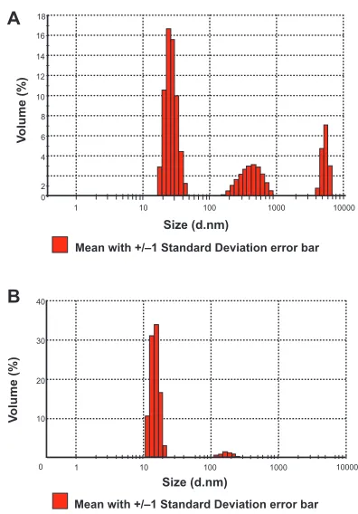
Particle size analysis
Figure 2A shows that average size of Fe3O4 particles used here is 201.2 nm. Polydispersity index value (PI) of Smart RISUG (Figure 2B) after Cuproferrogel formation done with the particle size analyzer illustrates that the average size of Smart RISUG particles is 628.2 nm.
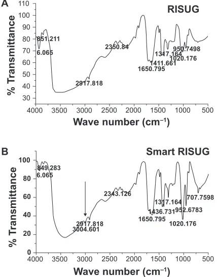
Transmittance peaks
FTIR peaks of the Fe3O4–Cu–SMA–DMSO (Smart RISUG)
compound found at 952 cm−1, 1020 cm−1, 1317 cm−1,
1650 cm−1, 2917 cm−1, etc. indicated presence of RISUG
mol-ecule in the ‘Smart RISUG’ (Figure 3B). In addition to parent constituents SMA and DMSO, the signifi cant peaks observed at 707 cm−1, 1436 cm−1 indicated presence of nanomicro
International Journal of Nanomedicine downloaded from https://www.dovepress.com/ by 118.70.13.36 on 23-Aug-2020
Smart RISUG: A potential new contraceptive
magnetic particles (Fe3O4) and 3004 cm−1 peak indicated
presence of copper particle in the new compound.
External magnetic
fi
eld-mediated
control and interaction of Smart
RISUG with sperm cell
Transport mechanism
HRTEM showed that Smart RISUG particles which could enter sperm cell membrane were of 50 nm to 200 nm size, the range suitable for delivery in a reproductive tube as it neither enters the blood circulation nor gets away with the reproductive tube fl uid. Figure 4A shows attachment of new compound particles to the sperm cell membrane at head region. Figures 4B and 4C track Smart RISUG particles inside the sperm cell. It is evident that particles either attach to cell membrane or enters only nearby cytoplasm without causing damage to the genetic material. This point is of con-siderable signifi cance in terms of safe genotoxic potential of the drug in clinical application.
Effect of external pulsed magnetic fi eld exposure
on Smart RISUG compound
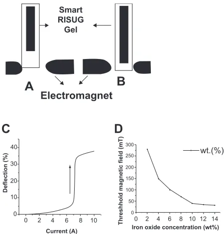
In physical magnetophoretic mobility experiments, the overall lengthening of the material kept in a test tube as a percentage of the pre PMF exposure length versus the magnetic fi eld strength is shown. The relative displacement induced by nonuniform magnetic fi eld can be 40% of the initial length (Figures 5A and 5B). The Smart RISUG drug mobility initiates above PMF of 80 mT. An abrupt increase in the lengthening in fi eld strength of about 280 mT provides a general guideline for the choice of optimal fi eld strengths for biophysical experiments. Thus a signifi cant magnetostriction took place and hysteresis was observed (Figure 5c).
One other experiment was conducted to determine optimum concentration of iron oxide. Taking different concentration (2% to 15% by weight) of iron oxide particles in the new compound wherein the threshold magnetic fi eld for mobility for each concentration was estimated. It was observed that with increasing iron oxide concentration the threshold magnetic fi eld required for movement initiation decreases. Suitable concentration of the iron oxide was found to be 8% to 12% by weight and threshold magnetic fi eld strength required for its lengthening initiation was found to be 80 mT (Figure 5D).
Interaction of sperm cell with Smart RISUG in the absence and presence of pulsed magnetic
fi eld produced by the impulse magnetizer
RISUG precipitate when made to attach to top of the test tube, it could not be transported into sperm cells kept at the bottom of tube either in presence or absence of PMF. Similarly, no signifi cant change in the sperm count or sperm motility percentage occurred when Smart RISUG precipitate and diluted sperm cells are kept at the two ends of the test tube in the absence of PMF as in this case the Smart RISUG is not able to reach or enter the sperm cell membrane in order to facilitate any changes thereof.
Figure 1 X-ray microanalysis of the Fe3O4–Cu–SMA–DMSO (Smart RISUG) indicating arrangement of drug particles in the Cuproferrogel compound.
0 2
A
B
V
olume (%)
V
olume (%)
Size (d.nm)
Mean with +/–1 Standard Deviation error bar
Mean with +/–1 Standard Deviation error bar
1 10 100 1000 10000
Size (d.nm)
1 10 100 1000 10000 10
20 30 40 0 4 6 8 10 12 14 16 18
Figure 2 Particle size analysis. A) Fe3O4 particles are in three ranges; nanosize (50 nm–100 nm), microsize (1 μm) and macrosize (5–7 μm) B) Smart RISUG particles size Z- average is 628 nm.
International Journal of Nanomedicine downloaded from https://www.dovepress.com/ by 118.70.13.36 on 23-Aug-2020
Jha et al
In the present study no signifi cant variation in the sperm count and percentage of motile sperm were found when RISUG interacted with sperm cell either in presence or absence of PMF given for 30 s to 50 s. The new contraceptive molecule started moving at 80 mT but showed abrupt transformation at 280 mT only. Table 1 reveals that Smart RISUG achieved abrupt transformation at about 280 mT and it could be transported into sperm cell membrane at 760 mT only. The interaction of rat sperm with Smart RISUG caused 30% decrease in sperm count and 70% decrease in sperm motility (p 0.05) (Table 1).
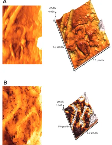
Drug-induced topological alteration
AFM image of untreated rat sperm cells (Figure 6A) gave evidence of smooth surface topography throughout the length of the sperm cell membrane. A cluster of sperm cells with prominent head were observed with AFM. In contrast, sperm cells treated with Smart RISUG (Figure 6B) gave disintegrated surface topology with a complete loss of smoothness. The three-dimensional AFM image of the same showed curved tail and damage to the middle piece. A signifi cant reduction in the cell height had been observed for treated sperm cells with AFM. An interesting observation here was the pattern of attachment of Smart
RISUG particles to sperm cell membrane complimenting HRTEM observation.
Discussion
The present study reveals magnetic fi eld-directed Fe3O4– Cu–SMA–DMSO Cuproferrogel drug interaction with sperm cells that may open new dimensions in the fi eld of biomagnetic technology. RISUG action on sperm cells is not altered by PMF. PMF in the fi eld strengths of 80–800 mT applied for 30 s to 50 s does not have signifi cant effect on the sperm. The change in the form of RISUG to that of Smart RISUG too does not alter the effect on sperm. That is, the concentrations of iron oxide and copper are suffi ciently low as not to affect sperm function by their presence. Only the combination of the iron oxide–copper–PMF enhances the spermicidal action and hence the contraceptive effi cacy of the parent RISUG compound. Moreover there is a threshold
110 100 90 80 70 60 50 40 30
4000 3500 3000 2500 2000 1500 1000 500 Wave number (cm–1)
4000 3500 3000 2500 2000 1500 1000 500 Wave number (cm–1)
% T
ransmittance
% T
ransmittance
A
B
RISUG
Smart RISUG
851.211
6.065
2917.818 2350.84
1650.7951411.661 1317.164
1020.176
100
80
60
40
20
0
849.283 6.065
2917.818 3004.601
2343.126 707.7598
1317.164 1436.731952.6783 1650.795
1020.176 950.7498
Figure 3 Fourier transform infrared spectroscopy. A) FTIR peaks for RISUG gel.
B) FTIR peaks for Smart RISUG Cuproferrogel indicate presence of Fe3O4 and Cu
particles embedded in matrix of parent compound RISUG (SMA–DMSO)
A
B
C
100 nm
50 nm
50 nm
Figure 4 High resolution transmission electron microscopic (HRTEM) study of Smart RISUG compound’s transport mechanism into sperm cell membrane. A) Surface of the rat sperm showing Smart RISUG particles attached to it. B, C) Mechanism of reaching of Smart RISUG particles into the sperm membrane. Arrows indicate passage of entry of drug into cell membrane.
International Journal of Nanomedicine downloaded from https://www.dovepress.com/ by 118.70.13.36 on 23-Aug-2020
Smart RISUG: A potential new contraceptive
iron oxide concentration above which the phenomena are manifest. Furthermore PMF of 760 mT or above is required to induce the spermicidal enhancement potential. The about 30% decrease in the sperm count and about 70% decrease in the sperm motility percentage demonstrated in the present study imply that good contraceptive action may be realized with lower drug dose than the conventional RISUG.
HRTEM elucidates the entry mode of Smart RISUG particles into the sperm cell. Figures 4A and 4B illustrates that particles are not endocytosed by the sperm cells rather they attach to the sperm head surface or enters cytoplasm in vicinity of the sperm cell plasma membrane (Figure 4C) and thus inactivate the sperm cells. It is previously hypothesized that plasma membrane is directly involved in the mechanisms by which nonthermal extremely low frequency (ELF) electromagnetic fi elds exert their action on cell functions.31 Membrane disintegration of the sperm
at the acrosomal region leads to leaching out of enzymes
acrosin and hyaluronidase, thus rendering the sperm infertile, a mechanism similar to RISUG observed with AFM (Figure 6B).
Previous work on RISUG also reports that as sperm come into contact with the SMA–DMSO complex, the combina-tion of positive and negative charges on the polymer surface
causes the sperm membrane to burst.11 When co-polymer
of styrene and maleic anhydride dissolved in DMSO was injected into the vas deferens of rats the morphological changes detected were confi ned to the mucosa.9 Just after
injection, RISUG lowers the pH suffi ciently to kill the sperm passing through.32,33 RISUG is presently undergoing extended
phase III clinical trials in India2,34 after successfully
complet-ing phase I and phase II trials.33,35–37 The presence of common
parent component SMA in Smart RISUG ensures long-term safety and safe reversibility of the drug.
For Smart RISUG drug synthesis the SMA copolymer is dissolved in solvent medium DMSO. Thereafter, magnetic material Fe3O4 is added to the polymer to impart magnetic properties and copper is added to obtain electrical conductivity and charge transfer capability that in turn affect structure of sperm/ovum by displacing specifi c molecules. Here, Fe3O4 added to SMA-DMSO facilitates noninvasive imaging as well as control of distribution of the drug in the reproductive tube under guidance of external magnetic fi eld by enhancing contrast of drug with the body tissue. Use of magnetic particle iron oxide in Smart RISUG exploit two major advantages; their low toxicity to human beings and the possibility to use their outstanding properties by allowing the high magnetiza-tion of the iron oxide to target drugs to/from a specifi c cell through an external magnetic fi eld.38,39 The addition of copper
particles displaces specifi c molecules like zinc from sperm cell, reduces sperm motility and increases sperm abnormality. Both additives are uniformly dispersed in the base polymer and aggregation is prevented by suitable coating with cross-linked SMA and continuous stirring for 45–50 hrs.
The contraceptive preparation is taken in a 250 pl syringe provided with a 23 gauge needle for injection into male/ female reproductive tube. In male a puncture is made in anterior scrotum to elevate vas deferens and then the needle is inserted into vas lumen. Smart RISUG has properties of electrical charge producing, pH lowering, molecular exchange property, charge transfer property and residual magnetism. The drug after injection swells and invaginates into the folds of the lumen to help retention but does not adhere to the tissue. When the pure polymeric RISUG is injected into the vas deferens the sperms present in the vas deferens at the time of injection are subjected to RISUG action and break-up. The fragments of the sperm are voided at the time of ejaculation. If on the other hand a compound such as Smart RISUG is injected into the vas deferens and concurrently PMF is applied the iron oxide enters the sperm membrane. A mosaic architecture of iron oxide and with it copper micro-strands corresponding to the sperm shape are formed (Guha, Pers Comm). These micro-strands enveloped
Smart RISUG
Gel
Electromagnet
A
C
D
B
40 30 20 10
0 0
2 4 6 8 10 0 2 4 6 8 10 12 14
Deflection (%)
Current (A) Iron oxide concentration (wt%)
Threshhold magnetic field (mT)
300
250
200
150
100
50
0
wt.(%)
Figure 5 Response of Smart RISUG. A) No magnetic fi eld. B) Electromagnet at the lower end caused Cuproferrogel elongation. C) Smart RISUG defl ection ∆h/h0 (%)
versus current (A) given –a hysteresis graph obtained (D) Threshold magnetic fi eld vs iron oxide concentration. The presence of 10% iron oxide causes threshold magnetic
fi eld effect in presence of external magnetic fi eld.
International Journal of Nanomedicine downloaded from https://www.dovepress.com/ by 118.70.13.36 on 23-Aug-2020
Jha et al
by sperm proteins and lipids persist and create a large surface of electrical charge interaction for the destruction of sperms passing later on even without PMF. In this manner the spermicidal action is enhanced in a sustained manner. The removal as proposed in present paper is easily possible by using magnetic properties of the contraceptive to propel the drug for fertility restoration by external magnetic fi eld.
This study explains that the new contraceptive molecule
Fe3O4–Cu–SMA–DMSO (Smart RISUG) starts moving as
a system in the presence of external PMF. The magnetic
fi eld exposure system developed for this study produced fl ux density up to 800 mT. As each particle is the elemen-tary carrier of a magnetic moment, similar is the case for Smart RISUG particles that couple the shape of the gel to the PMF. On application of varying PMF, as the current of the electromagnet increases there is small movement of the new compound followed by an abrupt rise in the transport properties (Figure 5C). At the threshold magnetic field 80 mT given for periods of 50 s, the Smart RISUG compound starts movement and at about 760 mT, 50 s, the compound
A
B
μm/div
μm/div
5.0 μm/div
5.0 μm/div 5.0 μm/div
5.0 μm/div 0.058
0
0.041
0
Figure 6 Atomic force microscopy (AFM) images of rat sperm. A) 2D and 3D images of untreated cells showing cluster cells with smooth surface. B) 2D and 3D images of Fe3O4–Cu–SMA–DMSO (Smart RISUG) treated cells showing blabs in plasma membrane, disintegration leading to enzyme leaching, shortened height, and curved tail.
International Journal of Nanomedicine downloaded from https://www.dovepress.com/ by 118.70.13.36 on 23-Aug-2020
Smart RISUG: A potential new contraceptive Table 1 Effect of external pulsed magnetic fi eld (PMF)-mediated Smart RISUG exposure on the sperm cell count and motility where respective drugs and sperm cells are kept at the two different ends of reproductive tube model for this noninvasive control experiment
Sperm cell count (106/ml)
Sperm cell motility (%)
1. C (absence of PMF) 25.05 ± 1.10 68.37 ± 3.97
2. C (presence of PMF) 24.89 ± 6.54 68.13 ± 2.89
3. RISUG (absence of PMF) 24.87 ± 1.13 67.90 ± 6.32
4. RISUG (presence of PMF) 24.32 ± 5.25 67.80 ± 4.66
5. Smart RISUG (absence of PMF) 24.87 ± 1.32 65.29 ± 2.24
6. Smart RISUG treated (presence of PMF) 17.74 ± 0.98 45.12 ± 4.13
Note: Data presents the mean ± SEM of fi ve samples (p 0.05).
Abbreviations: C, control; PMF, pulsed magnetic fi eld.
could enter the sperm cell membrane (Figure 4A). Transport occurs instantaneously and disappears abruptly when the external magnetic fi eld is removed.35%–45% lengthening of the Smart RISUG drug with respect to pre-PMF length could be produced by applying a nonuniform magnetic fi eld (Figure 5B).
EDS–X ray spectroscopy analysis observed the proper dispersion of Smart RISUG Cuproferrogel particles (Figure 1). FTIR analysis transmittance peaks of the Fe3O4–Cu–SMA–DMSO (Smart RISUG) found at 952 cm−1,
1020 cm−1, 1317 cm−1, 1650 cm−1, and 2917 cm−1 indicates the
presence of SMA–DMSO molecule in the ‘Smart RISUG’. In addition to these, the peaks at 707 cm−1, 1436 cm−1, and
3004 cm−1 indicate that the nanomicro-sized Fe
3O4 and
Cu particles has been encapsulated by the SMA–DMSO molecule (Figures 3A, 3B).
Our experimental and theoretical studies indicated that in the absence of magnetic fi eld neither RISUG nor Smart RISUG is able to interact with the sperm cells. Signifi cant transport properties of Smart RISUG compound can only be observed when the applied magnetic fi eld is above the threshold value. Present fertility control technology of magnetic fi eld-mediated sperm interaction of the new con-traceptive molecule Smart RISUG needs further study for any possible toxicity chances before applying this technol-ogy to human subjects even though the moderate amount of Fe3O4 being used here does not affect sperm functionality.27,40
However, initial studies of Smart RISUG on both gonad
weight analysis and sperm anomaly assay proves in vivo
safety and effi cacy of the new drug in rat.17 Present study
suggest use of new drug Smart RISUG in order to facilitate better spermicidal action, control of distribution of the fertility control agent implanted inside the male or female reproductive tube and noninvasive detection by application of external magnetic fi eld.
Acknowledgments
This work was supported by the Ministry of Health and Family Welfare, Government of India (M11011/2/2008-FP). The authors report no confl icts of interest in this work. We acknowledge Central Research Facility, Indian Institute of Technology, Kharagpur and Mr SN Jha for their experimental contribution.
References
1. Trussell J. Contraceptive efficacy. In: Hatcher RA, Trussell J,
Nelson AL, Cates W, Stewart FH, Kowal D, editors. Contraceptive
technology. 19th revised edition. New York, NY: Ardent Media; 2007; p. 747–826.
2. Sharma RS, Rajanna A, Singh BK, Mathur AK, Mukherjee AK. Current status of development of RISUG: an intravasal injectable male contraceptive. In: Sharma RS, Rajanna A, Rajalakshmi M, editors. Proceedings of the Conference on Recent Advances and Challenges in Reproductive Health Research. New Delhi, India: Indian Society for the Study of Reproduction and Fertility (ISSRF); 2007 Feb. 19–21. 2007; p. 127–140.
3. Sharma S, Sen P, Guha SK, Mukhopadhyay SN. Microbicidal male
contraceptive-RISUG induced morphostructural damage in E coli
Colloids Surf B Biointerfaces. 2003;32:43–50.
4. Guha SK. Contraceptive for use by male. United States Patent: 1996. 5,488,075.
5. Manivannan B, Mishra PK, Lohiya NK. Ultrastructural changes in
the vas deferens of langur monkeys Presbytis entellus entellus after
vas occlusion with styrene maleic anhydride and after its reversal. Contraception. 1999;59:137–144.
6. Manivannan B, Bhande SS, Panneerdoss S, Sriram S, Lohiya NK. Safety evaluation of long-term vas occlusion with styrene maleic anhydride and its non-invasive reversal on accessory reproductive organs in langurs. Asian J Androl. 2005;7:195.
7. Sethi N, Srivastava RK, Singh RK. Safety evaluation of a male injectable
antifertility agent styrene maleic anhydride, in rats. Contraception.
1989;39:217–226.
8. Sethi N, Srivastava RK, Nath D, Singh RK. Preclinical toxicity study of a male injectable antifertility agent (styrene maleic anhydride) in
rhesus monkeys, Macaca mulatta. J Med Primatol. 1991;20:89–93.
9. Verma K, Misro MM, Singh H, Mahajan S, Ray AR, Guha SK. Histology of the rat vas deferens after injection of a non-occlusive
chemical contraceptive. J Reprod Fertility. 1981;63:539–542.
10. Chaudhury K, Sharma U, Jagannathan NR, Guha SK. Effect of a new injectable male contraceptive on the seminal plasma amino acids studied
by proton NMR spectroscopy. Contraception. 2002;66:199–204.
International Journal of Nanomedicine downloaded from https://www.dovepress.com/ by 118.70.13.36 on 23-Aug-2020
Jha et al
11. Chaudhury K, Bhattacharya AK, Guha SK. Studies on the membrane integrity of human sperm treated with a new injectable male
contraceptive. Hum Reprod. 2004;19:1826–1830.
12. Sharma U, Chaudhury K, Jagannathan NR, Guha SK. A proton NMR study of the effect of a new intravasal injectable male contraceptive RISUG on
seminal plasma metabolites. Reproduction. 2001;122:431–436.
13. Sethi N, Srivastava RK, Nath D, Singh RK. Teratological evaluation of an injectable male antifertility agent, styrene maleic anhydride, in
rat. Int J Fertil. 1992;37:183–187.
14. Koul V, Srivastava A, Guha SK. Reversibility with sodium bicarbonate of styrene maleic anhydride, an intravasal injectable contraceptive, in
male rats. Contraception. 1998;58:227–231.
15. Lohiya NK, Manivannan B, Mishra PK. Repeated vas occlusion and non-invasive reversal with styrene maleic anhydride for male
contraception in langur monkeys. Int J Androl. 2000;23:36–42.
16. Lohiya NK, Manivannan B, Mishra PK, Sriram S, Bhande SS, Panneerdoss S. Preclinical evaluation for noninvasive reversal following long-term vas occlusion with styrene maleic anhydride in langur
monkeys. Contraception. 2005;71:214–226.
17. Jha RK, Jha PK, Rana SVS, Guha SK. Spermicidal action of styrene maleic anhydride polyelectrolyte in combination with magnetic and
electrically conductive particles. Int J Pharmacol. 2009;5(1):1–12.
18. Guha SK. An improved reversible contraceptive for male and female. Canada Patent: 2007. 2367414.
19. Gaganidze E, Esquinazi P, Ziese M. Vibrating ferromagnets in a magnetic
fi eld. Journal of Alloys and Compounds. 2000;310:144–152.
20. Zrínyi M, Barsi L, Büki A. Ferrogel: a new magneto-controlled elastic
medium. Polymer Gels and Networks. 1997;5:415–427.
21. Raikher YL, Stolbov OV. Magnetodeformational effect in ferrogel
samples. JMMM. 2003;258–259, 477–479.
22. Lebedev VT, Torok G, Cser L, et al. Magnetic phase ordering in
ferrogels under applied fi eld. JMMM. 1999;201:136–139.
23. Zhang Y, Kohler N, Zhang M. Surface modifi cation of superpara-magnetic magnetite nanoparticles and their intracellular uptake. Biomaterials. 2002;23:1553–1561.
24. Gruttner C, Rudershausen S, Teller J. Improved properties of magnetic particles by combination of different polymer materials as particle
matrix. J Magn Magn Mater. 2001;225:1–7.
25. Gupta AK, Curtis A. Surface modifi cation of superparamagnetic iron
oxide nanoparticles and their intracellular uptake. Eur Cells Mater.
2002;4:101–102.
26. Schopf B, Neuberger T, Schulze K, et al. Methodology description for detection of cellular uptake of PVA coated superparamagnetic
iron oxide nanoparticles (SPION) in synovial cells of sheep. JMMM.
2005;293:419.
27. Hofmann H, Juillerat J, Petri-Fink A, Chastellain M. Development of biocompatible functionalized superparamagnetic iron oxide
nanoparticles for human cancer cell uptake. Eur Cell Mater.
2004;7:14.
28. Roblero L, Guadarrama A, Lopez T, Zegers-Hochschild F. Effect of copper ion on the motility, viability, acrosome reaction and
fertilizing capacity of human spermatozoa in vitro. Reprod Fertil Dev.
1996;8:871–874.
29. Gravance CG, Garnen DL, Miller MG, Berger T. Fluorescent probes and fl ow cytometry to assess rat sperm integrity and mitochondrial
function. Reprod Toxicol. 2001;15:5–10.
30. Younglai EV, Holt D, Brown P, Jurisicova A, Casper RF. Sperm
swim-up techniques and DNA fragmentation. Hum Reprod.
2001;16:1950–1953.
31. Marinelli F, Bersani F, Ognibene A, Matteuci A, Santi S, Maraldi NM. Effect of the extremely low frequency (ELF) magnetic fi elds on cell
membrane. Eng Med Biol Soc. 1992;1:309–311.
32. Guha SK, Ansari S, Anand S, Farooq A, Misro MM, Sharma DN. Contraception in male monkeys by intra-vas deferens injection of a
pH lowering polymer. Contraception. 1985;32:109–118.
33. Misro M, Guha SK, Singh H, Mahajan S, Ray AR, Vasudevan P.
Injectable non-occlusive chemical contraception in the male-I.
Con-traception. 1979;20:467–473.
34. Guha SK. Biophysical mechanism-mediated time-dependant effect on sperm of human and monkey vas implanted polyelectrolyte
contraceptive. Asian J Androl. 2007;9:221–227.
35. Chaki SP, Das HC, Misro MM. A short-term evaluation of semen and accessory sex gland function in phase III trial subjects receiving
intravasal contraceptive RISUG. Contraception. 2003;67:73–78.
36. Guha SK, Singh G, Anand S, Ansari S, Kumar S, Koul V. Phase I
clinical trial of an injectable contraceptive for the male. Contraception.
1993;48:367–375.
37. Guha SK, Singh G, Ansari S, et al. Phase II clinical trial of a vas
deferens injectable contraceptive for the male. Contraception.
1997;56:245–250.
38. Marchal G, Van Hecke P, Demaerel P, Decrop E, Kennis C, Schueren E. Detection of liver metastases with superparamagnetic iron oxide in
15 patients: results of MR imaging at 1.5T. Am J Roentgen. 1989;
152:771–775.
39. Lubbe AS, Bergemann C, Riess H, Schriever F, Reichardt P, Possinger K. Preclinical experiences with magnetic drug targeting:
tolerance and effi cacy. Cancer Res. 1996;56:4694–4701.
40. Makhluf SBD, Qasem R, Rubinstein S, Gedanken A, Breitbart H. Loading magnetic nanoparticles into sperm cells does not affect their
functionality. Langmuir. 2006;22:9480–9482.
International Journal of Nanomedicine downloaded from https://www.dovepress.com/ by 118.70.13.36 on 23-Aug-2020