Efficacy of Edge Detection Methods in Hough
Transform for Segmentation of Cervical
Vertebrae
N.B.Sambre1, V.R.Udupi2
Associate Professor ,Department of E&TC,KIT,s College of Engineering, Kolhapur, Maharashtra, India 1
Principal, Maratha Mandal College of Engineering, Belgavi, Karnataka, India 2
ABSTRACT: X rays are the most common imaging test that helps a doctor diagnose, monitor, and treat many medical
illnesses. In the identification of degenerative spinal diseases the knowledge of the vertebrae location, contour and
alignment is necessary. The X-rays in many instances have poor subject contrast making it difficult to identify vertebrae from adjacent tissues. This paper presents segmentation method, the Hough transform as coarse segmentation. In this work Hough Transform based segmentation using different edge detection methods are investigated for digitized x ray images of cervical spine.
KEYWORDS: Cervical vertebrae, Segmentation, Edge detection, Hough transform.
I.INTRODUCTION
The segmentation of bony structures i n x-ray images is very vital in the identifying of abnormalities of the spine. X-ray images are affected by poor contrast of the subject, making it difficult to distinguish the subject from the background. An appropriate technique for object extraction is one of the most important investigation topic in the field of image processing. In medical applications, segmentation and analysis in the absence of computer facility is carried out manually by an expert .The X-ray images are marked by landmark points and analysis carried by observing different views of the image. Manual seg m en t a t i on is a tedious work and most likely to have to errors due to inter an d intra-subject variabilities. Segmentation of the region of interest by an algorithm run on a computer will facilitate the expert work and can automate the work of interpretation of medical images. The development of computer v i s i o n method f o r giving an automatic s o l u t i o n for segmenting v e r t e b r a e and detecting abnormalities is a challenging work.
The Generalized Hough Transform [1, 2, 3, 4] is normally used to identify straight lines in images. A random shape can also be identified by the generalisation of the Hough transform [4]. A Tezmol and A Gururajan[1,2] in their work have applied the Generalized Hough Transform (GHT) on the digitized x-rays of the spine along with the template of the lumbar spine .The GHT locates the best match in the vertebrae image to the template. The process of matching is a complete search of the entire image, using the accumulator “bin-counting” method. As the Generalized Hough Transform (GHT) is a template matching technique, the matching is limited by that obtained through variations in the pose, range, and orientation provided by the input template. The detection of vertebrae edges by an edge detection technique can also affect the result of segmentation. This work investigates the Prewitt, Sobel, and Canny edge detection methods for Hough transform segmentation of cervical vertebrae.
II. HOUGH TRANSFORM
associate points in a previously formed template and matches them in the target image by means of local gradient information [1].So the Hough Transform is basically a template matching process, based on an confirmation gathering approach, the confirmation are the votes collected in an array termed as accumulator. The implementation of GHT outlines a mapping from the spatial domain to the accumulator space. This is attained in a computationally efficient manner, related with the attributes of the intended shape. “The Hough transform detects curves by exploiting the duality between points on a curve and parameters of that curve” [4]. GHT methods are even generalized to detect analytical as well as non analytical shapes in grey level images [1].GHT is a transformation which is used to find arbitrary complex shapes. The GHT can find non-analytical curves [4], by determining an reference origin for shape,
and the orientation given by θ and two orthogonal scale factors on the x and y direction. An R-Table is constructed representing the variation of r for the arbitrary shape. The entries in the R-table are formed by selecting a reference point y, for the shape and for every boundary point x, the gradient direction are calculated and r is stored as a function of the gradient direction [4]. The R-table is used to detect occurrence of shapes in an image. All entries into the
columns of R table are length, r, and direction, α, from the reference point for each edge point in the shape. The
remaining part of segmentation is selecting the best approximation of the reference point. This is done by search
process to analyse every edge point in the target image and, based on the local value of θ, find the corresponding (r, α)
pairs in the R-table to update the accumulator. The accumulator collects the spatial location of the reference point. The accumulator having the highest value of this peak is selected to be the one that corresponds to the true value of scale and rotation of the vertebrae. Then the location having the highest peak in this accumulator is computed, and this is taken to be the reference point. Thus the pose estimation of the vertebrae is accomplished
A Tezmol and A Gururajan [1, 2] have indicated the importance of three parameters which contribute to the success of the Hough transform; they are gradient information associated with the edge, proper formation of the template, and reckoning of the votes.
III. GRADIENT INFORMATION: OBTAINING AN EDGE IMAGE
An edge image is used for correlating points in the template formed to those in the test image using local gradient information [7]. A superior edge image is required for consensus on the reference point by securing maximum votes. A clear edge image is required for implementation of the GHT, from which gradient information can be obtained.
An edge in an image is a significant local change in image intensity followed with discontinuity in image intensity or first derivative of image intensity [1]. Prewitt, Sobel, Canny operator are the generally used gradient operators. Following is the description of the edge operators
A) Prewitt: The Prewitt operator detects vertical and horizontal edges. It uses 3x3 mask which is convolved with the image to find the edges. The values of constant are as shown. The first mask will find the edges in vertical direction and the second in the horizontal direction.
B) Sobel: The Sobel operator uses a 3x3 neighbourhood to calculate the gradient. The3x3 convolution mask for Sobel in the horizontal and vertical direction denoted by Gx, Gy respectively is given as -
-1 0 +1
-2 0 +2
-1 0 +1
Gx Gy
The gradient magnitude is given by G = + .The angle of orientation of the edge giving the spatial gradient is
= arctan Gy/Gx. The Prewitt operator also uses these equations for computing gradient and orientation. -1 0 +1
-1 0 +1
-1 0 +1
+1 +1 +1
0 0 0
-1 -1 -1
+1 +2 +1
0 0 0
C) Canny: Canny edge detector [8] is the first derivative of a Gaussian and closely approximates the operator that optimizes the product of signal-to-noise ratio and localization. The steps for implementation for Canny edge detection are
1. Smooth the image with a Gaussian blur.
2. Determine the gradient magnitude and orientation using finite-difference approximations for the partial derivatives. 3. Apply nonmaxima suppression to the gradient magnitude.
4. Use the double thresholding algorithm to detect and link edges.
The following section describes the comparison between these three edge detection methods.
IV.METHODOLOGY
A set of 20 cervical x-ray images are taken from the NHANES –II database. The images are pre-processed using the three steps
1) A Gaussian filter is used to obtain a smoothed version of the original x-ray image. This operation will cancel any minor variation in the greyscale, keeping only abrupt changes.
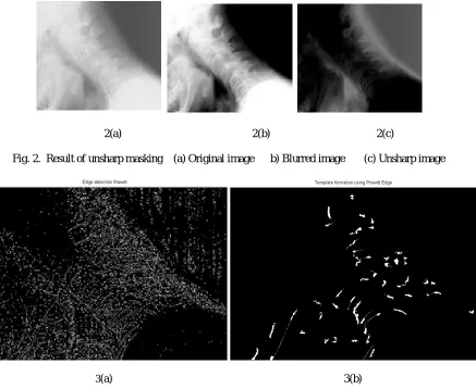
2) Unsharp masking is carried out on the smoothed image. The procedure for unsharp masking is subtracting the
blurred image from the original image. The resulting image after unsharp masking is represented [3] as Fu (m, n) = F (m, n) - Fb (m, n) ,where the original image is F (m, n) shown in Fig 2(a), Fb (m, n) is the Gaussian
blurred image shown in Fig 2(b), and Fu(m, n) is the unsharp masked image shown in Fig 2 (c). The unsharp mask provides only abrupt variations in contrast (edges) and removes large amount of unwanted information (background). 3) An averaging filter having mask of 5x5 is applied for reduces the unwanted high frequency components .The Prewitt, Sobel and Canny edge detection are implemented on the resulting image to extract the gradient information for segmentation using Hough transform. Morphological operation of opening and closing on the edge detected image is performed. The resulting filled image is used for matching with the template.
A) TEMPLATE FORMATION: The cervical vertebra, C3-C6, in the image is marked by 24 points describing the shape geometry of every vertebra. These points are called as land mark points. A total of 96 land mark points are marked per image and is called as template shown in Fig.1.Likewise 20 templates are formed representing all the variability in shape across the data base. A mean template is also formed from the twenty templates. The mathematical
expression for the mean template is, Mean template = ∑ ( )
Fig.1: Samples of Template
The following experiments are performed
i) The GHT with Prewitt edge, Sobel edge, and Canny edge is tested on all the images in the data set with its
related template and measure the number of land mark points that fall at their final location.
ii) The GHT with the three edge detection methods is tested by using the mean template on the images in the
V. RESULT AND DISCUSSION
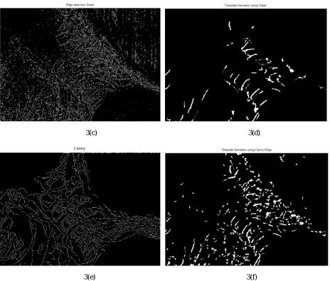
The results of experiment i) and ii) are shown in Fig. 2-5.The results of experiment number i) indicate a) an average of 67 landmark points are measured that fall on the template boundary for GHT with Prewitt edge detection method. b) Average of 80 landmark points for GHT with Sobel and 92 points for GHT with Canny edge method are measured. The Prewits edge based matching misses on the landmark points that are significant to recognize the shape of vertebrae. The Sobel operator also misses few land mark points on the anterior side and detects points outside the boundary of vertebrae.
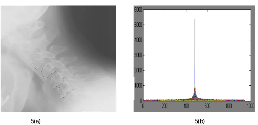
The results of experiment number ii) show that a) the accumulator does not record a single peak of highest count in reckoning of votes for either Prewitt or Sobel based edge detection when GHT is applied on vertebrae image using the mean template. These two edge detection do not correctly segment the vertebras in the image. b) The Canny edge based GHT shows that 74 land mark points fall on the boundary and the accumulator count shows a single peak of the highest votes for the reference point selected on the edge image as indicated by Fig. 5(b) .
2(a) 2(b) 2(c)
Fig. 2. Result of unsharp masking (a) Original image b) Blurred image (c) Unsharp image
3(c) 3(d)
3(e) 3(f)
4(a) 4(b) 4 (c)
Fig. 4 Sample results of template matching. 4(a) Prewitt 4(b) Sobel 4(c) Canny
5(a) 5(b)
Fig. 5. Segmentation result by GHT. Fig. 5 (a) Black dots show the GHT segmented vertebra using mean template and canny edge. 5(b) shows the plot of accumulator bin showing the highest peak for reference point
VI.CONCLUSION
The experiment result suggest that the Generalised Hough Transform with Canny edge detection and correct templates representing the changes in shape and position of vertebrae , provide a valid solution for segmentation of vertebrae in x-ray images.
REFERENCES
[2] A. Tezmol. “Customized Hough transform for robust segmentation of cervical vertebrae from X-ray images”, Master’s thesis, Texas Tech University, 2001.
[3] A Gururajan, “Coarse segmentation of cervical and lumbar vertebrae using a customized version of the generalized Hough transform”, Master thesis, Texas tech university,2001
[4] D.H. Ballard , “Generalizing the Hough Transform to detect arbitrary shapes”, Pattern Recognition, 13(2), pp. 111-122, 1981. [5] G Thoma, SAntani, “Content-Based Image Retrieval (CBIR) of Biomedical Images, A report to the Board of Scientific Counselors”, September 26-27, 2002.
[6] G Zamora-Camarena , “Automatic segmentation of vertebrae from digitized x-ray images”, PhD thesis, Texas Tech University,2002.
[7] G Zamora, H. Sari-Sarraf, S. Mitra, and R. Long, “Estimation of Orientation and Position of Cervical Vertebrae for Segmentation with Active Shape Models”, Proceedings of SPIE, San Diego, CA, USA, 2000.
[8] J. Canny. “ A computational approach to edge detection”. IEEE Transactions on Pattern Analysis and Machine Intelligence,vol.8,no.6, 1986.
[9] M Benjelloun, S Mahmoudi ,MA Larhmam “Template Matching Approaches Applied to Vertebra Detection, Advances in image segmentation”,Intech,2012.
[10] M A LARHMAM, Saïd MAHMOUDI and Mohammed BENJELLOUN, “Semi-Automatic detection of Cervical Vertebrae in X- ray images using Generalized Hough Transform”, Image Processing Theory, Tools and Applications (IPTA), 2012 3rd International Conference on 15-18 Oct. 2012.
[11] T. F. Cootes and C. J. Taylor. “Statistical models of appearance for computer vision”, Tech. Rep., Manchester, UK, University of Manchester, 2004.