Exogenous Slit2 does not affect ureteric branching or nephron
formation during kidney development
MICHAEL PIPER
1,2, VICTOR NURCOMBE
3, LORINE WILKINSON
1and MELISSA LITTLE
1,*
1Institute for Molecular Bioscience, University of Queensland, Brisbane, 2Department of Biochemistry, University of Queensland, Brisbane and 3Department of Anatomy and Developmental Biology, University of Queensland, Brisbane, Australia
ABSTRACT In an attempt to elucidate the role of Slit2 in vertebrate kidney development, the effect of adding exogenous human Slit2 protein (hSlit2) to developing murine metanephric kidney explants was examined. To confirm the activity of the recombinant Slit2 protein, neurons from 8 day old chick sympathetic nerve chain dorsal root ganglia were cultured with hSlit2 protein, which induced significant neurite branching and outgrowth. Using kidney explants as a model system, metanephric development in the presence of hSlit2 protein was examined. Addition of hSlit2 up to a final concentration of 1 µg/ml had no detectable effect on the formation of nephrons or on branching morphogenesis of the ureteric tree after 2 or 4 days in culture, as assessed via immunofluorescence for the markers WT1 and calbindin 28K respectively. Similarly, maturation of the nephrogenic mesenchyme occurred in a phenotypically normal fashion. In situ analysis of the Slit receptors, Robo1 and Robo2, the vasculogenic markers VEGFA and Flk-1, and the stromal cell marker BF2 displayed no difference in comparison to controls.
KEY WORDS:
Slit2, metanephros, mesenchyme, ureteric tree, Robo
0214-6282/2002/$25.00
© UBC Press Printed in Spain
www.ijdb.ehu.es
*Address correspondence to: Dr. Melissa Little. Institute for Molecular Bioscience, University of Queensland, St. Lucia, 4072, Australia. Fax: +61-7-3365-4388. e-mail: [email protected]
Abbreviations used in this paper: CNS, central nervous system; dpc, days post
coitum; NGF, nerve growth factor.
Introduction
The vertebrate metanephros develops via reciprocal interac-tions occurring between two heterologous tissues, the ureteric bud and the metenephric mesenchyme (Lipschutz, 1998). In the mouse, the ureteric bud forms as an evagination from the caudal end of the Wolffian duct at approximately 11 days post coitum (dpc). The bud subsequently invades the tissue proximal to it, a block of the intermediate mesoderm known as the metanephrogenic mesen-chyme, and begins to bifurcate, branching repeatedly to form the ureteric tree. As this occurs, a dynamic interplay of inductive events is conducted between the invading tree and the mesenchyme. The ureteric tree induces the mesenchyme to aggregate, condense and epithelialise, which is the first step in the process of nephron formation. The mesenchyme, on the other hand, signals to the ureteric tree, stimulating it to continue dichotomous branching. These reciprocal interactions are the hallmark of kidney develop-ment, and make the vertebrate kidney an excellent model to study the inductive interactions that underlie many aspects of develop-ment.
The central nervous system (CNS) has traditionally been a leading model for studying development and specification of cellu-lar fate. Interestingly, many molecules which play a role in devel-opment of the CNS also play an essential role throughout develop-ment of the metanephros, and provoke dramatic kidney
pheno-types when disrupted. These include glial cell-line derived neu-rotrophic factor (GDNF, Sanchez et al., 1996), c-Ret (Schuchardt et al., 1996) and Pax2 (Dressler et al., 1990). Gene targeting experiments have shown mice homozygous for Pax2 have a complete absence of kidneys (Torres et al., 1995), while mice homozygous for GDNF or c-Ret exhibit renal agenesis or severe dysgenesis (Pichel et al., 1996; Schuchardt et al., 1994).
1999), retinal ganglion cell layer (Erskine et al., 2000; Niclou et al., 2000; Ringstedt et al., 2000), hippocampus and dentate gyrus (Nguyen Ba-Charvet et al., 1999). In an interesting twist, Slit2 has also been shown to promote branching and elongation of spinal sensory neurons of the dorsal root ganglia (Wang et al., 1999), indicating that Slit2 is a bifunctional molecule capable of mediating multiple signalling events. A large secreted extracellular matrix associated protein, Slit2 transduces these signals through interac-tions with members of the Robo family of transmembrane recep-tors (Brose et al., 1999; Kidd et al., 1999). Subsequent phospho-rylation of the cytoplasmic domain of Robo is thought to initiate downstream events (Bashaw et al., 2000). The role of Slit2 is not confined solely to axonal guidance. Slit2 has been demonstrated to mediate other forms of cellular guidance both in invertebrates and vertebrates. Examples of this include migration of ventral muscle precursors towards the body wall in Drosophila (Kramer et al., 2001), mesodermal migration during gastrulation in zebrafish (Yeo et al., 2001) and tangential migration of interneurons in the mouse telencephalon (Hu, 1999; Zhu et al., 1999).
As Slit2 has been shown to play a vital role both in guidance of various populations of cells from both the CNS and those of mesodermal origin, we were interested to assess the role that the Slit genes, in particular Slit2, were playing during metanephric development. All three vertebrate Slit homologs are expressed in distinct patterns during development of the murine metanephros, as are their receptors, Robo1 and Robo2 (Piper et al., 2000). Specifically, Slit2 is expressed throughout kidney development at the tips of the ureteric tree, as well in the proximal end of the comma and S-shaped bodies after epithelialisation of the metanephric
mesenchyme has occurred. Addition of Slit1 and Slit2 to explant cultures of neuronal tissues has provided a convenient method for assessing the role of these secreted ligands (Erskine et al., 2000). Therefore, in order to analyse the role Slit2 plays during meta-nephric development, kidney explants were grown in the presence of recombinant hSlit2 protein, and analysed for differences in ureteric tree morphology, nephron endowment and maturation, and expression of its receptors, Robo1 and Robo2.
Results
Production of hSlit2 Protein
Brose et al. (1999) have observed that the full length Slit2 is cleaved into 2 fragments, a 140 kDa N-terminal and a 50-60 kDa C-terminal fragment. Cleavage occurs between the fifth and sixth EGF-like repeat. Wang et al. (1999) further demonstrated that the activity promoting elongation and branching of isolated sensory neurons of the dorsal root ganglia resided in the 140 kDa N-terminal fragment. More recently, Nguyen Ba-Charvet et al. (2001) determined that N-terminal Slit2 is responsible for both axon repulsion and growth cone collapse in neurons from the olfactory bulb in vitro. To analyse the role Slit2 is playing in metanephric development, a construct encoding the 140 kDa N-terminal frag-ment of the Slit2 protein was cloned into a mammalian expression vector. This construct was transfected into COS-7 cells, and the media collected after 48 hr. Recombinant hSlit2 was purified using an anti-myc antibody covalently attached to an agarose resin (Fig. 1A). The purified product was run on a SDS gel and silver stained (Fig. 1B). Mock-transfected media which had undergone the same Fig. 1. Production of recombinant N-terminal hSlit2 protein. (A) Western blot analysis of progressive steps involved in purification of 140 kDa hSlit2. A monoclonal anti-myc epitope antibody was used to detect the recombinant protein. (1) 10 µL pooled media. (2) 5 µL pooled eluate. (3) 10 µL spin through. (4) 5 µL pre-dialysis. (5) 5 µL post-dialysis. (6) 10 µL spin through. (7) 1 µL recombinant N-terminal hSlit2 protein. (B) Silver stain of recombinant hSlit2. M, purified mock-transfected media; Slit2, Recombinant N-terminal hSlit2 protein. (C) The N-terminal hSlit2 protein is stable over a period of 96 hours when diluted in culture medium and incubated at 37°C. Numbers to the left of the blots represent size in kDa.
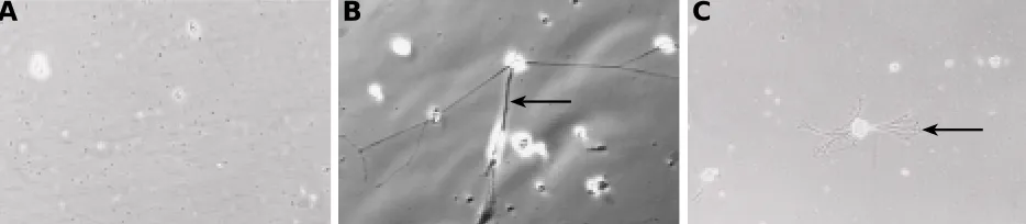
Fig. 2. hSlit2 promotes branching of neurites from sensory neurons. (A) Neurons of the dorsal root ganglia essentially all die within the 24 h culture period when treated with mock-transfected media (arrowhead). (B) NGF (5 ng/ml) treated neurons exhibit neurite outgrowth (arrow). (C) Recombinant N-terminal hSlit2 (1 µg/ml) promotes branching of neurites from sensory neurons of the dorsal root ganglia (arrow).
A
B
C
250
150
100
75
50
1 2 3 4 5 6 7
250
150
100
75
50
50 40 30 20 10 M Slit2
ngBSA
250
150
100
75
50
0 24 48 72 96
Hours in culture medium
purification protocol did not demonstrate a corresponding band at 140 kDa (Fig. 1B). This media was used as a negative control in subsequent experiments. Incubation of the purified protein in explant culture medium at 37°C over a period of 4 days did not show significant degradation of hSlit2 (Fig. 1C).
Activity of the N-Terminal hSlit2 Protein
In order to assess whether the N-terminal hSlit2 protein gener-ated was active, it was assayed against isolgener-ated 8 day old chick spinal sensory neurons. Isolated sensory neurons from the dorsal root ganglia were incubated for 24 hr in media containing dilutions of recombinant hSlit2, mock-transfected media or nerve growth factor (NGF, 5 ng/ml). Slit2 has previously been shown to be involved in mediating axonal elongation and branching of these neurons in rats (Wang et al., 1999). It was expected that the hSlit2 protein would perform a similar function in this assay. As predicted, the N-terminal hSlit2 protein (1 µg/ml) promoted branching and elongation of neurites from isolated spinal sensory neurons (Fig. 2C). Neurons cultured with the NGF exhibited elongation of neurites, whilst neurons grown with mock-transfected media died within the culture period (Fig. 2 A,B). This result verified the functional activity of the hSlit2 protein.
Addition of hSlit2 to Kidney Explants
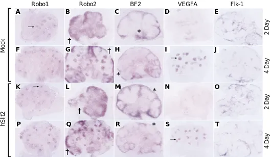
Kidney explants were isolated at 11.5 dpc, when the ureteric tree was invading the metanephric mesenchyme. hSlit2 (1 µg/ml) or mock-transfected media was added to the culture medium and the kidney explants were allowed to grow for 2 or 4 days. To ascertain whether addition of Slit2 affected the expression of the receptors of Slit, Robo1 and Robo2, in situ hybridisation was performed. Robo1 expression is seen in the aggregating mesen-chyme and epithelial condensates, and persists in the distal end of the comma and S-shaped bodies, which will eventually fuse with the ureteric tree. Robo2 is expressed in the induced metanephric mesenchyme and in the proximal end of the S-shaped bodies. The expression of both Robo1 (Fig. 3 A,F,K,P) and Robo2 (Fig. 3 B,G,L,Q) was not affected by the addition of hSlit2 protein to the culture medium. Expression of the stromal cell marker BF2 was also investigated. Again, no difference between explants grown in the presence of hSlit2 or purified mock-transfected media was observed (Fig. 3 C,H,M,R). Finally, expression of the vascular growth factor VEGFA and its receptor Flk-1 was also compared. VEGFA expression, seen diffusely in the metanephric mesen-chyme after 2 days in culture and strongly upregulated in the comma and S-shaped bodies after 4 days, was unaffected by the
Fig. 3. In situ analysis of hSlit2 treated kidney explants. 2 day E, K-O) and 4 day (F-J, P-T) kidney explants grown with mock-transfected media (A-J) or 1 µg/ml N-terminal hSlit2 protein (K-T). Expression of Robo1 in both hSlit2 and control treated explants is comparable. After 2 days in culture, expression is observed in the mesenchymal aggregates (arrows, A,K), and after 4 days of culture, expression is confined to the distal ends of the comma and S-shaped bodies (arrowheads, F,P). Robo2 expression is also unaffected by the presence of hSlit2 protein. Expression can be seen in the metanephric mesenchyme at both ages (B,G,L,Q) and in the comma and S-shaped bodies of 4 day explant cultures (arrowhead, G,Q). Expression of the stromal cell marker BF2 (* in C,H,M,R) in hSlit2 treated explants is consistently similar to explants grown with the mock control. VEGFA production is diffuse in 2 day explants (D,N) and upregulated in the comma and S-shaped bodies of 4 day explants (arrows, I,S). Expression of VEGFA is comparable between sample groups. Finally, the filigree-like expression of Flk-1 is equivalent in both mock and hSlit2 treated explants (E,J,O,T).
hSlit2
Mock
BF2
VEGFA
Flk-1
Robo2
Robo1
2 Day
4 Day
2 Day
4 Day
a
b
c
d
e
f
h
i
j
k
g
l
m
n
o
p
q
r
s
t
†
†
†
†
*
*
*
*
F
G
H
I
J
K
L
M
N
O
P
Q
R
S
T
addition of hSlit2 (Fig. 3 D,I,N,S). The filigree-like expression of Flk-1 in the mesenchyme was also comparable between mock treated and hSlit2 treated explants (Fig. 3 E,J,O,T).
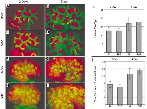
As the expression of Robo1, Robo2, VEGFA, Flk-1 and BF2 was not altered by the addition of recombinant hSlit2 protein, we looked at various markers of kidney development to assess whether Slit2 was influencing other aspects of kidney development. Using immu-nofluorescence, antibodies against the proteins Pax2 and WT1 were employed to analyse development of the metanephric mes-enchyme and its epithelial derivatives, and antibodies against calbindin 28K were used to assess development of the ureteric tree. WT1, which is expressed diffusely in the metanephric mesen-chyme and later upregulates in the comma and S-shaped bodies, did not show any change in expression upon addition of hSlit2 (Fig. 4 A-D; F-I). Numbers of comma and S-shaped bodies between treated explants and controls did not differ (Fig. 4J), indicating nephron endowment was not altered. Similarly, expression of Pax2, a protein expressed by all cells in the metanephric kidney,
was identical between sample groups (Fig. 4 F-I). Pax2 expression also gave an indication of overall size and development of kidney explants. At both time points assayed, both morphology and relative development of explants was indistinguishable between control and hSlit2 treated groups (Fig. 4 F-I). Finally, expression of calbindin 28K, which is produced exclusively by cells of the ureteric tree, did not alter (Fig. 4 A-D). Counts of the total number of ureteric tree tips between hSlit2 and control treated explants demonstrated no significant difference (Fig. 4E). Slit2 did not cause any other discernible alterations to the morphology of the tree, including number of branch points or length between branch points, and the overall architecture of the explants was unaffected.
Discussion
The Slit family of ligands, including Slit2, play a variety of roles in development, including repulsive guidance of retinal ganglion cell axons (Niclou et al., 2000) and spinal neuron axons (Brose et al., Fig. 4. Expression of markers of kidney development in hSlit2 treated explants. Expression of the ureteric tree marker calbindin 28K (green, A-D), and WT1, a marker of the metanephric mesenchyme and the comma and S-shaped bodies (red, A-D) as seen in 2 day (A,C) and 4 day (B,D) kidney explant cultures. As demonstrated in (E), explants grown with 1 µg/ml N-terminal hSlit2 (C,D) had no significant difference in the number of ureteric tree tips present when compared to those grown in mock controls (A,B). Pax2, which is expressed by all cells of the developing metanephros (red, F-I) and WT1 (green, F-I) were also investigated together. The number of WT1 positive comma and S-shaped bodies was counted. No significant difference between control explants and those grown with 1 µg/ml hSlit2 could be observed after 2 days (F,H) or 4 days (G,I) in culture (J). Data shown are representative of at least 3 separate experiments.
b
c
d
f
g
h
a
i
2 Days 4 Days
Mock
Slit2
Mock
Slit2
Ureteric T
ree T
ips
0 5 10 15 20 25
5 10 15 20 25
2 Day 4 Day
M Slit2 M Slit2
T
otal Comma and S-shaped Bodies
0 5 10 15 20 25 30
5 10 15 20 25 30
2 Day 4 Day
M Slit2 M Slit2
A
B
C
D
E
F
G
H
I
1999), repulsive guidance of neuronal precursors (Wu et al., 1999), leucocyte chemotaxis (Wu et al., 2001) and mesodermal migration during gastrulation in zebrafish (Yeo et al., 2001). With regard to its diverse and integral role in such a broad range of mechanisms, we decided to investigate the role Slit2 was playing in murine meta-nephric development. Slit2 is expressed throughout metanephric development, both in the tips of the ureteric tree and in the proximal end of the comma and S-shaped bodies (Piper et al., 2000). Postnatally Slit2 is expressed in the epithelial cells of the Bowman’s capsules (Wu et al., 2001). To try and elucidate the function that Slit2 plays in metanephric development, kidney explants were cultured in the presence of recombinant N-terminal Slit2 protein and subse-quently analysed via in situ hybridisation and immunofluorescence. Why the addition of Slit2 to kidney explants did not alter their morphogenesis is a difficult, yet interesting question. As degradation of the protein in culture medium at 37°C was not significant, Slit2 must not have been impairing normal development. Endogenous Slit2 is expressed strongly throughout kidney development (Holmes et al., 1998; Piper et al., 2000), so we initially hypothesized that Slit2 would be involved in the interplay of signals occurring between the ureteric tree and the metanephric mesenchyme. Its expression in the tips of the ureteric tree would make it an ideal candidate as a ligand to regulate the behaviour of the Robo2 expressing mesenchyme. By adding more Slit2 we hoped to disrupt this potential interaction and start to understand the role of Slit2 via the subsequent phenotype. Although the addition of exogenous protein to explant cultures of CNS origin has allowed the role of the Slit family to be investigated, this approach does not seem to be an effective way of educing the role of Slit2 in kidney development. The addition of Slit2 at a concentration of 1 µg/ml to cultures of kidney explants displayed no significant effect on a number of aspects of kidney morphogenesis. Slit2 did not alter the pattern of branching of the ureteric tree, nephron endowment, nor the expression of the proteins Pax2, WT1, the vasculogenic markers VEGFA and Flk-1, or the stromal cell marker BF2. The expression of the Slit receptors Robo1 and Robo2 was also unaffected by the addition of Slit2. This result was somewhat surprising as we had shown that the protein diluted to this concentra-tion was funcconcentra-tionally active in promoting neurite branching behaviour using cultured spinal sensory neurons.
The lack of altered phenotype in treated explants may be simply due to the fact that the endogenous source of Slit2 is sufficient to saturate the receptors available, and adding more Slit2 in excess of this has no deleterious effect. Alternatively, it is possible that it is either the full-length protein or the C-terminal cleavage product which is mediating the activity of the Slit2 gene in vivo, rather than the 140 kDa N-terminal protein. Recombinant Slit2 might also have been immobilised by antagonistic interactions with molecules in the kidney extracellular matrix, masking its activity. Finally, whilst it has been conclusively shown that Slit binds Robo in the CNS, it cannot be assumed that this interaction occurs in the kidney. Slit may be working through another receptor, either independent from, or in conjunction with the Robo proteins. This may have resulted in a very subtle phenotype in our experiments which could not be appreciated at the gross morphological level. Indeed, studies in other laboratories have shown Slit2 is able to bind laminin, glypican-1 and netrin 1, although the significance of these interactions is still unclear (Brose et al., 1999; Liang et al., 1999; Ronca et al., 2001).
An alternative explanation for the apparent inability of Slit2 to alter kidney morphogenesis stems from the shortfalls of the explant culture technique itself. Kidneys were removed at 11.5 dpc for
culture, before the precursors for vasculogenesis had invaded the mesenchyme. While kidney explants do exhibit expression of various factors essential for vasculogenesis, such as VEGFA and its receptor Flk-1, the process of vasculogenesis itself is severely curtailed. So whilst kidney organ culture is a very good model system for studying inductive interactions between tissues, it is not an optimal tool for studying vascular development. Importantly, Wu et al. (2001) have shown Slit2 to be expressed in the parietal epithelial cells of the Bowmans capsule and the vascular endothe-lial cells of the human kidney via in situ hybridisation. Slit2 may thus play a role in development of blood vessels in the kidney, which would not be detected using this system. In light of the data demonstrated here, it is evident that bringing to light the role of Slit2 in kidney development will require an efficient and specific means of decreasing the amount of Slit2 protein. Whist we have antibodies raised against the Slit2 protein, they do not decrease the efficacy of the endogenous protein when explants are cultured in media containing them (data not shown). However, we do not know if this is due to low binding efficiency of the antibodies, or to the epitope they recognise not being involved in Slit/Robo interactions. Gene disruption studies aimed at the Slit2 locus will hopefully cast a light on what role this intriguing molecule is playing during the process of metanephrogenesis.
Materials and Methods
Initial experiments on kidney explants were performed using a full length hSlit2 construct. Whilst we could successfully transfect COS-7 cells with this construct, growth of kidney explants near aggregates of Slit expressing cells was phenotypically normal (data not shown). In addition to this, purification of the full length product proved to be difficult to achieve in the quantities needed for functional studies. Therefore it was decided to clone the portion of the hSlit2 gene responsible for encoding the N-terminal cleavage product of the Slit2 protein and to use the purified protein directly in explant studies. The hSlit2 expression construct was generated via PCR using a full length human Slit2 construct as a template, and a myc tag was incorporated in the C-terminal end. The tag was designed to be between the fifth and sixth EGF-like repeat, immediately after the residue where cleavage of the full length protein is predicted to occur (Brose et al., 1999). This PCR fragment was cloned into pGEM-T Easy before being shuttled into the final expression vector, pcDNA 3.1. COS-7 cells were transfected with this construct using Lipofectamine 2000 according to the manufactur-ers instructions. The transfection media was left on for 48 hr, after which it was pooled. The cells were washed twice in 1 M NaCl in PBS for 1 min. These wash solutions were added to the pooled media. The pooled media was centrifuged to remove cellular debris. The media was then incubated with an agarose resin linked covalently to an anti-myc antibody. The resin was incubated overnight at 4°C. The Slit2 was eluted from the resin using 0.2 M glycine buffer, pH 2.3. 25 µg of BSA was added. The media was then concentrated through a 15 000 MW cut off centrifugal device (Millipore, Bedford, USA), and dialysed for 48 hr in PBS at 4°C in a 10 000 MW cut off dialysis cassette (Pierce Chemical Company, Rockford, USA). A final concentration step using an Amicon 10 000 MW cut off Concentrator (Millipore, Bedford, USA) was performed. Samples were run on SDS-polyacrylamide gels to assess the efficacy of the purification, using both silver stains and western blots with an anti-myc primary antibody. Media from mock-transfections containing no DNA was purified in the same manner and was used for negative controls.
with 10% foetal calf serum (DMEM/FCS) was added to the cells to stop the trypsin reaction. A 24 well plate was treated with 0.01% poly-lysine for at least 5 min. The wells were then rinsed three times with PBS. Wells were allowed to dry before use. Slit2 protein (1 µg/ml), mock-transfected media and NGF (5 ng/ml, Sigma Chemical Company, St. Louis, USA) were all diluted in DMEM/FCS. 2×10⁴ cells were plated per well. Cultures were incubated for 24
hr at 37°C, 5% CO2, then fixed in 4% paraformaldehyde in PBS.
Metanephric kidney explants were isolated and grown as described in Piper et al. (2000), except that the culture medium used was DMEM/F12 + 20 mM glutamine + 5 µg/ml holo-Transferrin. Recombinant hSlit2 protein was added at concentrations ranging from 0.1-1 µg/ml. Only data from explants treated with the highest concentration of hSlit2 protein is de-scribed, as it was representative of all treatments. Expression patterns were determined via in situ hybridisation using digoxigenin-labelled sense and antisense riboprobes. Probes were synthesised as described in Piper
et al. (2000). For immunofluorescence, kidney explants were fixed in 100% methanol, washed with PBS and permeabilised with 1% Triton X-100 in PBS. Kidney explants were incubated for 1 hr at 37°C in primary antibody diluted in 1%BSA in PBS, washed, then incubated for 1 hr at 37°C in secondary antibody diluted in 1%BSA in PBS. Dilutions used were as follows: WT1 1/100, calbindin 28K 1/100, Pax2 1/200.
Acknowledgments
The authors would like to thank S. Boyd for providing tissue culture support and M. Tessier-Lavinge for providing the Robo1 and Robo2 probes. Our work is supported by the National Health and Medical Re-search Council (NHMRC) of Australia (Grant No. 990605). M.P. holds an Australian Postgraduate Award. M.L. is a Sylvia and Charles Viertel Senior Research Fellow.
References
BASHAW, G.J., KIDD, T., MURRAY, D., PAWSON, T. and GOODMAN, C.S. (2000). Repulsive axon guidance: Abelson and Enabled play opposing roles downstream of the roundabout receptor. Cell 101: 703-715.
BROSE, K., BLAND, K.S., WANG, K.H., ARNOTT, D., HENZEL, W., GOODMAN, C.S., TESSIER-LAVIGNE, M. and KIDD, T. (1999). Slit proteins bind Robo receptors and have an evolutionarily conserved role in repulsive axon guidance.
Cell 96: 795-806.
DRESSLER, G.R., DEUTSCH, U., CHOWDHURY, K., NORNES, H.O. and GRUSS, P. (1990). Pax2, a new murine paired-box-containing gene and its expression in the developing excretory system. Development 109: 787-795.
ERSKINE, L., WILLIAMS, S.E., BROSE, K., KIDD, T., RACHEL, R.A., GOODMAN, C.S., TESSIER-LAVIGNE, M. and MASON, C.A. (2000). Retinal ganglion cell axon guidance in the mouse optic chiasm: expression and function of robos and slits. J. Neurosci. 20: 4975-4982.
HOLMES, G.P., NEGUS, K., BURRIDGE, L., RAMAN, S., ALGAR, E., YAMADA, T. and LITTLE, M.H. (1998). Distinct but overlapping expression patterns of two vertebrate slit homologs implies functional roles in CNS development and organogenesis. Mech. Dev. 79: 57-72.
HU, H. (1999). Chemorepulsion of neuronal migration by Slit2 in the developing mammalian forebrain. Neuron 23: 703-711.
KIDD, T., BLAND, K.S. and GOODMAN, C.S. (1999). Slit is the midline repellent for the robo receptor in Drosophila. Cell 96: 785-794.
KRAMER, S.G., KIDD, T., SIMPSON, J.H. and GOODMAN, C.S. (2001). Switching repulsion to attraction: changing responses to slit during transition in mesoderm migration. Science 292: 737-740.
LI, H.S., CHEN, J.H., WU, W., FAGALY, T., ZHOU, L., YUAN, W., DUPUIS, S., JIANG, Z.H., NASH, W., GICK, C., ORNITZ, D.M., WU, J.Y. and RAO, Y. (1999). Vertebrate slit, a secreted ligand for the transmembrane protein roundabout, is a repellent for olfactory bulb axons. Cell 96: 807-818.
LIANG, Y., ANNAN, R.S., CARR, S.A., POPP, S., MEVISSEN, M., MARGOLIS, R.K. and MARGOLIS, R.U. (1999). Mammalian homologues of the Drosophila
slit protein are ligands of the heparan sulfate proteoglycan glypican-1 in brain.
J. Biol. Chem. 274: 17885-17892.
LIPSCHUTZ, J.H. (1998). Molecular development of the kidney: a review of the results of gene disruption studies. Am. J. Kidney Dis. 31: 383-397.
NGUYEN BA-CHARVET, K.T., BROSE, K., MA, L., WANG, K.H., MARILLAT, V., SOTELO, C., TESSIER-LAVIGNE, M. and CHEDOTAL, A. (2001). Diversity and specificity of actions of Slit2 proteolytic fragments in axon guidance. J. Neurosci. 21: 4281-4289.
NGUYEN BA-CHARVET, K.T., BROSE, K., MARILLAT, V., KIDD, T., GOODMAN, C.S., TESSIER-LAVIGNE, M., SOTELO, C. and CHEDOTAL, A. (1999). Slit2-mediated chemorepulsion and collapse of developing forebrain axons. Neuron 22: 463-473.
NICLOU, S.P., JIA, L. and RAPER, J.A. (2000). Slit2 is a repellent for retinal ganglion cell axons. J. Neurosci. 20: 4962-4974.
PICHEL, J.G., SHEN, L., SHENG, H.Z., GRANHOLM, A.C., DRAGO, J., GRINBERG, A., LEE, E.J., HUANG, S.P., SAARMA, M., HOFFER, B.J., SARIOLA, H. and WESTPHAL, H. (1996). Defects in enteric innervation and kidney development in mice lacking GDNF. Nature 382: 73-76.
PIPER, M., GEORGAS, K., YAMADA, T. and LITTLE, M. (2000). Expression of the vertebrate Slit gene family and their putative receptors, the Robo genes, in the developing murine kidney. Mech. Dev. 94: 213-217.
RAJAGOPALAN, S., VIVANCOS, V., NICOLAS, E. and DICKSON, B.J. (2000). Selecting a longitudinal pathway: Robo receptors specify the lateral position of axons in the Drosophila CNS. Cell 103: 1033-1045.
RINGSTEDT, T., BRAISTED, J.E., BROSE, K., KIDD, T., GOODMAN, C., TESSIER-LAVIGNE, M. and O’LEARY, D.D. (2000). Slit inhibition of retinal axon growth and its role in retinal axon pathfinding and innervation patterns in the diencephalon. J. Neurosci. 20: 4983-4991.
RONCA, F., ANDERSEN, J.S., PAECH, V. and MARGOLIS, R.U. (2001). Character-ization of Slit protein interactions with glypican-1. J. Biol. Chem. 276: 29141-29147.
ROTHBERG, J.M., HARTLEY, D.A., WALTHER, Z. and ARTAVANIS-TSAKONAS, S. (1988). slit: an EGF-homologous locus of D. melanogaster involved in the develop-ment of the embryonic central nervous system. Cell 55: 1047-1059.
ROTHBERG, J.M., JACOBS, J.R., GOODMAN, C.S. and ARTAVANIS-TSAKONAS, S. (1990). slit: an extracellular protein necessary for development of midline glia and commissural axon pathways contains both EGF and LRR domains. Genes Dev. 4: 2169-2187.
SANCHEZ, M.P., SILOS-SANTIAGO, I., FRISEN, J., HE, B., LIRA, S.A. and BARBACID, M. (1996). Renal agenesis and the absence of enteric neurons in mice lacking GDNF. Nature 382: 70-73.
SCHUCHARDT, A., D’AGATI, V., LARSSON-BLOMBERG, L., COSTANTINI, F. and PACHNIS, V. (1994). Defects in the kidney and enteric nervous system of mice lacking the tyrosine kinase receptor Ret. Nature 367: 380-383.
SCHUCHARDT, A., D’AGATI V., PACHNIS, V. and COSTANTINI, F. (1996). Renal agenesis and hypodysplasia in ret-k- mutant mice result from defects in ureteric bud development. Development 122: 1919-1929.
SIMPSON, J.H., BLAND, K.S., FETTER, R.D. and GOODMAN, C.S. (2000). Short-range and long-Short-range guidance by Slit and its Robo receptors: a combinatorial code of Robo receptors controls lateral position. Cell 103: 1019-1032.
TORRES, M., GOMEZ-PARDO, E., DRESSLER, G.R. and GRUSS, P. (1995). Pax-2 controls multiple steps of urogenital development. Development 121: 4057-4065.
WANG, K.H., BROSE, K., ARNOTT, D., KIDD, T., GOODMAN, C.S., HENZEL, W. and TESSIER-LAVIGNE, M. (1999). Biochemical purification of a mammalian slit protein as a positive regulator of sensory axon elongation and branching. Cell 96: 771-784.
WU, J.Y., FENG, L., PARK, H.T., HAVLIOGLU, N., WEN, L., TANG, H., BACON, K.B., JIANG, Z., ZHANG, X. and RAO, Y. (2001). The neuronal repellent Slit inhibits leukocyte chemotaxis induced by chemotactic factors. Nature 410: 948-952.
WU, W., WONG, K., CHEN, J., JIANG, Z., DUPUIS, S., WU, J.Y. and RAO, Y. (1999). Directional guidance of neuronal migration in the olfactory system by the protein Slit.
Nature 400: 331-336.
YEO, S.Y., LITTLE, M.H., YAMADA, T., MIYASHITA, T., HALLORAN, M.C., KUWADA, J.Y., HUH, T.L. and OKAMOTO, H. (2001). Overexpression of a slit homologue impairs convergent extension of the mesoderm and causes cyclopia in embryonic zebrafish. Dev. Biol. 230: 1-17.