Dottorato di Ricerca
NEUROSCIENZE CLINICO-SPERIMENTALI E PSICHIATRIA
XXIX ciclo
Coordinatore Prof. Alfredo Berardelli Dipartimento di Neurologia e Psichiatria
Tesi di Dottorato
Frequency-dependent habituation deficit of the nociceptive blink
reflex in aura with migraine headache. Can migraine aura play a
modulating role?
Relatore: Candidata: Prof. Francesco Pierelli Anastasio Maria Grazia
Correlatore:
Dott. Armando Perrotta
2
INDEX
INTRODUCTION ... 3
1. PHENOMENON OF HABITUATION ... 6
2. NOCICEPTIVE BLINK REFLEX ... 9
3. MATERIALS AND METHODS ...11
3.1 Study population ...11
3.2 Neurophysiological measurements ...16
3.2.1 Nociceptive blink reflex measurement ...16
3.2.2 Habituation of the nociceptive blink reflex ...18
3.3 Statistical methods ...19
3.4 Experimental procedure ...21
4. RESULTS ...22
4.1 Nociceptive blink reflex baseline parameters ...23
4.2 Nociceptive blink reflex habituation ...23
5. DISCUSSION ...34
6. CONCLUSIONS ... 40
6.1 Clinical implications ...40
3
INTRODUCTION
Migraine is a neurovascular disorder affecting more than 10% of population worldwide with a relevant interference on the quality of life and high social costs (Stovner et al., 2007). In a percentage of subjects suffering by migraine headache, headache is announced by sensory, usually visual, disturbances, the migraine aura, now referred as aura with migraine headache (AwMH) by the current international classification (ICHD 3rd edition, 2013).
The phenomenon of the cortical spreading depression (CSD) that propagates across the brain surface is supposed to be the underlying mechanism of the migraine aura, however, its role in migraine symptoms remains still incompletely clear (Gorji, 2001; Charles and Baca, 2013; Noseda and Burstein, 2013). The possibility of imaging the typical visual aura with blood oxygenation level dependent (BOLD) functional magnetic resonance imaging (MRI) has revealed multiple neurovascular events in the occipital cortex within a single attack that closely resemble CSD. As transient synchronized neuronal excitation precedes CSD, changes in cortical excitability underlie the migraine attack (Shibata, 2007). In particular, the relationship between the neurophysiological abnormalities at the basis of the susceptibility to develop the CSD and the interictal neurophysiological abnormalities detected in migraine without aura (MWoA), are poorly understood.
It is well known that one of the most reproducible and endophenotypic abnormality in MWoA is the neurophysiological evidence of an interictal habituation deficit of cortical and subcortical responses, as consequence of multimodal (nociceptive
4 and non-nociceptive) repeated stimulations (Schoenen 2006; de Tommaso et al., 2014). Habituation is speculated to be part of a dual process, in balance with the sensitization phenomenon in order to determine the behaviour of a neural system underlie to a repeated stimulation (Groves and Thompson, 1970). In turn, both habituation and sensitization of a neural system are modulated through supraspinal structures including endogenous antinociceptive system (Mesulam, 1990). A large amount of data documented a clear deficit of habituation of both non-nociceptive (De Marinis et al., 2003; Perrotta et al., 2008) and nociceptive-specific (de Tommaso et al., 2002; Katsarava et al., 2003; Di Clemente et al., 2005; Di Clemente et al., 2007) trigeminal responses in MWoA subjects during the interictal period, however, the habituation of trigeminal pain responses has been poorly studied in AwMH subjects. Previous animal studies demonstrated that experimental CSD is able to modify the habituation of brainstem responses (van der Staak, 1976; van der Staak and Fischer, 1976) and to drive the trigeminovascular system activation (Zhang et al., 2010 and 2011), the underlying mechanism of migraine headache (Moskowitz, 1993).
We hypothesize that in human subjects with AwMH the habituation of the trigeminal nociceptive responses could be differently modulated than in subjects affected by MWoA.
Methods exploring the nociceptive processing at trigeminal level, such as the study of the nociceptive blink reflex (nBR) responses, appear suitable for revealing functional changes in the excitability of the nociceptive component of the trigeminal system in migraine subjects, including the habituation deficit (Kaube et al., 2002; de
5 Tommaso et al., 2002; Katsarava et al., 2003; Di Clemente et al., 2005; Di Clemente et al., 2007). The nBR is elicited by a concentric stimulation electrode that predominantly stimulates cutaneous superficial nociceptive Aδ-fibres without depolarizing non nociceptive Aβ-fibres in deeper layers of the skin (Kaube et al., 2000) and it is considered highly sensitive to changes in trigeminal nociception (Katsarava et al., 2002).
The study of the habituation of the nBR responses in AwMH could improve our knowledge of the relationship between migraine aura and migraine pain. As the habituation is a frequency-dependent phenomenon and we previously demonstrated that the conventional blink reflex response habituation rate is influenced by the stimulation frequency (Perrotta et al., 2008), we aimed at investigating the habituation of the nBR R2 responses across a wide range of stimulation frequencies in a group of AwMH. We analysed comparatively a group of MWoA subjects and a group of healthy subjects (HS).
6
1. PHENOMENON OF HABITUATION
The notion of habituation is as old as humankind.As Ctesippus says in Plato’s Lysis:
“Indeed, Socrates, he has literally deafened us and stopped our ears with the praises of Lysis; and if he is a little intoxicated, there is every likelihood that we may have our sleep murdered with a cry of Lysis.”
To take an even older example:
“A fox who had never yet seen a lion, when he fell in with him for the first time in the forest was so frightened that he was near dying with fear. On his meeting with him for the second time, he was still much alarmed, but not to the same extent as at first. On seeing him the third time, he so increased in boldness that he went up to him and commenced a familiar conversation with him.” (Esop’s Fables) (Thompson, 2009).
The habituation is an ubiquitous phenomenon observed in different experimental settings and in neuronal circuits of a wide range of complexity, from the withdrawal reflex of the gill and siphon in Aplysia to the autonomic and behavioral component of the whole-of-body reflex called the “orienting response” in humans (Thompson and Spencer, 1966; Groves and Thompson, 1970; Glanzman, 2009).
The habituation is defined as a behavioral response decrement that results from repeated stimulation and that does not involve sensory adaptation/sensory fatigue or motor fatigue (Rankin et al., 2009). Habituation to a repeated stimulation is considered a form of non-associative learning and represents a kind of neural plasticity
7 observed in both humans and animals, which can be detected in all sensory modalities and could be considered a frequency-dependent mechanism devoted to avoid an overcharging in useless information (Thompson and Spencer, 1966).
The phenomenon of habituation is considered useful for studyng the neuronal substrates of behaviour, the mechanisms of learning processes, or information processing in the central nervous system both in health and in disease (Coppola et al., 2013).
Rennefeld et al. (2010) show a central component involved in the long-term habituation to pain. The pattern of heterotopic habituation induced by repetitive stimulation strongly suggests the contribution of a central mechanism involving the supraspinal central nervous system underlying this effect. Whether this central component involves a change in cognitive or affective processing of pain over time still has to be investigated. The pharmacological intervention with the opioid antagonist naloxone speaks against the involvement of the endogenous opioid system in pain habituation. Habituation to painful stimulation takes place both short term – within one stimulation period, as has been demonstrated for electrical stimulation of the skin or the tooth pulp (Condes-Lara et al., 1981; Ernst et al., 1986; Milne et al., 1991), as well as long term – with gradually decreasing behavioural responses to daily repeated painful stimulation (Neisser, 1959; Strempel, 1976; Taylor et al., 1993; Greenspan and McGillis, 1994).
Bingel et al. (2007) demonstrates significant attenuation of pain-ratings to identical painful stimuli over the 8 day stimulation epoch, are thus consistent with these previous reports on pain habituation over time.
8 The habituation to different sensory stimuli is reduced in migraine patients.
Lack of habituation is the principal and most reproducible abnormality found interictally on evoked potential studies in migraineurs (Schoenen et al., 2003), and may be considered as an endophenotypic marker of migraine (Sándor et al., 1999). Pattern reversal-visual evoked potentials (PR-VEP) in particular are characterized by a deficit in habituation, or even a potentiation, in migraine patients between attacks (Schoenen et al., 1995; Afra et al., 1998 and 2000; Wang et al., 1999; Bohotin et al., 2002; Ambrosini et al., 2016) but normalize during an attack (Afra et al., 2000). This deficit of habituation has been also highlighted in recent years through the laser evoked potentials (Valeriani et al., 2003; de Tommaso et al., 2016) and the trigeminal reflexes (Perrotta et al., 2008).
9
2. NOCICEPTIVE BLINK REFLEX
An eyelid closure in response to some stimulus is a blink reflex (BR), which is normally isolated. In humans and primates, the closing is bilateral while in other animals, mostly those with eyes set laterally, the closure is frequently unilateral (Esteban, 1999). The BR is a trigeminofacial brain-stem reflex. After electrical stimulation of the supraorbital nerve 3 components can be distinguished: an oligosynaptic ipsilateral pontine R1 component (onset latency 11 ms) and two polysynaptic bilateral medullary components R2 and R3 (onset latencies 33 and 84 ms) (Ellrich and Hopf, 1996).
Kaube and colleagues (2000) developed a novel concentric electrode to allow specific study of nociceptive components of the R2 component of the reflex, the nociception-specific blink reflex (nBR). By virtue of its concentric design and small anode–cathode distance, a high current density is achieved that allows low current intensities to be used such that depolarization is limited to the superficial layer of the dermis containing nociceptive fibers but does not reach the deeper, non-nociceptive fiber containing, layers. The R2 response of this modified BR has been shown to be nociception specific (Kaube et al., 2000). The nBR can be highly sensitive to changes in trigeminal nociception. Using the nBR it detect a selective impairment of the trigeminal A-δ fibers, suggesting that nBR is a useful tool for neurological clinical assessment (Katsarava et al., 2002; Giffin et al., 2004).
10 The nociception specific blink reflex offers a reproducible, quantifiable window with which to examine the trigeminal nociceptive system in humans (Marin et al., 2015).
11
3. MATERIALS AND METHODS
The study was approved by the IRCCS Neuromed Ethics Committee and was carried out following the guidelines for proper human research conduct in accordance with the Helsinki Declaration of the World Medical Association and its revisions (1997). All the participants gave their written consent.
3.1 Study population
According to the International Classification of Headache Disorders 3rd edition beta version (ICHD-III beta version, 2013), fifty-four
consecutive subjects diagnosed as suffering from AwMH (1.2.1-ICHD-III beta version) and MWoA (1.1-(1.2.1-ICHD-III beta version), were recruited among those seeking treatment at the Headache Clinic of the IRCCS Neuromed, Pozzilli, Isernia, Italy. In order to study groups of comparable clinical severity, inclusion criteria included 3 to 6 AwMH or MWoA mean attacks per month and disease duration longer than five years. Subjects were studied during the interictal period and they had to be attack-free for three days prior to and after the recording sessions to avoid inclusion of patients investigated during a migraine attack (they were interviewed to verify this). Of the recruited subjects, eight were discarded because experienced a migraine attack within three days after the experimental session. Forty-six subjects were finally enclosed in the study. Seventeen subjects were diagnosed as suffering from AwMH and twenty-nine from MWoA. Multiple
12 diagnoses were not allowed. Subjects had a higher attack prevalence on one side (AwMH 14 right side, 3 left side; MWoA 21 right side, 8 left side). All patients completed the Headache Impact Test (HIT-6, Table 1) and Migraine Disability Assessment Scale (MIDAS, Table 2). Migraine subjects were compared with thirty age- and sex-matched healthy subjects (HS) without personal or family (first- or second-degree relatives) history of primary headaches or aura-like symptoms. They were studied in parallel with migraine subjects. For all participants exclusion criteria included secondary headaches, neurological disorders or clinical history (including family history) of neurological disorders, any systemic or psychiatric disorder, Beck Depression Inventory (BDI) scale score higher than 9, current use of prophylactic agents for migraine, anti-depressive and anti-epileptic medications (in the previous three months) or analgesics (in the previous 3 days); clinical or instrumental evidence of any central or peripheral disease potentially causing sensory impairment; fibromyalgia, neuropathic pain, complex regional pain syndrome, chronic low back pain and other pain conditions, accordingly with current guidelines. Clinical data of the participants are summarized in Table 3.
13 Table 1. Headache Impact Test HIT-6 Questionnaire
This questionnaire was designed to help you describe and communicate the way you feel and what you cannot do because of headaches.
INSTRUCTIONS : To complete, please circle one answer for each question.
1. When you have headaches, how often is the pain severe? Never Rarely Sometimes Very often Always
2. How often do headaches limit your ability to do usual daily activities including household work, work, school, or social activities?
Never Rarely Sometimes Very often Always
3. When you have a headache, how often do you wish you could lie down? Never Rarely Sometimes Very often Always
4. In the past 4 weeks, how often have you felt too tired to do work or daily activities because of your headaches?
Never Rarely Sometimes Very often Always
5. In the past 4 weeks, how often have you felt fed up or irritated because of your headaches?
Never Rarely Sometimes Very often Always 6. In the past 4 weeks, how often did headaches limit your ability to concentrate on work or daily activities?
Never Rarely Sometimes Very often Always Column1 Column2 Column3 Column4 Column5 6 points 9 points 10 points 11 points 13 points each each each each each __ __ __ __ __
To score, add points for answers in each column. Total Score: ____
Class I: 36-49, Class II: 50-55, Class III: 56-59, Class IV: 60 and more. It suggested to talk to your physician for class II and more.
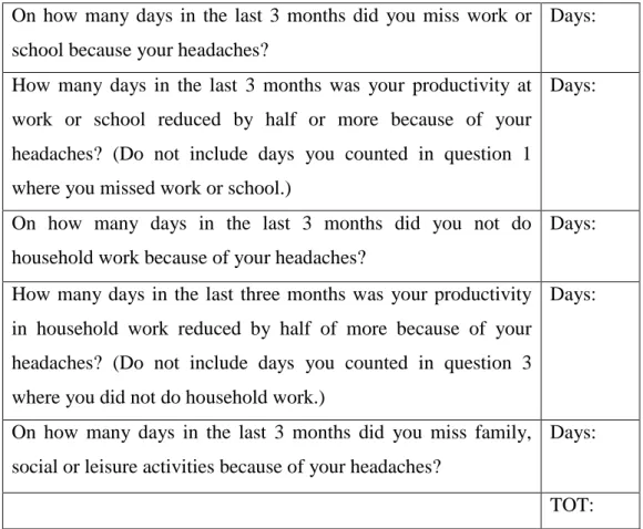
14 Table 2. Migraine Disability Assessment Questionnaire
On how many days in the last 3 months did you miss work or school because your headaches?
Days: How many days in the last 3 months was your productivity at
work or school reduced by half or more because of your headaches? (Do not include days you counted in question 1 where you missed work or school.)
Days:
On how many days in the last 3 months did you not do household work because of your headaches?
Days:
How many days in the last three months was your productivity in household work reduced by half of more because of your headaches? (Do not include days you counted in question 3 where you did not do household work.)
Days:
On how many days in the last 3 months did you miss family, social or leisure activities because of your headaches?
Days:
TOT:
Enter the total number of days you entered in questions 1-5. This is your MIDAS level of disability.
0 to 5- MIDAS Grade I, Little or no disability 6 to 10- MIDAS Grade II, Mild disability 11 to 20- MIDAS Grade III, Moderate disability 21+- MIDAS Grade IV, Severe disability
15 Table 3. Demography and clinical characteristics of the study population.
N ♀ Age, years Attack duration (hours) Duration of disease (years) Attack frequency (month) HIT-6 MIDAS AwMH 17 11 34.53 ± 10.91 (19-60) 27.59 ± 18.35 (4-72) 11.65 ± 4.64 (4-20) 3.71 ± 0.85 (3-6) 61.83 ± 11.88 (41-70) 17.58 ± 9.50 (11-20) MWoA 29 21 37.34 ± 10.56 (18-59) 39.34 ± 22.23 (4-72) 14.95 ± 6.88 (5-32) 3.93 ± 0.92 (3-6) 68.51 ± 10.29 (48-72) 20.70 ± 12.29 (11-23) HS 30 20 35.16 ± 7.40 (20-47) F (2,73) = 2.07 p = 0.134 t = 1.84 p = 0.072 t = 1.76 p = 0.086 t = 0.82 p = 0.416 t = 1.69 p = 0.233 t = 1.73 p = 0.225
AwMH = aurawith migraine headache; MWoA = migraine without aura; HS = healthy subjects; HIT-6 = headache impact test; MIDAS = migraine disability assessment scale.
16 3.2 Neurophysiological measurements
3.2.1 Nociceptive blink reflex measurement
The subjects were comfortably settled in an armchair in a quiet, temperature controlled room and were asked to sit back and relax, keeping their eyes open.
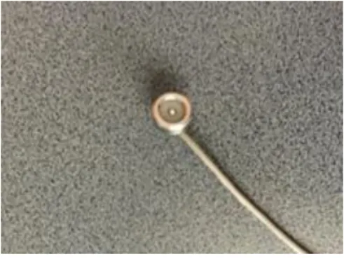
The nBR R2 response was elicited by a planar concentric electrode (Bionen, Florence, Italy) (Figure 1) ten mm above the emergence of the supraorbital nerve.
Figure 1: Planar concentric electrode (Bionen, Florence, Italy).
The stimuli (monopolar square-wave pulse with a duration of 0.3 ms delivered by a constant current stimulator - electric stimulator DS7A, Digitimer, UK) were applied on the usual headache side in migraine subjects, and on the right side in HS.
Electromyographic signals were recorded from both orbicularis oculi muscles via a standard pair of Ag/AgCl surface electrodes placed on the midline of the lower eyelid. The position of the reference electrodes is lateral to the eye. The ground electrode was
17 placed on the subject’s forehead. The filter bandpass settings were between 3 Hz and 3 kHz, sampling rate 2.5 kHz. The analysis time was 200 ms, sensitivity set at 100 mV. The signals were amplified and full-wave rectified (CED Powerlab interface 1401, Cambridge Electronic Design, UK; electronic amplifier BM623, Biomedica Mangoni, Italy).
In each participant, the sensory threshold (ST) were determined on the basis of a sequence of stimuli of increasing intensity (increased in 0.1 mA steps) delivered at unpredictable intervals (+/-10 sec). Subjects were asked to indicate verbally the stimulation levels at which they became aware of sensory sensations.
The staircase method was used to evaluate the reflex threshold (RT) for the R2 component of the BR by raising the stimulus intensity (in 0.1 mA steps) until a stable reflex response, with an amplitude exceeding 50 µV for more than 20 ms in the time interval 30-50 ms, appeared and persisted over a series of five stimuli. The subjective pain sensation elicited by supraorbital nerve stimulation at RT was graded on an 11-item numerical rating scale (NRS) for pain (0 = no pain; 10 = severe pain). For the RT assessment, in order to avoid R2 response habituation, the stimuli were delivered at pseudorandom frequencies between 0.033 and 0.025 Hz (Perrotta et al., 2008). The stimulation intensity was then fixed at 1.5 times the RT to ensure an affordable persistence/reproducibility of the reflex response. The latency (L), visually determined as the take-off point from the baseline, and area under the curve (AUC) of the R2 component were automatically measured and expressed in ms and µV x s, respectively. For each component, the time window to calculate the AUC was defined according to the measurable latencies of the best defined template, both at the beginning and at
18 the end of the component, and was then kept constant in each subject. For the L and AUC basal assessment, at least three to five successful responses were recorded and averaged in all participants.
3.2.2 Habituation of the nociceptive blink reflex
To evaluate the habituation phenomenon of the R2 component of the nBR, a series of electrical stimuli delivered at different, randomly chosen stimulation frequencies (SF) (0.05, 0.1, 0.2, 0.3, 0.5 and 1 Hz) were used. The stimulus intensity was set at 1.5 times the R2 RT. A sequence of 26 consecutive rectified EMG responses was recorded for each SF. The first sweep of each sequence of responses was excluded from further analysis to avoid contamination with a startle response. In off-line analysis, the sequence of responses for each SF was subdivided into five blocks of five and the R2 AUC values were calculated and averaged for each block of responses. The mean AUC values of the second to the fifth block expressed as the percentage of the mean AUC value of the first block, were taken as an index of habituation for each SF.
19 3.3 Statistical methods
A priori power analysis was conducted to determine the minimal sample size needed to obtain a statistical power of 0.80 at an alpha level of 0.05. In a previous study (Perrotta et al., 2008) evaluating the difference in BR R2 response habituation rate at 1 Hz SF between MWoA and HS, we calculated a standardized effect size of 1.69 for this variable. The a priori power analysis estimated a minimum total sample size of fourteen participants and a minimum sample size per group of seven participants.
Mean values of demographic and clinical features as well as of neurophysiological (ST, RT, R2 L, R2 AUC,) and related psychophysical values (NRS) clustered for group of participants (AwMH, MWoA and HS) were considered in statistical analysis. Distribution of variables was tested by Kolmogorov-Smirnov analysis and considered normal for p value > 0.05. Parametric tests were used as all variables considered passed the test.
T-test for independent samples were used to compare clinical characteristics between AwMH and MWoA subjects.
One-way analysis of variance (ANOVA) was used to compare the mean values of neurophysiological and psychophysical measurements detected at baseline between the different groups of participants.
In order to verify the effect of the clinical condition (AwMH, MWoA and HS) on the habituation rate (the percentage change of the mean nBR R2 AUC value of the second to fifth block with respect to the first), a two-way ANOVA for repeated measures was performed for each SF, with factors GROUPS (3 levels: AwMH, MWoA and HS) and BLOCKS (5 levels: first, second, third, fourth,
20 fifth block) to evaluate the differences between groups at each habituation block from the second to the fifth. Subsequently, a one-way ANOVA for repeated measures was run on each group to compare the percentage change of the mean nBR R2 AUC value with regard to the blocks from the first to the fifth; within subject factor was the percentage change of the AUC on the different blocks from the first to the fifth at each SF.
Student’s t-tests with Bonferroni’s correction for multiple comparisons were used for post-hoc analysis. The level of significance was set at 0.05. All values were reported as means ± SD.
Pearson’s correlation was used to search for correlations among electrophysiological parameters and clinical variables. Values of p < 0.05 were considered statistically significant.
The Statistical Package for the Social Sciences (SPSS) for Windows, version 19.0, was used for all analyses (SPSS Inc., Chicago, IL, USA).
21 3.4 Experimental procedure
The experimental session was conducted between 09.00 and 11.00 to minimize any possible effect of diurnal variation. Participants were required to be nicotine-, caffeine- and drug-free in the 8h (including sleep time) before the experiments. Female participants were investigated during the follicular phase in order to avoid differences in pain processing due to the hormonal phase. Each participant underwent two experimental consecutive sessions consisting of a baseline neurophysiological and psychophysical recording followed by the evaluation of the nBR habituation rate at different SF. In order to avoid any carry-over effect from one SF to the next, participants rested between each SF for not less than 20 min.
To guarantee the blinded condition, enrollment (A.P.), neurophysiological acquisitions (R.D.I.) and data analysis (M.G.A.) are made by different physicians.
22
4. RESULTS
No significant differences emerged between AwMH, MWoA and HS in physiological and clinical parameters (Table 4).
Table 4. Mean values standard deviation of the
neurophysiological (ST, RT, R2 L) and psychophysical parameters (NRS).
AwMH MWoA HS One-way
ANOVA ST (mA) 0.57 ± 0.26 0.53 ± 0.20 0.56 ± 0.25 F (2,73)=0.18 p = 0.831 RT (mA) 2.01 ± 1.00 2.50 ± 2.81 1.77 ± 0.62 F (2,73)=1.06 p = 0.353 R2 L (ms) 37.6 ± 3.95 42.1 ± 3.63 38.6 ± 3.72 F (2,73)=1.09 p = 0.285 NRS RT 5.00 ± 1.22 5.03 ± 1.82 4.40 ± 1.54 F (2,73)=0.86 p = 0.428
AwMH = aurawith migraine headache; MWoA = migraine without aura; HS = healthy subjects; ST = sensory threshold; RT = reflex threshold; L = latency; NRS =numeric rating scale: 0-10
23 4.1 Nociceptive blink reflex baseline parameters
The nBR R2 response was elicited in all subjects. No statistically significant differences emerged at baseline in ST, RT, L and AUC of the nBR R2 component (ipsi- and contralateral) between the groups of subjects (AwMH, MWoA and HS). Mean values ± SD are reported in Table 4.
4.2 Nociceptive blink reflex habituation
Clear differences emerged between the three groups of subjects in the habituation of the ipsilateral R2 component of the nBR. Since no ipsilateral vs contralateral differences in habituation rate of the R2 component were detected in any group at any SF, we reported only the ipsilateral responses.
No significant differences emerged in nBR R2 AUC mean values of the first block of responses at every SF between the groups (AwMH, MWoA and HS) (Table 5).
24 Table 5. Mean area under curve values ± standard deviation of the first block of then nBR R2 responses at different
stimulation frequencies on the symptomatic side in migraine subjects and on the right side in healthy subjects. First block nBR R2 AUC (μV x s)
Stimulation
Frequencies AwMH MWoA HS One-way ANOVA
1 Hz 0.53 ± 0.22 (0.18 – 1.06) 0.44 ± 0.21 (0.15 – 1.17) 0.60 ± 0.27 (0.12 – 1.12) F (2,73) = 3.34, p = 0.410 0.5 Hz 0.61 ± 0.41 (0.14 – 1.53) 0.71 ± 0.45 (0.17 – 1.94) 0.55 ± 0.27 (0.19 – 1.12) F (2,73) = 1.44, p = 0.244 0.3 Hz 0.67 ± 0.54 (0.15 – 2.16) 0.82 ± 0.68 (0.18 – 2.92) 0.75 ± 0.48 (0.26 – 2.34) F (2,73) = 0.35, p = 0.703 0.2 Hz 0.66 ± 0.49 (0.13 – 1.66) 0.78 ± 0.57 (0.13 – 2.73) 0.88 ± 0.48 (0.32 – 2.12) F (2,73) = 0.98, p = 0.382 0.1 Hz 0.77 ± 0.56 (0.24 – 1.99) 0.83 ± 0.66 (0.17 – 2.76) 0.77 ± 0.40 (0.13 – 1.46) F (2,73) = 0.13, p = 0.880 0.05 Hz 0.84 ± 0.58 (0.10 – 1.98) 1.07 ± 0.57 (0.23 – 2.49) 0.90 ± 0.40 (0.27 – 1.75) F (2,73) = 1.33, p = 0.270 nBR = nociceptive blink reflex; AUC = area under the curve; AwMH = aurawith migraine headache; MWoA = migraine without aura; HS = healthy subjects.
25 The two-way ANOVA for repeated measures analysis revealed a significant effect for factor GROUP for 1, 0.5, 0.3, 0.2 Hz SF; for factor BLOCK for all SF studied; for BLOCK x GROUP interaction for 1, 0.5, 0.3, 0.2 Hz SF (Table 6). No significant effect has been detected for both factor GROUP and BLOCK x GROUP interaction for the lower 0.1 and 0.05 SF (Table 6).
26 Table 6. Results of two-way repeated measures ANOVAs (with F and p values) with factors GROUPS (3 diagnosis: AwMH, MWoA, HS) and HABITUATION (5 levels: five blocks of five responses of the R2 component of the nBR) for six stimulation frequencies. Stimulation Frequencies GROUPS F (2, 73)p HABITUATION F (4, 292) p INTERACTION F (8, 584) p 1 Hz 12.52 0.001 272.14 0.001 6.87 0.001 0.5 Hz 11.28 0.001 201.75 0.001 6.32 0.001 0.3 Hz 11.05 0.001 171.46 0.001 4.22 0.001 0.2 Hz 13.30 0.001 73.57 0.001 5.19 0.001 0.1 Hz 1.71 0.189 23.72 0.001 0.83 0.581 0.05 Hz 0.22 0.800 25.48 0.001 1.10 0.367
27 As expected, post-hoc analysis of the BLOCK x GROUP interaction revealed that, depending on the habituation block, MWoA showed a significant effect on habituation rate in almost all the SF (1, 0.5, 0.3, 0.2 Hz) studied when compared to HS, while AwMH showed a significant effect on habituation rate at 0.5, 0.3, 0.2 Hz SF when compared to HS. No significant effect emerged at 0.1 and 0.05 Hz SF between groups (Figure 2 A, B, C, D, E, F). In details, the post-hoc analysis revealed a significant deficit in habituation rate in all blocks from second to fifth at 1, 0.5, 0.3, 0.2 Hz in MWoA when compared to HS (Figure 2 A, B, C, D); no differences were detected in habituation rate at 1 Hz SF between AwMH and HS (Figure 2 A); a significant deficit in habituation rate was detected in second, fourth and fifth block at 0.5 and 0.3Hz (Figure 2 B, C) and from second to fifth at 0.2 Hz in AwMH when compared to HS (Figure 2 D).
One-way ANOVA for repeated measures revealed a significant habituation rate of the mean nBR R2 AUC across the five blocks of responses (from second to fifth compared to the first one) at any SF (1, 0.5, 0.3, 0.2, 0.1 and 0.05 Hz, all p<0.0001) in all groups of participants, with the exception of the second block compared to first one in MWoA at 0.1 Hz SF (Figure 2 E) and of the second block compared to the first one SF in all groups of participants and the third block compared to the first one in AwMH and HS at 0.05 Hz (Figure 2 F).
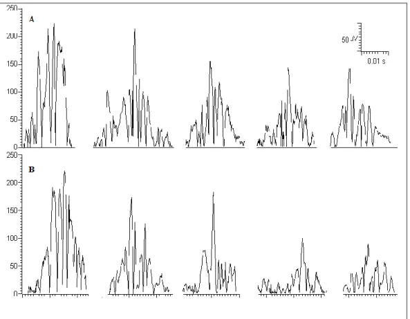
The sequence of the twenty-five nBR R2 consecutive responses at 0.3 Hz SF, grouped into five blocks of five averaged and rectified responses each in a representative AwMH (A) and HS (B) subject is reported in Figure 3.
28 Figure 2. Habituation of the ipsilateral nociceptive blink reflex (nBR) R2 area under the curve (AUC) in five blocks of five averaging at increasing stimulation frequencies (SF) (A, 1 Hz; B, 0.5 Hz; C, 0.3 Hz; D, 0.2 Hz; E, 0.1 Hz; F, 0.05 Hz) expressed as a percentage of the first block. Data are shown as mean values and standard deviations of the mean.
A
MWoA = migraine without aura; AwMH = aura with migraine headache; HS = healthy subjects.
Bonferroni’ test: * = p< 0.05 vs. baseline; # = p< 0.05 vs. HS
* * * * * * * * * *# *# *# *#
29
B
C
MWoA = migraine without aura; AwMH = aura with migraine headache; HS = healthy subjects.
Bonferroni’ test: * = p< 0.05 vs. baseline; # = p< 0.05 vs. HS * * * * * *# *# *# *# *# *# *# * * * * * *# *# *# *# *# *# *#
30
D
E
MWoA = migraine without aura; AwMH = aura with migraine headache; HS = healthy subjects.
Bonferroni’ test: * = p< 0.05 vs. baseline; # = p< 0.05 vs. HS
*# *# *# *# *# *# *# *# *# * * * * * * * * * * * * * * *
31 F
MWoA = migraine without aura; AwMH = aura with migraine headache; HS = healthy subjects.
Bonferroni’ test: * = p< 0.05 vs. baseline
* * * * * * *
32 Figure 3. The sequence of the twenty-five nociceptive blink reflex R2 consecutive responses at 0.3 Hz SF, grouped into five blocks of five averaged and rectified responses each in a representative migraine with aura (A) and healthy(B) subject.
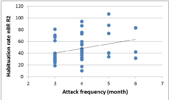
33 Pearson’s test disclosed in the MWoA subjects a positive correlation between the habituation rate of the nBR R2 responses at 0.3 Hz SF and the frequency of migraine attacks per month (r=0.474, p=0.009) (Figure 4).
Figure 4. Correlation between the habituation rate of the nBR R2 (0.3 Hz SF) and the frequency of migraine attacks per month in the MWoA subjects (r=0.474, p=0.009).
34
5. DISCUSSION
The main results of this study were that 1) both AwMH and MWoA subjects showed a clear frequency-dependent deficit of habituation of the nBR R2 responses between 1 and 0.2 Hz SF when compared to HS, whereas no differences between groups were found at slower 0.1 and 0.05 Hz SF; 2) however, AwMH subjects showed, as opposed to MWoA subjects, a less pronounced and not significant deficit of habituation of the nBR R2 responses at faster 1Hz SF when compared to HS, as well as a less homogeneous deficit of habituation between second to fifth block at 0.5 and 0.3 Hz SF when compared to HS; 3) AwMH and MWoA showed a complete overlap of the deficit of habituation from second to fifth block at 0.2 Hz when compared to HS; 4) all groups showed a frequency-dependent habituation of the nBR R2 responses from second to fifth block in almost all SF when compared to first block; 5) in MWoA subjects, the mean frequency of migraine attacks per month correlates positively with the habituation rate of the nBR R2 responses at 0.3 Hz SF.
Our results confirmed the lack of habituation of trigeminal nociceptive reflex responses in MWoA after repeated stimulations, as previously observed by other studies (de Tommaso et al., 2002; Katsarava et al., 2003; Di Clemente et al., 2005; Di Clemente et al., 2007). In addition, we demonstrated that in MWoA, the habituation deficit of the nociceptive component of the BR R2 responses is diffusely detectable in a very wide range of stimulation frequencies, including 1, 0.5, 0.3, and 0.2 Hz, making MWoA subjects clearly distinguishable from HS. The habituation deficit obtained by
35 nociceptive stimulations, match those obtained by using non-nociceptive stimulations evoking conventional BR R2 responses in a similar range of stimulation frequencies (Perrotta et al., 2008). Taking together, our data revealed the habituation deficit of the trigeminal responses (nociceptive and non-nociceptive) as a highly representative neurophysiological abnormality of MWoA subjects during the interictal period.
As novel aspect, our study demonstrated in AwMH subjects during the interictal period a deficit of habituation of nociceptive trigeminal responses largely resembling that observed in MWoA subjects of comparable clinical severity. These data are in line with previous observations of deficit of habituation of evoked responses derived from repeated stimulation of visual cortex in subjects with migraine aura during the interictal phase (Coppola et al., 2013 and 2015). The habituation deficit of nociceptive trigeminal reflex responses in both MWoA and AwMH permits to hypothesize that these two forms of migraine share a common, probably genetically determined, pathogenic substrate. However, it must be noticed that, in AwMH subjects the habituation deficit of the trigeminal nociceptive reflex responses was less pronounced than that observed in subjects with MWoA, although without reach a significant statistical level. In particular, at 1 Hz SF AwMH group showed a less noticeable deficit of habituation of the nBR R2 responses when compared to MWoA group, which resulted not significantly different from both HS and MWoA. A less consistent deficit of habituation in AwMH was also detectable at 0.5 and in part at 0.3 Hz SF, while an indistinguishable deficit of habituation between MWoA and AwMH was only demonstrated at 0.2Hz SF.
36 This less pronounced deficit of habituation in AwMH rises the question about a possibly modulating role of the underlying migraine aura pathophysiology on the habituation and in turn on the clinical behaviour of the AwMH, including the frequency of the attacks, usually less pronounced than in MWoA. However, we are aware that the lack of a statistical significant difference between AwMH and MWoA habituation rate makes our hypothesis highly speculative.
The pathophysiological mechanism underlying the habituation deficit in interictal migraineurs is not completely clear. It has been hypothesized that, an interictal hypoactivity of brainstem-cortical monaminergic pathways (low level of cortical pre-activation) may cause a functional disconnection of the thalamus in migraine leading to an abnormal intracortical short-range lateral inhibition and/or an abnormal rhythmic activity between thalamus and cortex, namely thalamocortical dysrhythmia (Coppola et al., 2009 and 2013). These interictal abnormal information processing between brainstem, thalamus and cortex could contribute to the habituation deficit observed during stimulus repetition in migraine (Coppola et al., 2013).
From a physiological point of view, the habituation to repeated external stimulation, including that nociceptive, is hypothesized to be in balance with the sensitization of the pain pathways in a dual control process operating to determine the behaviour of a neural system underlie to a repeated stimulation (Groves and Thompson, 1970), both, in turn, modulated by a supraspinal control system (Mesulam, 1990). Interestingly, subjects with episodic MWoA during the interictal period showed a subclinical facilitation in
37 temporal processing of nociceptive stimuli to different extracephalic stimulation modalities (Weissman-Foegel et al., 2003; Perrotta et al., 2010 and 2011). Temporal summation of pain sensation is considered to be the human counterpart of the animal wind-up phenomenon and it is strictly related to the sensitization of the pain pathways (Li et al., 1999; Eide 2000; Herrero et al., 2000). On these bases could be hypothesized that in migraineurs a common pathogenetic substrate, possibly of genetic predetermined origin, is responsible for a loss of balance between habituation and sensitization leading to both a deficit of habituation and a subclinical facilitation in pain processing. As consequence, in migraineurs, the abnormal balance between habituation and sensitization cyclically lead to migraine attack during which the habituation deficit normalizes and sensitization of the pain pathways increases (Kaube et al., 2002; Katsarava et al., 2003). The hypothesis of a genetic predetermined abnormal balance between habituation and sensitization in migraine is further supported by the evidence of a reduced habituation and increased facilitation of nociceptive cortical responses in children with migraine during the interictal period (de Tommaso et al., 2016) and of a reduced habituation of the nociceptive trigeminal responses in asymptomatic first-degree relatives of migraine subjects (Di Clemente et al., 2007). A further evolution of these abnormal balance between habituation and sensitization can be observed in chronic forms of migraine, including medication overuse headache, where in a sort of never-ending attack, has been observed a late reduction in habituation of evoked potentials (Coppola et al., 2010) coupled with a marked sensitization of the pain pathways (Ayzenberg et al., 2006; Perrotta et al., 2010 and 2011). In this sense could be
38 explained the positive correlation, although of moderate size, observed in MWoA group between the mean frequency of migraine attacks per month and the habituation rate at 0.3 Hz, one of the SF in which is more evident the habituation deficit in migraineurs. An interesting evidence emerged from our study is the less pronounced deficit of nBR R2 responses in AwMH subjects when compared to MWoA subjects of comparable clinical severity. The neurophysiological basis of this difference in habituation rate between MWoA and AwMH subjects can be only speculative, as other data about the habituation phenomena of the trigeminal responses in AwMH subjects are lacking. Similarly, previous study demonstrated a deficit of habituation of cortical evoked responses after visual stimulation in migraine with aura (MWA) (Coppola et al., 2015), however a direct comparison with MWoA is not known. Furthermore, a critical point should be taken into account when interpreting the results of our study. The less pronounced habituation deficit of nBR R2 responses in MWA when compared to MWoA subjects is clearly detectable only at the highest studied SF, while is inconstantly observed at 0.5 and 0.3 Hz SF. Further studies with larger samples and by using different stimulation modalities are needed to clarify if the observed differences in nociceptive trigeminal excitability between MWA and MWoA represent a pathognomonic feature of subjects with migraine aura. The role of migraine aura underlying pathophysiology as modulating factor of the excitability at brainstem level has been hypothesized in previous animal studies in which experimental bilateral CSD significantly reduced the long-term habituation to intense acoustic stimulation (van der Staak and Fischer, 1976) and
39 induced a significant effect on short-term habituation of the acoustic startle response (van der Staak 1976). More recently, has been demonstrated that experimental CSD induced by focal stimulation of rat visual cortex induces a delayed facilitation in meningeal pain processing after trigeminal neurons activation (Zhang et al., 2010 and 2011). In healthy humans, the modulation of the excitability of the visual cortex is linked to a change in the excitability of the trigeminal nociceptive system assessed by nBR. In particular, the excitatory flash light stimulation lead to an increase in trigeminal pain threshold and to an increase in habituation rate of the nBR (Sava et al., 2014). From an anatomical point of view, the visual cortex projects downward on brainstem nuclei involved both in migraine as well as in top-down inhibitory control system, exerting an inhibitory effect (Noseda et al., 2010). Could be hypothesized that MWA subjects could have a higher excitability level of the visual cortex (Brighina et al., 2015) with respect to MWoA subjects (Ambrosini et al., 2016) which influences the excitability of the nociceptive trigeminal system counterbalancing in part the habituation deficit observed in MWoA.
40
6. CONCLUSIONS
In conclusion, as aspect of novelty, our study demonstrated first, that subjects with typical aura with migraine headache showed a clear deficit of habituation of trigeminal nociceptive reflex responses as demonstrated in MWoA, supporting the hypothesis of a common pathogenetic substrate; second that in subjects with migraine aura the habituation deficit is less significantly recognizable at higher SF with respect to MWoA subjects of comparable clinical severity, permitting to speculate on a modulating role of the migraine aura susceptibility on excitability of the nociceptive trigeminal pathways.
6.1 Clinical implications
The excitability of the nociceptive trigeminal system in typical aura with migraine headache subjects has been poorly explored. In animals, experimental cortical spreading depression modulates habituation to external stimuli and excitability of trigeminal neurons.
We studied the habituation of the nociceptive blink reflex (nBR) R2 responses in migraine with (MWA) and without (MWoA) aura subjects and both groups showed a frequency-dependent habituation deficit when compared to healthy subjects. However, MWA subjects showed a less pronounced habituation deficit when compared to MWoA subjects.
41
Our results support the hypothesis that these two form of migraine share a common, probably genetically determined, pathogenic substrate.
We hypothesized that migraine aura underlying pathophysiology could modulate the excitability of the nociceptive trigeminal system in MWA subjects during the interictal period.
42
REFERENCES
Afra J, Cecchini AP, De Pasqua V, Albert A, Schoenen J. Visual evoked potentials during long periods of pattern-reversal stimulation in migraine. Brain 1998; 121:233–241.
Afra J, Proietti Cecchini A, Sandor PS, Schoenen J. Comparison of visual and auditory evoked cortical potentials in migraine patients between attacks. Clin Neurophysiol 2000; 111:1124–1129.
Afra J, Sandor PS, Schoenen J. Habituation of visual and intensity dependence of auditory evoked cortical potentials tends to normalize just before and during the migraine attack. Cephalalgia 2000; 20:714–719.
Ambrosini A, Iezzi E, Perrotta A, Kisialiou A, Nardella A, Berardelli A, Pierelli F, Schoenen J. Correlation between
habituation of visual-evoked potentials and
magnetophosphene thresholds in migraine: A case-control study. Cephalalgia 2016; 36:258–264.
Ambrosini A, Coppola G, Iezzi E, Pierelli F, Schoenen J. Reliability and repeatability of testing visual evoked potential habituation in migraine: A blinded case-control study. Cephalalgia 2016; May 6. pii: 0333102416648656. [Epub ahead of print].
43
Ambrosini A, Kisialiou A, Coppola G, Finos L, Magis D, Pierelli F, Schoenen J. Visual and auditory cortical evoked potentials in interictal episodic migraine: An audit on 624 patients from three centres. Cephalalgia 2016; Aug 31 pii:0333102416665224. [Epub ahead of print].
Ayzenberg I, Obermann M, Nyhuis P, Gastpar M, Limmroth V, Diener HC, Kaube H, Katsarava Z. Central sensitization of the trigeminal and somatic nociceptive systems in medication overuse headache mainly involves cerebral supraspinal structures. Cephalalgia 2006; 26:1106–1114.
Bingel U, Schoell E, Herken W, Büchel C, May A.
Habituation to painful stimulation involves the
antinociceptive system. Pain 2007; 131:21-30.
Bohotin V, Fumal A, Vandenheede M, Gérard P, Bohotin C, Maertens de Noordhout A, Schoenen J. Effects of repetitive transcranial magnetic stimulation on visual evoked potentials in migraine. Brain 2002; 125:912–922.
Brighina F, Bolognini N, Cosentino G, Maccora S, Paladino P, Baschi R, Vallar G, Fierro B. Visual cortex hyperexcitability in migraine in response to sound-induced flash illusions. Neurology 2015; 84:2057–2061.
Charles A, Baca S. Cortical spreading depression and migraine. Nat Rev Neurol 2013; 9:637–644.
44
Condes-Lara M, Calvo JM, Fernandez-Guardiola A. Habituation to bearable experimental pain elicited by tooth pulp electrical stimulation. Pain 1981; 11:185–200.
Coppola G, Pierelli F, Schoenen J. Habituation and migraine. Neurobiol Learn Mem 2009; 92:249–259.
Coppola G, Currà A, Di Lorenzo C, Parisi V, Gorini M, Sava SL, Schoenen J, Pierelli F. Abnormal cortical responses to somatosensory stimulation in medication-overuse headache. BMC Neurol 2010; 10:126.
Coppola G, Parisi V, Di Lorenzo C, Serrao M, Magis D, Schoenen J, Pierelli F. Lateral inhibition in visual cortex of migraine patients between attacks. J Headache Pain 2013; 14:20.
Coppola G, Di Lorenzo C, Schoenen J, Pierelli F. Habituation and sensitization in primary headaches. J Headache Pain 2013; 14:65.
Coppola G, Bracaglia M, Di Lenola D, Di Lorenzo C, Serrao M, Parisi V, Di Renzo A, Martelli F, Fadda A, Schoenen J, Pierelli F. Visual evoked potentials in subgroups of migraine with aura patients. J Headache Pain 2015; 16:92.
45
De Marinis M, Pujia A, Natale L, D’Arcangelo E, Accornero N. Decreased habituation of the R2 component of the blink reflex in migraine patients. Clin Neurophysiol 2003; 114:889–893.
de Tommaso M, Murasecco D, Libro V, Guido M, Sciruicchio V, Specchio LM, Gallai V, Puca F. Modulation of trigeminal reflex excitability in migraine: effects of attention and habituation on the blink reflex. Int J Psychophysiol 2002; 44:239–249.
de Tommaso M, Ambrosini A, Brighina F, Coppola G, Perrotta A, Pierelli F, Sandrini G, Valeriani M, Marinazzo D, Stramaglia S, Schoenen J. Altered processing of sensory stimuli in patients with migraine. Nat Rev Neurol 2014; 10:144–155.
de Tommaso M, Sciruicchio V, Ricci K, Montemurno A, Gentile F, Vecchio E, Barbaro MG, Simeoni M, Goffredo M, Livrea P. Laser-evoked potential habituation and central sensitization symptoms in childhood migraine. Cephalalgia 2016; 36:463–473.
Di Clemente L, Coppola G, Magis D, Fumal A, De Pasqua V, Schoenen J. Nociceptive blink reflex and visual evoked potential habituations are correlated in migraine. Headache 2005; 45:1388–1393.
46
Di Clemente L, Coppola G, Magis D, Fumal A, De Pasqua V, Di Piero V, Schoenen J. Interictal habituation deficit of the nociceptive blink reflex: an endophenotypic marker for presymptomatic migraine? Brain 2007; 130:765–770.
Eide PK. Wind-up and the NMDA receptor complex from a clinical perspective. Eur J Pain 2000; 4:5–15.
Ellrich J, Hopf HC. The R3 component of the blink reflex: normative data and application in spinal lesions. Electroencephalogr Clin Neurophysiol 1996; 101:349-354.
Ernst M, Lee MH, Dworkin B, Zaretsky HH. Pain perception decrement produced through repeated stimulation. Pain 1986; 26:221–231.
Esteban A. A neurophysiological approach to brainstem reflexes. Blink reflex. Neurophysiol Clin 1999; 29:7–38.
Giffin NJ, Katsarava Z, Pfundstein A, Ellrich J, Kaube H. The effect of multiple stimuli on the modulation of the 'nociceptive' blink reflex. Pain 2004; 108:124–128.
Glanzman DL. Habituation in Aplysia: the Cheshire cat of neurobiology. Neurobiol Learn Mem 2009; 92:147–154.
Gorji A. Spreading depression: a review of the clinical relevance. Brain Res Brain Res Rev 2001; 38:33–60.
47
Greenspan JD, McGillis SL. Thresholds for the perception of pressure, sharpness, and mechanically evoked cutaneous pain: effects of laterality and repeated testing. Somatosens Mot Res 1994; 11:311–317.
Groves PM, Thompson RF. Habituation: a dual-process theory. Psychol Rev 1970; 77:419–450.
Headache Classification Committee of the International Headache Society (IHS). The International Classification of Headache Disorders, 3rd edition (beta version). Cephalalgia 2013; 33:629–808.
Herrero JF, Laird JMA, Lopez-Garcia JA (2000), Wind-up of spinal cord neurones and pain sensation: much ado about something? Prog Neurobiol 61:169–203.
Katsarava Z, Ellrich J, Diener HC, Kaube H. Optimized stimulation andrecording parameters of human 'nociception specific' blink reflex recordings. Clin Neurophysiol 2002; 113:1932–1936.
Katsarava Z, Giffin N, Diener H, Kaube H. Abnormal habituation of ‘nociceptive’ blink reflex in migraine-evidence for increased excitability of trigeminal nociception. Cephalalgia 2003; 23:814–819.
48
Kaube H, Katsarava Z, Käufer T, Diener H, Ellrich J. A new method to increase nociception specificity of the human blink reflex. Clin Neurophysiol 2000; 111: 413–416.
Kaube H, Katsarava Z, Przywara S, Drepper J, Ellrich J, Diener HC. Acute migraine headache: possible sensitization of neurons in the spinal trigeminal nucleus? Neurology 2002; 58:1234–1238.
Li J, Simone DA, Larson AA. Windup leads to characteristics of central sensitization. Pain 1999; 79:75–82.
Marin JC, Gantenbein AR, Paemeleire K, Kaube H, Goadsby PJ. Nociception-specific blink reflex: pharmacology in healthy volunteers. J Headache Pain 2015; 16:87.
Mesulam MM. Large-scale neurocognitive networks and distributed processing for attention, language, and memory. Ann Neurol 1990; 28:597–613.
Milne RJ, Kay NE, Irwin RJ. Habituation to repeated painful
and non-painful cutaneous stimuli: a quantitative
psychophysical study. Exp Brain Res 1991; 87:438–444.
Moskowitz MA. Neurogenic inflammation in the pathophysiology and treatment of migraine. Neurology 1993; 43:16–20.
49
Neisser U. Temperature thresholds for cutaneous pain. J Appl Physiol 1959; 14:368–372.
Noseda R, Constandil L, Bourgeais L, Chalus M, Villanueva
L. Changes of meningeal excitability mediated by
corticotrigeminal networks: a link for the endogenous modulation of migraine pain. J Neurosc 2010; 30:14420– 14429.
Noseda R, Burstein R. Migraine pathophysiology: anatomy
of the trigeminovascular pathway and associated
neurological symptoms, cortical spreading depression, sensitization, and modulation of pain. Pain 2013; 154:S44– S53.
Perrotta A, Serrao M, Sandrini G, Bogdanova D, Tassorelli C, Bartolo M, Coppola G, Pierelli F, Nappi G. Reduced Habituation of Trigeminal Reflexes in Patients with Episodic Cluster Headache During Cluster Period. Cephalalgia 2008; 28:950–959.
Perrotta A, Serrao M, Sandrini G, Burstein R, Sances G, Rossi P, Bartolo M, Pierelli F, Nappi G. Sensititation of spinal cord pain processing in medication overuse headache involves supraspinal pain control. Cephalalgia 2010; 30:272– 284.
50
Perrotta A, Serrao M, Ambrosini A, Bolla M, Coppola G, Sandrini G, Pierelli F. Facilitated temporal processing of pain and defective supraspinal control of pain in cluster headache. Pain 2013; 154:1325–1332.
Rankin CH, Abrams T, Barry RJ, Bhatnagar S, Clayton DF, Colombo J, Coppola G, Geyer MA, Glanzman DL, Marsland S, McSweeney FK, Wilson DA, Wu CF, Thompson RF. Habituation revisited: an updated and revised description of the behavioral characteristics of habituation. Neurobiol Learn Mem 2009; 92:135–138.
Rennefeld C, Wiech K, Schoell ED, Lorenz J, Bingel U. Habituation to pain: further support for a central component. Pain 2010; 148:503–508.
Sándor PS, Afra J, Proietti-Cecchini A, Albert A, Schoenen J. Familial influences on cortical evoked potentials in migraine. Neuroreport 1999; 10:1235–1238.
Sava SL, de Pasqua V, Magis D, Schoenen J. Effects of visual cortex activation on the nociceptive blink reflex in healthy
subjects. PLoS One 2014; 9(6): e100198.
doi:10.1371/journal.pone.0100198.
Schoenen J, Wang W, Albert A, Delwaide PJ. Potentiation instead of habituation characterizes visual evoked potentials in migraine patients between attacks. Eur J Neurol 1995; 2:115–122.
51
Schoenen J, Ambrosini A, Sandor PS, De Noordhout A. Evoked potentials and transcranial magnetic stimulationin migraine: Published data and viewpoint on their pathophysiologic significance. Clin Neurophysiol 2003; 114:955–972.
Schoenen J. Neurophysiological features of the migrainous brain. Neurol Sci 2006; 27:S77–S81.
Shibata K. Neurophysiology of visual aura in migraine. Brain Nerve 2007; 59:961–970.
Stovner LJ, Hagen K, Jensen R, Katsarava Z, Lipton R, Scher A, Steiner T, Zwart JA. The global burden of headache: a documentation of headache prevalence and disability worldwide. Cephalalgia 2007; 27:193–210.
Strempel H. Adaptive modifications of cold pain (author’s transl). Eur J Appl Physiol Occup Physiol 1976; 36:19–25.
Taylor DJ, McGillis SL, Greenspan JD. Body site variation of heat pain sensitivity. Somatosens Mot Res 1993; 10:455–465.
Thompson RF, Spencer WA. Habituation: a model phenomenon for the study of neuronal substrates of behavior. Psychol Rev 1966; 73:16–43.
52
Thompson RF. Habituation: a history. Neurobiol Learn Mem 2009; 92:127–34.
Valeriani M, de Tommaso M, Restuccia D, Le Pera D, Guido M, Iannetti GD, Libro G, Truini A, Di Trapani G, Puca F, Tonali P, Cruccu G. Reduced habituation to experimental pain in migraine patients: a CO(2) laser evoked potential study. Pain 2003; 105:57–64.
Van der Staak C. Habituation and sensitization of the acoustic startle response during cortical spreading depression in rats. Physiol Behav 1976; 16:681–687.
Van der Staak C, Fischer W. Habituation to intense acoustic stimulation during cortical spreading depression in rats. Physiol Behav 1976; 17:29–33.
Wang W, Wang GP, Ding XL, Wang YH. Personality and response to repeated visual stimulation in migraine and tension-type headaches. Cephalalgia 1999; 19:718–724.
Weissman-Fogel I, Sprecher E, Granovsky Y, Yarnitsky D. Repeated noxious stimulation of the skin enhances cutaneous pain perception of migraine patients in-between attacks: clinical evidence for continuous sub-threshold increase in membrane excitability of central trigeminovascular neurons. Pain 2003; 104:693–700.
53
World Medical Association Declaration of Helsinki. Recommendations guiding physicians in biomedical research involving human subjects. JAMA 1997; 277:925–926.
Zhang X, Levy D, Noseda R, Kainz V, Jakubowski M, Burstein R. Activation of meningeal nociceptors by cortical spreading depression: implications for migraine with aura. J Neurosci 2010; 30:8807–8814.
Zhang X, Levy D, Kainz V, Noseda R, Jakubowski M, Burstein R. Activation of central trigeminovascular neurons by cortical spreading depression. Ann Neurol 2011; 69:855– 865.