Vol.86,pp.3828-3832, May 1989 MedicalSciences
Human
umbilical
cord
blood as a
potential source of
transplantable
hematopoietic stem/progenitor
cells
HAL
E.
BROXMEYER*tt§,
GORDON W. DOUGLAS¶,GIAo
HANGOC*t, SCOTT COOPER*t, JUDITH BARD", DENISENGLISH*t**,
MARGARET ARNY¶, LEWISTHOMASIItt,
AND EDWARD A.BOYSE11
Departments of *Medicine(Hematology/Oncology),tMicrobiologyandImmunology, **Pathology,and thetWalther Oncology Center, Indiana University School ofMedicine, Indianapolis,IN46223;I'Memorial Sloan-KetteringCancer Center, New York, NY10021; $Departmentof Obstetrics andGynecology,
NewYorkUniversityMedical Center, New York, NY 10016; andttComellUniversity Medical Center, New York, NY10021 Contributed by EdwardA.Boyse,February9, 1989
ABSTRACT The purpose of this study was to evaluate humanumbilical cord blood as analternative to bone marrow in the provision oftransplantable stem/progenitor cells for hematopoietic reconstitution. Although no direct quantitative assay for humanhematopoietic repopulating cells is at present available, thegranulocyte-macrophageprogenitor cell (CFU-GM)assay has been used withsuccess as avalid indicator of engrafting capability.Weexamined >100collectionsofhuman umbilical cordblood for their content of nucleated cells and granulocyte-macrophage,erythroid (BFU-E), and multipoten-tial(CFU-GEMM)progenitor cells, in many cases both before and after cryopreservation. First it was determined that granulocyte-macrophage, erythroid, and multipotential pro-genitor cells remainedfunctionally viable in cord blood un-treatedexcept for addition ofanticoagulant for at least 3 days
at
40C
or 250C (room temperature), though not at370C,
implying that these cells could besatisfactorily studied and usedorcryopreserved for therapy after transport of cord blood by overnight air freight carriage fromaremoteobstetricalservice. Granulocyte-macrophage progenitorcells from cord blood so receivedresponded normally to stimulation by purified recom-binantpreparationsofgranulocyte-macrophage,granulocyte, andmacrophagecolony-stimulatingfactors and interleukin 3. The salient finding, based on analysis of 101 cord blood collections, isthat thenumbers ofprogenitorcellspresent inthe
low-density (<1.077
gm/ml)
fraction afterFicoll/Hypaque
separation typically fell withintherange that hasbeenreported for successful engraftment by bone marrow cells. Another observation of practical importance is that procedures toremove erythrocytes or granulocytes prior to freezing, and washingof thawed cells beforeplating,entailedlargelossesof progenitorcells, theyield of unwashedprogenitor cellsfrom unfractionated cord blood being many times greater. The
provisionalinference is that humanumbilical cord blood from
a single individual is
typically
a sufficientsource of cells for autologous(syngeneic)and formajorhistocompatibility
com-plex-matched allogeneichematopoieticreconstitution.
Circulating blood cellsare derived and
replaced
byacate-nated systemoriginating from
hematopoietic
stem and pro-genitor cells,recognized
mostly by functional tests rather thanbymorphology (1-4).Theprimarysite ofproductionofstem/progenitor
cells in human adults is the bonemarrow(1,
2). Henceautologousormajor histocompatibility
complex-matched bonemarrowtransplantationis the usual therapeu-tic vehicle forhematopoietic
reconstitution. Althoughadult blood has had some use as an alternative source(5),
in practicethe contentofstem/progenitor
cells issolow(1,2) that multiple leukopheresis is necessary and has been re-portedprimarily
inpatients
undergoing intense temporaryrebound hematopoiesis resulting from recentchemotherapy (5-17).
Inhuman ontogeny, hematopoieticstem/progenitorcells occurfirstin theyolk sac, later in fetal liver, and then in fetal bonemarrow (1, 17), andtransplantation offetal liver cells has been used in a limited setting to correcthematopoietic deficiencies (18, 19). Stem/progenitor cells occur in fetal blood(1, 17),andhumanumbilical cord blood contains stem cells (20, 21), socalled because in colony assay in vitro they exhibitreplatingefficiency indicative of self-renewal, as well as multipotential (CFU-GEMM), erythroid (BFU-E), and granulocyte-macrophage (CFU-GM) progenitor cells (20-26). Thefrequencyofcord-bloodprogenitors (no. of colonies
formed/no.
of cellsplated)equals or exceeds that of marrow andgreatly surpasses that of adult blood. Progenitor cells fromhumanumbilicalcord blood can be maintained for many weeks inlong-term liquid culture systems, suggesting their productionfrom more primitivecells (22, 27).The use of human umbilical cord blood for therapeutic reconstitution was proposed by one of us (E.A.B., unpub-lishedwork), whosubsequently observed successful hema-topoietic reconstitution of lethally irradiated inbredmice with syngeneic neonatal blood.The prospectof storing cordblood cells(normally discarded) for potentialfuturemedicaluseby thedonorhas manyadvantages, notably indisposing ofthe need for bone marrow donors, in providing a disease-free sourceofhematopoietic cells, andinallowingreconstitution ofpatients who lack a clinically approved donor, freedom from graft-versus-host disease, and freedom from the in-creasedmortality that may accompanynonautologous bone marrowtransplantationincircumstanceswhere thedegree of accidentalhematopoietic injury is uncertain (seerefs.28-30). The main questions addressed in the following study concern comparative determinations of the
reconstituting
cellularcontentsofcordblood andbonemarrow,by
measureof
hematopoietic progenitor
cells(31-34)and thecollection,
transport, andoptimalcryopreservation
of cord blood.MATERIALS
ANDMETHODS
Cells. Umbilical cord blood cells were obtained
mainly
from Bellevue Hospital Center (New York), New York University Hospital (New York), Booth Memorial Center (Flushing, NY), and the IndianaUniversity
MedicalCenter (Indianapolis). Thecellswereobtainedfromumbilical cord andplacentaltissues scheduledfordiscard,afterdeliveryofAbbreviations:CSF,colony-stimulatingfactor;G,granulocyte; M,
macrophage; Epo, erythropoietin; CFU-GM, granulocyte-macro-phage colony-forming unit(s); BFU-E,
erythroid
burst-formingunit(s); CFU-GEMM,
granulocyte/erythrocyte/macrophage/mega-karyocytecolony-forming unit(s); CM,conditioned medium. tTowhomreprintrequestsshould beaddressedat: DepartmentofMedicine, Indiana University School of Medicine, 541 Clinical Drive,Indianapolis, IN46223.
3828 Thepublicationcostsof this articleweredefrayedinpartbypagecharge payment.This article mustthereforebeherebymarked"advertisement"
the infant and after
prior
needs,
if any, forsamples
for clinicalstudy
had been satisfied. The source of each collectionwas not identifiedby
name or otherdesignation,
and in vitrohematologic
studiesweremadeonly
asdescribedherein. The InstitutionalBoard of Research Associates ofthe NewYorkUniversity
Medical Center and the Institutional Review Board of IndianaUniversity
ruled thecollectionof blood in thesecircumstancestobeexempt fromtheconsentprocess.Immediately
upondelivery
of theinfant,
the umbilicalcord,
in mostcases, wasdoubly clamped
5-7 cmfrom the umbilicus andtransected
betweentheclamps.
The infantwasremoved from the field. Blood was collected from the
maternal
(placental)
end of thetransected
cord while theplacenta
remainedin situ(to
takeadvantage
ofthe enhanced bloodflowgenerated by
uterinecontraction).
Insomecases, blood was obtained from the removedplacenta by
needleaspiration
ofexposed,
engorged
vesselsonthe fetal surface. In allsamples
collected outside ofIndiana,
collection wasmade into a sterile
(A-irradiation;
MedicalSterilization,
Syosset,
NY)
wide-mouthglass
bottle(no. 25625-200;
Corn-ing) containing acid-citrate/dextrose
(CPD,
20ml; Sigma)
withpenicillin
(0.03
mg/ml)
andstreptomycin (0.05
mg/ml)
(Sigma)
and sent at ambient temperatureby overnight
ex-press serviceto the Indiana
University
SchoolofMedicine forstudy.
Inall except 2 of >100samples,
bloodsamples
were received within 24 hr after harvest from the donor. Blood collections in Indiana were made inCPD,
ACD(Sigma),
orheparin.
Cells were leftunseparated,
were sed-imentedby gravity
orinmethylcellulose,
orwereseparated
into alow-density
fraction(<1.077
gm/ml
and,
in a fewcases, <1.070
gm/ml)
by using
Ficoll/Hypaque
(Pharmacia).
Analysis
ofHematopoietic Progenitor
CellsinVitro.Growthfactors.
Recombinantpreparations
ofinterleukin3(specific
activity, 109 units/mg,
Immunex,
Seattle,
WA), granulocyte
(G)-macrophage
(M)
colony-stimulating
factor(CSF)
(108
units/mg;
Immunex),
G-CSF(108
units/mg;
Cetus),
M-CSF(5
x107
units/mg;
Cetus) (35), erythropoietin
[Epo; Toyoba(New
York)
orAmgen],
and medium conditionedby
thehuman
urinary
bladder carcinomacell line 5637(CM 5637)
(36,
37)
wereusedasfactorstosupportthe clonalgrowth
ofhematopoietic
progenitor
cells.CFU-GM. The assay for CFU-GM was
performed
asdescribed
(35, 36)
in 0.3% agarculturemedium(Difco)
that includedMcCoy's
SA mediumsupplemented
with essential and nonessential aminoacids, glutamine,
serine, asparagine,
vitamins,
andsodiumpyruvate(GIBCO)
with10%(vol/vol)
prescreened
heat-inactivated(56°C
for 0.5hr)
fetal bovineserum
(HyClone)
in the absence or presence ofCM 5637,interleukin
3,
GM-CSF, G-CSF,
or M-CSF. Colonies(>40
cells peraggregate)
and clusters(3-40
cells per aggregate)were scoredafter7
days
and 14days
ofincubation,
asthesetwo
scoring days recognize
differentCFU-GM(38, 39). Large
colonies formed(>1000
cells)
but resultsareexpressed
as coloniesplus
clusters as this moreaccurately
reflects the total CFU-GMcompartment(38).
BFU-E,
CFU-GEMM,
andCFU-GM.The assayforBFU-E,
CFU-GEMM,
and CFU-GM wasperformed
in a 1-ml mixture of Iscove's modified Dulbecco's medium(GIBCO),
0.8%methyl
cellulose,
30%(vol/vol)
prescreenednon-heat-inactivated fetal bovine serum
(HyClone),
50AM
2-mercaptoethanol,
0.5-1.0 unit ofEpo,
with or without 0.1mMhemin
(Eastman
Kodak)
or10%(vol/vol)
CM 5637(37).BFU-E,
CFU-GEMM,
andCFU-GM colonies werescored after 14days
of incubation.BFU-E-1,
CFU-GEMM,
and CFU-GM assays were scored from the sameplates,
which includeEpo, hemin,
and CM 5637. BFU-E-2werecultured asBFU-E-1but without CM 5637. BFU-E-2 colonies contain-ed at least 50 cells or werecomposed
ofat least three sub-coloniescontaining
a minimum of10cells each butwereus-ually
muchlarger.
Colonies derived from BFU-E-1 weremuchlargerthan those derived fromBFU-E-2. BFU-E-1 may represent a more immature
progenitor
thanBFU-E-2,
al-though
this has not been proven.Overlap
in BFU-E-1 and BFU-E-2 colonies between the two assay conditions arelikely.
Plating
conditions. Cells wereplated
atconcentrations of 0.25, 0.5, and 1.0 x10⁵
cells per ml for each assay and incubated in a humidified environment with 5%CO2
atlowered (5%) oxygen tension. Low oxygen tension was
maintained using an oxyreducer (Reming
Bioinstruments,
Redfield,NY)because loweredoxygen tension increases the incidence ofdetectableprogenitorcells(22,
40).
Cryopreservation. Cellswere
cryopreserved
understerile
conditions in two ways. In the initialstudies,
cells weresuspendedat aconcentration of 4 x 10⁶cellsper ml
using
amixture of cold (40C) 50% (vol/vol) autologous
plasma/
culture medium and placed on ice. A 1-ml
portion
of the above-mentionedcellsuspensionwascarefullylayered
on 1 ml of chilled sterile cryoprotective medium[20%
(vol/vol)
dimethyl sulfoxide/culture
medium]
in acryotube
(Nunc).
Approximately 10 minpriortofreezing,the 1:1mixturewas
slowly inverted to promote mixing, then
placed
on ice toallow equilibriumbetween the cells and the
cryoprotective
medium. Vialswereplacedinarack inamethanol bathat
40C
just deepenoughto coverthecellsuspension.Thiswasthen placed in the bottom ofafreezerat-800Cuntilcellsreached the frozen state. Within 24hr,
the vials werequickly
transferredto acontainerholdingliquidnitrogenand
placed
into the liquid phase. The cell suspension was thawed
by
gently agitatingthe vial in a 370C waterbath until a small amount of ice waspresent. A chilled mixtureof50%
(vol/vol)
autologous serum/culture medium was asceptically added dropwise with slight mixing between each drop until the suspension volumewasdoubled. Thissuspensionwastrans-ferredto alargertube with thedropwise addition ofthe50% (vol/vol) autologousserum/culture mixture continued untila
volume of 6-7 ml was reached. Diluent was then added dropwise, with mixing at every 0.5-ml incrementuntil the volume reached 9-10 ml.Cells were pelleted by centrifuga-tion at 4°C 200 x g for 10
min,
the supernatant wasaspirated
off, and 1 ml of chilled 20% (vol/vol) autologousserum/
culture mixturewasaddeddropwisewithgentle
mixing
of the solution. An additional 4 ml ofchilledserum/culture mediumwasaddedslowly. Cells were washed a secondtime. In laterstudies,larger volumes of cells were placed without separation,concentration, orwashingintofreezingbagsand cooled to -80°C in a liquid nitrogen programmed freezer (Cryomed, Mt. Clemens, MI) coded at an average rate of -1°C per
min,
asdescribed elsewhere (41), priortoplacing thebagsfor storage in the liquid phase of liquid nitrogen. The defrosting procedure was done without serum-containing medium as the cells were originally frozen in their ownplasma.
RESULTS
Survival ofHematopoietic Progenitor Cells. Before evalu-ating large numbers of umbilical cord blood collections for theircontentof hematopoietic progenitor cells, experiments
weresetup to determine whether thesecells could survive in cord blood withanticoagulantfor up to3days without added exogenous growth factors. Cord blood was collected and separatedinto 10 tubes (2 ml each). One tube was used at time 0 for separation of cells into a low-density fraction (<1.077 gm/ml) and for plating in semisolid medium for assessing progenitorcell numbers. Three tubeseach were left at
4°C,
-25TC(room
temperature), and370C
(in an incubator). Ondays
1(t
= 24hr),
2(t = 48 hr),and 3(t = 72hr), onetube from each temperature was used for separation ofexperiments with reproducibleresults areshownin Table1.
Overall, the various progenitor cells survivedwell after 1 day ateach of the three temperatureswith littleor noloss, and
some progenitor cell compartments increased in numbers.
Progenitorcells survived well also after2and 3 daysat4°C
androomtemperature, but appreciablydecreased numbers
were seen after 2 days, and no progenitors were apparent after3daysat37°C. The survival of hematopoietic progenitor cells mayreflect the endogenous production of interleukin1
byaccessorycells (42). Resultsweresimilar whether the cells were collected in CPD, ACD, or heparin. These results
demonstrated the feasibility of assessing cordblood samples received by overnightexpress mail serviceandimpliedthat samplescollectedeven2or3days previously would contain viable progenitor cells.
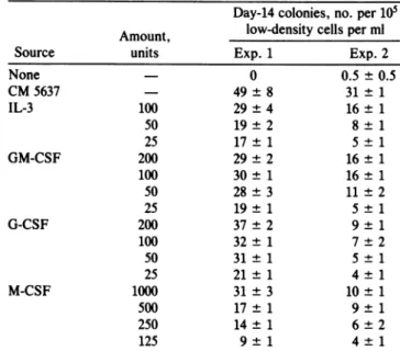
Responsiveness ofUmbilical Cord BloodtoStimulation by
Purified Preparations ofRecombinant CSF. Toevaluate the
types ofCFU-GMpresentin cord blood and whethercord
blood contained CFU-GM responsive tothe fourtypes of CSF, low-density cord blood cellswere platedin the pres-ence of various concentrations of interleukin 3, GM-CSF, G-CSF, and M-CSF. In twoexperiments, day-14CFU-GM responded in a dose-dependent manner to stimulation by each of the CSF preparations (Table 2). The number of colonies stimulated by each of the CSFwas less than that stimulated by CM 5637. CM 5637 contains a number of growth factors including GM-CSF and G-CSF (37) and the higher number of colonies detected with CM 5637 probably reflects additive orsynergistic actions between the growth
factors(43).
Numbers ofHematopoietic Progenitor Cells. One hundred
andonesamples received by overnightexpressmailservice wereassessed foranumber ofparameters,including volume
of blood collected, numbers of unfractionated nucleated cells, numbersoflow-density nucleated cells, and absolute numbers ofhematopoietic progenitor cells in the low-density fraction (Table 3). Although variability was noted between
cord blood samples, itwasclearthat cord blood contained
substantial numbers of hematopoietic progenitor cells. In
fact, theaverage number of CFU-GM collectedpersample waswithinthe lowerrangeof numbers ofCFU-GMindonor
marrowthat havebeen associated with successfulautologous engraftment(32, 34).
It became apparent, however, that the percent of cells
recoveredin thelow-density fraction, 15.1 ± 0.9o(n= 101
samples),wasmuch lower thanwasattained with adult bone marrow and circulating adult blood. This low recovery of
low-density cord blood cells did not reflect the number of cellsloaded onthe Ficoll/Hypaque gradient,andremoving
Table2. Responsiveness of CFU-GMin human umbilicalcord blood tostimulation by recombinanthuman CSF
Day-14 colonies,no. per 105 Amount, low-density cells per ml
Source units Exp. 1 Exp.2
None - 0 0.5 ± 0.5 CM5637 49±8 31±1 IL-3 100 29±4 16± 1 50 19±2 8± 1 25 17±1 5±1 GM-CSF 200 29±2 16± 1 100 30± 1 16± 1 50 28±3 11±2 25 19± 1 5± 1 G-CSF 200 37±2 9± 1 100 32± 1 7±2 50 31±1 5±1 25 21± 1 4± 1 M-CSF 1000 31±3 10± 1 500 17± 1 9± 1 250 14±1 6±2 125 9±1 4±1
None is the background value. CM-5637 was added at 10%
(vol/vol).
thered blood cellspriortothe densitycutdidnotimprove the
recovery.Mostorallof hematopoietic progenitor cells found
in adultbonemarrow orbloodareisolated in thelow-density fraction of cells (refs. 1 and 2; and H.E.B., unpublished observations). Experiments were set up to determine whether thedensity-cut procedure wasalsocausing loss of hematopoietic progenitor cells. Progenitor cells recovered
from unfractionatedcord blood (setup without removal of
redbloodcells)werecompared with numbers of progenitors isolatedinthe low-(<1.077 gm/ml) and high-(>1.077 gm/ml) density fractions. As few as 6-40% of the total number of day-7andday-14CFU-GM,BFU-E-2, BFU-E-1, and
CFU-GEMM cellswererecovered inthelow-densityfraction. The sum of progenitors in both the low- and heavy-density fractions did notequalthatof the unfractionatedgroupand wassometimesaslowas30-40% of theunfractionated blood
cell number.Collectingcells withadensityof <1.070gm/ml didnotimprove therecoveryofprogenitor cells.Attemptsto
removeerythrocytesfromunfractionated cord bloodby lysis
of the nonnucleated cells with ammonium chloride or by sedimenting erythrocytes by gravity, methylcellulose, or centrifugation prior to culturing them for assessment of
progenitor cell numbers resulted in losses of 50-90% of Table1. Influence of timeand temperatureonsurvival of nucleated cells andhematopoietic progenitorcells in human umbilical cordblood
Hematopoietic progenitorcells,total number
Low-density Agarculture Methylcelluloseculture
Time, nucleatedcells, Day-7 Day-14 Day-14
days Temp. no. x 10-6 CFU-GM CFU-GM CFU-GM BFU-E-2 BFU-E-1 CFU-GEMM
0 9.3 5952 8556 10,416 4,092 4464 744 1 40C 5.7 3420 7296 15,048 5,244 7980 684 1 250C 7.9 4108 8848 10,428 4,740 6004 632 1 370C 13.5 4320 7290 13,500 5,670 5940 810 2 40C 6.9 3588 6900 13,248 7,452 4140 552 2 250C 8.4 3912 7392 19,488 10,752 8400 1344 2 370C 0.9 144 324 1,152 360 360 0 3 40C 7.6 3648 6536 18,544 3,040 72% 152 3 250C 8.6 2752 5848 19,608 4,816 9976 688 3 370C 0 0 0 0 0 0 0
Cord bloodwascollectedandassessed for survival.Day-7and-14,CFU-GMinagarcultureswerestimulated with10%o(vol/vol)CM 5637. BFU-E-2cultureswerestimulatedwith 1 unitofEpoplus0.1mMhemin andBFU-E-1,CFU-GEMM,andday-14CFU-GM inmethylcellulose
Table 3. Nucleatedcellularityof unfractionated humanumbilical
cordbloodandnumbersof nucleated cells andhematopoietic
progenitorcells presentin thelow-densityfractionofcordblood
No.of
Parameterevaluated Value samples
Bloodcollected,ml 56.3± 2.4 101 Unfractionated nucleated cells,no. x 10-6 784 ± 59 101 Low-density nucleated cells,no. x 10-6 126± 14 101 Hematopoieticprogenitor cells,no. x 10-5 Agarculture Day-7CFU-GM 0.42 ± 0.06 101 Day-14CFU-GM 1.10± 0.16 101 Methylcelluloseculture Day-14CFU-GM 2.34± 0.39 85 BFU-E-2 0.94 ±0.15 91 BFU-E-1 0.86 ±0.13 91 CFU-GEMM 0.51 ±0.08 91
Progenitor cell assays weresetup asdescribed in text and in the legendtoTable 1. Valuesaremean ± 1SEM.
progenitorcells. These datasuggestedthat actual numbers of hematopoieticprogenitorcellsinhumanumbilicalcord blood had been largely underestimatedandthat themost accurate
way to make this estimate was to use unfractionated cord blood.
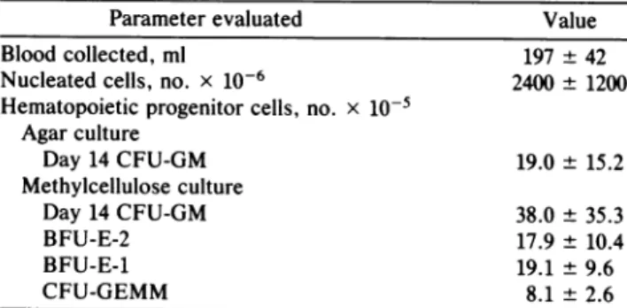
With this information in mind, attempts were made to
optimize the amount ofcord blood collected, and also to
recover blood from the placenta after the baby had been delivered. Results from three cord blood collections made in thisway, which assessed progenitorcell numbers in unsep-arated blood, areshown in Table 4. These studiesillustrate much greateryieldsof nucleated andhematopoietic progen-itor cells. The yields ofnucleated cells recovered are well within the range of cell numbers usually used forautologous andallogeneic bone marrow transplantation, and the
num-bers ofCFU-GMare in the upper range ofthe numbers of CFU-GMin donor marrows that have beenassociatedwith successful engraftment in autologous bone marrow trans-plantationand are in therange used forallogeneic transplan-tation (31-34).
Cryopreservation of Umbilical Cord Blood Cells. Twelve samples oflow-density(<1.077gm/ml)cordbloodcells were frozen in smalltubes in 2-mlaliquotsof 8 x 10⁶cells using the methanol freezing technique. Cells were thawed after 1-10 months and washedtwice, and viable nucleated cells were counted andplatedfor hematopoietic progenitor cell quan-titation. The recoveriesfor thawed samples (mean ± SEM) of nucleated cells, day-7 and -14 CFU-GM (agarcultures), Table4. Nucleatedcellularityandnumbers of hematopoietic progenitorcells present inunfractionated human umbilical cord blood
Parameter evaluated Value
Bloodcollected, ml 197 ±42
Nucleatedcells, no. x 10-6 2400± 1200 Hematopoieticprogenitor cells, no. x 10-5
Agar culture Day 14 CFU-GM 19.0 ± 15.2 Methylcelluloseculture Day 14CFU-GM 38.0± 35.3 BFU-E-2 17.9± 10.4 BFU-E-1 19.1 ±9.6 CFU-GEMM 8.1± 2.6
Valuesarefrom three donor
samples
andexpressedasmean± 1 SEM.Progenitorcellassaysweresetupasdescribedintextandin thelegendtoTable1.day-14 CFU-GM, BFU-E-2,
BFU-E-1,
and CFU-GEMM(methylcellulose
cultures)
were,respectively,
35.0 ±4.5%,
66.6± 10.1%,63.1 ±7.3%,
45.8 ±9.6%,
44.9 ±4.9%,
41.3±
6.2%,
and 30.4±2.6%.Addition of 40-100 units of DNaseto the
samples
during thawing
to reduce cellclumping,
slightly [but
notsignificantly (p
>0.05)]
enhancedrecoveryby
12-20%.Unseparated
cells thatwerefrozen, thawed,
and washed had the same percent recovery as the frozenlow-density
cells(three
experiments,
p >0.05). Recovery
wassimilar whether cells were thawed 1 or 10 months after freezing.
Infurther
experiments,
80-100% of nucleated cells wererecovered inaviablestate
immediately
afterthawing,
but any washing of the cellsto removedimethylsulfoxide decreased the recovery rate. Tofreezelarge
numbers ofunseparated
cells,
the cryopreservationprotocol
was modified to usefreezing bags
holding up to 100 ml ofthe cellpreservation
medium mixture and to use a timedfreezing
apparatus asdescribed elsewhere
(41).
Smallaliquots
of cells werethawed, counted,and platedwithout washing. The
starting
concentration ofunseparated cellsfrozen (8-20 x 106 cells perml)
allowed theplating
of cellsat aconcentration(2.5-5
x104
cellsperml)
such that the actualamount ofdimethyl
sulfoxide addedtothe 1-mlculture disheswerediluted below theconcentration that wouldinterferewith thegrowthof the
progenitor
cells. In three experiments, the recoveries ofday-14 CFU-GM,
BFU-E-1, andCFU-GEMM, as assessed in methylcellulose cultures, were, respectively, 100%,40-60%,
and75-100% from 1 monthto6months after the initialfreezing.
Theseresults indicatethatlargenumbers of nucle-ated and hematopoietic progenitorcells fromhuman umbil-ical cordbloodcanbecryopreservedandretrieved withgood
recovery.DISCUSSION
Weseethatblood collectedfrom theumbilical cord contains CFU-GMinnumberswell within the rangeofmarrow CFU-GM thathavebeen associated with successfulautologousand major histocompatibility complex-matched allogeneic bone
marrowtransplantation (31-34). Although the CFU-GMisnot astemcell,but ratheralineage-restricted progenitor cell,the speed ofhematologic recovery and survival of mice
trans-planted
with bonemarrowhave been correlateddirectlywith thenumber ofCFU-GMtransplanted (44),and theCFU-GM contentof humanbonemarrowalsocorrelatedwith therateof hematopoietic reconstitution after autologous bone marrow transplantation (45). Cord blood BFU-E and CFU-GEMMwere also within the range of these progenitors found in successful human marrowdonor grafts (31).
Unfortunately, there is not yet a direct assay for human hematopoieticrepopulatingcells. Pluripotentialhematopoietic stemcellsaredefined functionallyby their ability to self-renew (make moreof themselves) and to give rise to blood cells of multilineages (1). Murine colony assays in vivo detect what appeartobe subsets of myeloid stem cells, thecolony-forming units in spleen (CFU-S) (1, 3, 43). Murine CFU-S have self-renewalcapacity and give rise to multicell lineages, but it doesnotappearthat theCFU-S is a marrow-repopulating cell (1, 3, 43). The closest human equivalent to the murine CFU-S
in vitro is the stem (S) cell (46, 47). Human umbilical cord blood contains in vitro stem cells (20, 21), which were
identified in human cord blood before they were detected in human bonemarrow (48-50). The frequency of stem cells in both human cord blood and human marrow is essentially the same, but their numbers are very low (20, 21, 48-50). Also, human cord blood stemcellsand human bone marrow stem
cells donotshow the same degree ofreplating efficiency in vitroasdoesthemurinestemcell(20, 21, 46-50).Thehuman
of itslow frequency and also because colonies deriving from stem cells are detected retrospectively, after it has been shown that the colony scored as being stem cell-derived contains cells
capable upon replating of giving rise to secondary colonies
containing cells of multilineages. The human CFU-GEMM,
because it has little or no replating efficiency in vitro, is
considered to be a multilineage progenitor cell rather than a stem cell (1, 3, 43). There is no evidence that numbers of
CFU-GEMM present in a donor inoculum are a better
pre-dictor of
engraftment
potential than are the numbers ofCFU-GM (51).
Our results suggest that umbilical cord blood from a single
donor could serve as a source of autologous or major
histocompatibility complex-matched allogeneic transplant-able hematopoietic repopulating cells. These cells have been cryopreserved and hematopoietic progenitor cells, CFU-GM, BFU-E, and CFU-GEMM, have been retrieved in a
functionally viable form. In this context, it is clear that the cordbloodshould notbe separated to remove any cell types prior to freezing and should not be washed or otherwise manipulated after thawing since all such procedures caused severelosses of hematopoietic progenitor cells. Fortunately, there have already been clinical situations in which unsepa-rated marrow cells have been frozen, thawed, and infused into recipients without washing. Marrowaspirated for bone marrow transplantation,because of the large amounts of cells needed, is usually diluted with blood and, therefore, would
contain more erythrocytes than are normally present in
marrow. It therefore seems reasonable to assume that cord blood infused into donorsimmediately after thawing would not present serious problems.
Thefinal question of whether human cord bloodcells can
successfully reconstituteahuman subject is the subjectofan international and multiinstitutionalcollaboration (E. Gluck-man, H.E.B., A. D.Querbach, H. S.Friedman, G.W.D., A.
Devergie,H. Esperou, D.Thierry, G. Socie,P. Lehn, S.C., D.E., J.Kurtzberg,J.B.,andE.A.B.,unpublished research).
We thank LindaCheung for typing themanuscript. These studies were supported by agrant from the Biocyte Corporation (New York, NY) and by PublicHealth Service Grants CA36464 and CA36740 (to H.E.B.) from the National Cancer Institute.
1. Broxmeyer, H. E. (1982) in The Human Bone Marrow, eds. Trubowitz, S. & Davis,S. (CRC, Boca Raton, FL), pp. 77-123. 2. Broxmeyer, H. E. (1982) in The Human Bone Marrow, eds. Trubowitz, S. & Davis, S. (CRC, Boca Raton, FL), pp. 145-208.
3. Broxmeyer, H. E. (1983) CRC Crit. Rev. Oncol/Hematol. 1, 227-257.
4. Williams, D. E. & Broxmeyer, H. E. (1987) Immunol. Res. 6, 294-304.
5. To,L. B. & Juttner, C. A. (1987) Br. J.Haematol. 66, 285-288. 6. Kessinger, A., Armitage, J.O.,Landmark, J. D.,Smith,D. M.
&Weisenburger, D. D. (1988) Blood 71, 723-727.
7. Korbling, M., Dorken, B., Ho, A. D.,Pezzutto, A., Hunstein, W. &Fliedner, T. M. (1986) Blood 67, 529-532.
8. Kessinger, A., Armitage, J.O., Landmark, J. D. & Weisen-burger, D. D. (1986) Exp. Hematol. 14,192-1%.
9. Reiffers, S., Bernard, P., David, B., Vezon, G., Sarrat, A., Marit, G., Moulinier,J. & Broustet,A. (1986) Exp. Hematol. 14, 312-315.
10. Bell, A. J., Figes, A., Oscier, D.G. &Hamblin, T. J. (1986) Lanceti, 1027.
11. Tilly, H., Bastit, D., Lucet, J.-C., Esperou, H., Monconduit, M.& Piguet, H. (1986) Lancetii, 154-155.
12. Castaigne, S., Calvo, F., Dovoy, L.,Thomas, F.,Benbunant,
M.,Gerota,J.&Degos,L.(1986) Br. J. Haematol. 63,209-211. 13. Juttner, C. A., To, L.-B., Haylock, D. N., Branford, A. &
Kimer, R. J. (1985) Br. J. Haematol. 61, 739-745.
14. Juttner, C. A., Dyson, P. G., To, L.-B., Ho, J. Q. K., Hay-lock, D. N. &Roberts, M. M. (1985) Lancet i, 419-420.
15. Abrahms, R. A., Glaubiger, D., Appelbaum, F. R. & Deisse-roth, A. B. (1980) Blood56, 516-520.
16. Hershko, C., Ho, W. G., Gale, R. R. & Cline, M. J. (1979) Lancet i, 945-947.
17. Migliaccio, G., Migliaccio, A. R., Petti, S.,Mavillo, F., Russo, G.,Lazzaro, D., Testa, U., Marinucci, M.& Peschle, C. (1986) J. Clin.Invest. 78, 51-60.
18. Prummer, 0. & Fliedner, T. M. (1986)Int.J. Cell Cloning 4, 237-249.
19. Gale, R. P., Touraine, J.-L. &Lucarelli, G. (1985) Fetal Liver Transplantation (Liss,NewYork), pp. 237-342.
20. Nakahata, T. & Ogawa, M. (1982) J. Clin. Invest. 70, 1324-1328.
21. Leary, A. G., Ogawa, M., Strauss, L. C. & Civin, C. I. (1984) J. Clin. Invest. 74, 2193-2197.
22. Smith, S. & Broxmeyer, H. E. (1986) Br. J. Haematol. 63, 29-34.
23. Bodger, M. P. (1987)Exp. Hematol. 15, 869-876.
24. Hassan, M. W., Lutton, J. D., Levere, R. D., Rieder, R. F. & Cederqvist, L. L. (1979) Br. J. Haematol. 41, 477-484. 25. Vainchenker, W., Guichard, J. & Bretan-Gorius, J. (1979)
Blood Cells 5, 25-42.
26. Knudtzon, S. (1974) Blood 43, 357-361.
27. Salahuddin, S. Z.,Markham, P. D., Ruscetti, F. W. & Gallo, R. C. (1981) Blood 58, 931-938.
28. Gengozian, N. & Makinodan, T. (1957) Cancer Res. 17, 970-975.
29. Silobrcic, V. & Trentin, J. J. (1966) Transplantation4, 719-731.
30. Allegretti, N. (1968) J. Natl. Cancer Inst. 40, 431-440. 31. Ma, D. D. F., Varga, D. E. &Biggs, J. C. (1987) Leuk. Res. 11,
141-147.
32. Douay, L.,Gorin, N.-C., Mary, J.-Y.,Lemarie,E.,Lopez,M., Najman, A., Stachowiak, J., Giarratara. M.-C., Baillou, C., Salmon,C. &Duhamel,G. (1986) Exp. Hematol. 14, 358-365. 33. Faille, A., Maraninchi, D., Gluckman, E., Devergie, A., Balitrand, N., Ketels, F. & Dresch, C. (1981) Scand. J. Haematol. 26, 202-214.
34. Spitzer, G., Verma, D. S., Zander, A., Vellekoop, L.,Litam, J.,McCredie, K. B.&Dicke, K. A. (1980)Blood 55, 317-323. 35. Broxmeyer,H. E.,Lu, L., Copper, S., Tushinski,R., Mochi-zuki, D., Rubin, B. Y., Gillis, S. &Williams, D. E. (1988)J. Immunol. 141, 3852-3862.
36. Broxmeyer, H. E., Bognacki, J., Ralph, P., Dorner, M. H., Lu, L. & Castro-Malaspina, H. (1982)Blood60, 595-607. 37. Lu, L., Welte, K.,Gabrilove, J. L., Hangoc, G., Bruno, E.,
Hoffman, R.& Broxmeyer, H. E.(1986)Cancer Res.46, 4357-4361.
38. Jacobsen, N., Broxmeyer, H. E., Grossbard, E. & Moore, M. A. S. (1979) Cell Tissue Kinet. 12, 213-226.
39. Ferrero, D., Broxmeyer, H. E., Pagliardi, G. L., Venuta, S.,
Lange, B., Pessano, S. & Rovera,G. (1983) Proc.Natl.Acad. Sci. USA 80, 4114-4118.
40. Lu, L. & Broxmeyer, H. E. (1985) Exp.Hematol.13, 989-993. 41. English, D., Lamberson, R., Graves, V., Akard, L. P.,
Mc-Carthy, L. J. & Jansen, J. (1989) Transfusion 29, 12-16. 42. Williams, D. E. & Broxmeyer, H. E. (1988)Blood72,
1608-1615.
43. Broxmeyer, H. E. & Williams, D. E. (1988) CRC Crit. Rev. Oncol/Hematol. 8, 173-226.
44. Jones, R. J., Sharkis, S. J.,Celano,P.,Colvin,0. M.,Rowley,
S. D. &Sensenbrenner, L. L. (1987)Blood70, 1186-1192. 45. Rowley, S. D.,Zuehlsdorf, M., Braine,H. G.,Colvin,0. M.,
Davis, J., Jones, R. J., Saral, R., Sensenbrenner, L. L.,
Yeager, A. & Santos, G. W.(1987) Blood70, 271-275. 46. Nakahata, T. & Ogawa, M. (1982)Proc.Natl.Acad. Sci. USA
79, 3843-3847.
47. Williams, D. E., Boswell, H. S., Floyd, A. D. & Broxmeyer, H. E. (1985) J. Immunol. 135, 1004-1011.
48. Rowley, S. D., Sharkis, S. J., Hattenburg, C. & Sensenbren-ner, L. L. (1987) Blood 69, 804-808.
49. Leary, A. G. &Ogawa, M. (1987)Blood69,953-956. 50. Brandt, J.,Baird,N., Lu, L.,Srour, E.&Hoffman,R.(1988)
J. Clin. Invest. 82, 1017-1027.
51. To, L. B., Dyson, P. G., Branford, A. L.,
Haylock,
D. N.,Kimber, R. J. & Juttner, C. A. (1987) Exp. Hematol. 15, 351-354.