CC S.. = BY NC ND
http://doi.org/10.5114/bta.2019.90238
Textile azo dye decolorization and detoxification
using bacteria isolated from textile effluents
ARUN KARNWAL
School of Bioengineering and Biosciences, Lovely Professional University, Phagwara, Punjab, India
Abstract
Azo dyes, which are highly toxic, carcinogenic, and mutagenic for living organisms, are used as coloring chemicals in textile industries. Physicochemical methods used for removing azo dyes are expensive, can generate secondary waste, and are not very efficient. In this study, we used a biological approach to reduce the toxicity of three azo dyes, i.e., congo red, methyl orange, and reactive red 198, from textile effluents. Six dye-decolorizing bacteria were screened from waste water obtained from the textile industry (in the dyeing process) at a concentration 100 mg/l for each azo dye. Using a 16S rRNA approach, the bacteria were identified and assigned as Enterococcus faecalis VTK04, Staphylococcusaureus VTK013, Pseudomonas aeruginosa VTK018, Proteusmirabilis VTK024,
Bacilluscereus VTK035, and Enterococcusfaecium VTK054. These bacteria were tested for their ability to pro-duce biofilms on the abiotic surface. The adherence assay showed that VTK013, VTK054, and VTK024 had a po-tential to form stable biofilms on abiotic surfaces (OD570 = 1.37). Moreover, the dye decolorization popo-tential was spectrophotometrically measured after seven days with and without a carbon substrate. The results demonstrated that the absence of the carbon source had a negative impact on decolorization, whereas a carbon-supplemented medium showed a considerable increase in the decolorization of congo red (80% and 96% by VTK013 and VTK018, respectively), methyl orange (100% and 75% by VTK054 and VTK035, respectively), and reactive red 198 (90% and 86% by VTK04 and VTK024, respectively). The phytotoxicity study of the treated dye solutions showed lesser toxicity compared to the untreated dye solution. These results support the possibility of using bacterial isolates for the biodegradation of azo dye effluents.
Key words: azo reactive dye, chick pea, decolorization, phytotoxic study, textile effluent, wheat
Introduction
Usually, artificial dyes are recalcitrant elements that form the primary constituents identified in particular ef-fluents such as fabrics, papers, food, beauty products, and pharmaceutics industries (Jha et al., 2016). These types of coloring agents are polyaromatic molecules (Wang et al., 2018) that provide a permanent color to textile materials. Over 100,000 industrial synthetic che-mical dyes of various classes have been generated globally, of which 60!70% belong to azo dyes, and their yearly production is 7 × 10⁵ metric tons (Karim et al.,
2018). Phytotoxicity investigations demonstrated that most fabric dyes are not hazardous; however, a small fraction of dye in the drinking water supply (5–60 mg/l) is clearly noticeable and has an impact on taste, odor,
transparency, and gas mixability in water bodies (Karim et al., 2018; Neoh et al., 2015). However, their persis-tency and long-time exposure might lead to severe and undesirable outcomes such as mutagenic deterioration and carcinogenicity in living biota (Celebi et al., 2013; Mnif et al., 2016a; Vats and Mishra, 2017). For certain coloring operations, as much as 10–15% of the dyes used never affix to a fabric and are usually released in waste-water. The resulting colored refuse water generates a significant water toxicity issue/problem because of the color content and its hazardous components (Karim et al., 2018). Among all the synthetic chemical dyes, azo dyes contain a significant amount of synthetic organic colorants that are extensively applied (60–70%) owing to the various types of colour tones they offer, lesser
ding, better binding with fabric material, and reduced energy usage (Chaieb et al., 2016). Azo dyes are organic compounds that contain a number of colouring azo func-tional groups (R1-N = N-R2) and aromatic rings, which were simply substituted by sulfonate groupings. These dyes are considered to be electron-deficient xenobiotic substances because they contain electron-withdrawing groups that produce electron insufficiency in the chemi-cals, which makes them resistant to degradation (Liu et al., 2013). However, because of water pollution-rela-ted legislation Water Act 1974 and Water Cess Act (1977–1988), several restraints are being placed on dye release in industrial wastewaters/effluents because of concerns related to environment and human health. Such legislations have forced industries to remove co-louring chemicals from their effluents before they are released into water systems (Iqbal and Asgher, 2013). Several physicochemical methods, such as photocata-lysis, ozonation, electrochemical oxidation process, ad-sorption via activated carbon, coagulation/flocculation, membrane filtration, and precipitation, are usually used for treating textile wastewater but these strategies are expensive and are producing considerable amount of sludge, which has to then be removed safely (Karim et al., 2018). Biological methods (such as microbial de-colorization, microbial degradation, adsorption by (living or dead) microbial biomass, enzymatic degradation, and bioremediation) available for the biodegradation of dyes have been reported to yield better results compared to physicochemical methods. The biodegradation of dyes is effective and promising for the mineralization and de-toxification of azo dyes. Note that biodegradation asso-ciated with azo dyes may take place either aerobically, anaerobically, or even by a combination of both methods. Previously, studies have reported the effectiveness of bac-terial and fungi (such as Phanerochaete chrysosporium, Trametes (Coriolus) versicolor, Bjerkandera adusta, As-pergillus ochraceus, as well as species of Pleurotus and Phlebia, Irpex lacteus, Funalia trogii ATCC 200800, As-pergillus flavus, Alternaria solani, Lentinus polychrous,
Pycnoporus sanguineus, Trametes sp. SQ01, Chlorella
py-renoidosa, Chlorella vulgaris, and Oscillatoria tenuis,
Micrococcus glutamicus NCIM 2168, Enterobacter EC3, Citrobacter sp. CK3, and Pseudomonas aeruginosa) in the biodegradation of azo dyes (Ma et al., 2017; Paz et al., 2017). This study targeted the separation and recogni-tion of microbial strains that are effective for
decolori-zing commonly used azo dyes in textile industrial sectors of Baddi, Himachal Pradesh (India). Moreover, the phytotoxic effect of decolorized dye solution was evalua-ted on chick pea and wheat seeds.
Material and methods
Media and chemicals
Congo red, methyl orange, and reactive red 198 azo dyes were purchased from Sigma-Aldrich Co. and used for bio-decolorization analyses. The other chemicals and microbial media utilized in the study were of analytical grade and the highest purity. All chemicals were acqui-red from Sigma-Aldrich, USA, and Hi-Media, India.
Culture medium
Bushnell and Haas medium (BH) containing 0.2 g/l of MgSO4, 1.0 g/l of K2HPO4, 0.02 g/l of CaCl2, 0.05 g/l of FeCl3, and 1.0 g/l of NH4NO3 supplemented with or
without glucose (0.4% w/v) and yeast extract (0.05% w/v) was used for all experiments. The final pH of the me-dium was adjusted to 7.2.
Isolation and screening of bacterial isolates for dye decolourization
Bacterial isolation was performed using wastewater collected from a textile industry plant located in Baddi, Himachal Pradesh, India (30E56N39.4ON 76E49N10.6OE). The isolated bacterial cultures were screened for the highest dye decolorizing potential. Wastewater samples (10 ml) were added to 100 ml BH broth supplemented with 100 mg/l respective dye (methyl orange, reactive red 198, or Congo red) in 250 ml Erlenmeyer flasks. The mixtures were incubated at 37EC for 48 h at 150 rpm in a shaking incubator for proper agitation and aeration. After incubation, 1 ml of each bacterial culture was trans-ferred into a fresh dye-enriched media until dye decolori-zation occurred. Then, 1 μl of bacterial culture from the final treatment was serially diluted (10!6) and 100 μl was
Identification of bacterial species
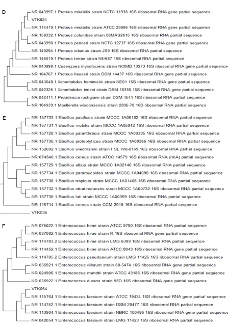
All six bacterial isolates (VTK013, VTK018, VTK035, VTK054, VTK04, and VTK024) were identified using microscopic (colony shape, color, margin, elevation, gram staining, and spore staining) and 16s rRNA gene sequencing methods. The bacterial isolates were cha-racterized through 16S rDNA sequencing and phyletic analysis. The universal bacterial primers 534r (5N!ATTA CCGCGGCTGCTGG!3N) and U1517R (5N!ACGGCTAC CTTGTTACGACTT!5N) were used for 16S rDNA ampli-fication using PCR conditions as described previously (Srinivasan et al., 2015). ProbeBase online software and BLAST (Genbank database) were used to verify the spe-cificity of the primers. For a multiple sequence align-ment (MSA) analysis of sequences, MUSCLE alignalign-ment algorithm was applied (Karnwal, 2017a). Both phylo-genetic analysis and tree creation were performed using PhyML (Dereeper et al., 2008). The isolates were tested for various biochemical assays (motility, starch lysis, citrate utilization, oxidation potential, casein hydro-lysis, urease production, catalase activity, gelatinase production, indole production, siderophore production, H2S production, and various sugar utilization) as repor-ted in Bergey’s Manual of Systematic Bacteriology (Krieg and Holt, 1984).
Biofilm production by adherence assay
The biofilm production assay for VTK04, VTK013, VTK018, VTK024, VTK035, and VTK054 strains was performed in 96-well microtitre plates using a previously reported method by Chaieb et al. (2016). All bacterial isolates were grown in a BH medium enriched with 2% (w/v) glucose for 24 h at 37EC. Then, an aliquot of 200 μl of bacterial suspension per well was dispensed into a 96-well flat bottom microplate and incubated at 37EC for 24 h. After incubation, the bacterial suspen-sions from all plates were removed by washing three times with phosphate buffer, followed by drying at room temperature. The bacterial adherence on the wells of microtitre plate was achieved by treatment with 95% ethanol. These wells were stained with a crystal violet solution (1% w/v). After 6 min of staining, the wells of microtitre plates were washed three times with sterile distilled water to remove the excess amount of dye from wells and air dried at room temperature for 2 h. The optical density for stained wells was recorded using an automated ELISA reader at 570 nm. The bacteria
ad-herence as biofilm formation was measured as described by Chaieb et al. (2016) to be highly positive (OD570
$ 1), low-grade positive (0.1 # OD570 < 1), or negative (OD570 < 0.1).
Azo dye decolorization assay with respective dye decolorizing bacterial isolates
The dye (color) removal assay was performed in 250 ml Erlenmeyer flasks containing 50 ml of pre-steri-lized Bushnell and Haas medium with or without co-sub-strate (0.4% w/v Glucose and 0.05% w/v yeast extract), which was supplemented with respective azo dye to a maximum strength (100 mg/l) and 10% (v/v) respective bacterial inoculum (1 McFarland). These mixtures were incubated for 7 days at 37EC with continuous agitation at 150 rpm. Moreover, the control was maintained with-out bacterial inoculation. At every 24-h intervals, the culture broth was collected and centrifuged at 10 000 g for 15 min in a cooling centrifuge at 4EC. Dye decoloriza-tion was spectrophotometrically measured using UV–vis spectra of the supernatant at a corresponding wavelength of the respective dye solution (Congo red: 540 nm; Methyl orange: 463 nm; Reactive red 198: 518 nm). The stan-dardized dye solution was prepared in BH medium with 0–100 mg/l dye concentration and stored at 4EC in dark after filtration through a 0.22-μm membrane filter. The color removal was recorded as a percentage of the de-colorization using the following equation:
% of dye decolorization = (initial dye strength – final dye strength) × 100 /initial dye strength
Phytotoxicity of the degraded dyessolution
An in vitro toxicity study of the dye solution before and after dye decolorization was performed as described by Chaieb et al. (2016) using chick pea (Cicer arietinum) and wheat (Triticum aestivum) seeds (10 seeds of each plate). During the in vitro study, 2 ml of initial (dye) and decolorized (100 mg/l) solution was added to the seeds. BH medium without dye and bacteria was used as a con-trol. After 10 days of incubation at room temperature, the plant growth parameters such as percentage of seed germination, radicle length, and plumule length were measured.
Statistical analysis
test, followed by the Wilcoxon signed-rank test. P values of < 0.05 were considered to be significant.
Results and discussion
Isolation and screening of bacterial isolates for dye decolorization
Depending on the dyeing activities (stock dyeing, top dyeing, yarn dyeing, piece dyeing, dope dyeing, garment dyeing, beck, jig, pad, and jet dyeing), the textile in-dustry refuse contains an array of different levels of va-rious chemical dyes (Miranda Rde et al., 2013). There-fore, the microbial cultures that can be used for elimina-ting dyes coming from these types of effluents should have the ability to neutralize varied quantities of che-mical dyes (Khandare and Govindwar, 2015; Miranda Rde et al., 2013; Muhammad Nasir Iqbal and Asgher, 2013). In this study, a total of 86 morphologically dis-tinct bacterial colonies were isolated from a wastewater sample collected from the textile colouring industry. Out of these 86 isolates, 62 various bacterial strains showed dye decolorization ability on BH medium supplemented with variable concentrations of different dyes (congo red, methyl orange, and reactive red 198). The presence of microorganisms in the textile dye effluents indicates their natural adaptation and survival abilities in the pre-sence of toxic dyes (Haq et al., 2018). On BH plates, all 62 isolates generated decolorization zones around their colonies but only six isolates had maximum zones of dye colour removal in the plate assay (from 40 to 56 mm). These six isolates were designated as VTK013, VTK018 for congo red; VTK035, VTK054 for methyl orange; and VTK04, VTK024 for reactive red 198. All six bacterial isolates were characterized as described in Bergey’s Ma-nual of Systematic Bacteriology using microscopic and biochemical assays (Krieg and Holt, 1984).
Colony and biochemical characteristics
Bacteria grow fast when supplied with an abundance of nutrients. Different types of bacteria will produce dif-ferent-looking colonies, some colonies may be colored, some are circular in shape, and others are irregular. The characteristics of a colony (such as shape, size, and pig-mentation) are termed the colony morphology. The mor-phology (shape, color, elevation, and margin) of the six isolated bacterial strains is listed in Table 1. The shape
of all six bacterial colonies varied from irregular to circu-lar. As shown in Table 1, the colony margin varied be-tween all bacterial isolates. The color of VTK024, VTK035 was pale yellow; that of VTK018, VTK054, VTK013 was whitish; and that of VTK04 was light orange.
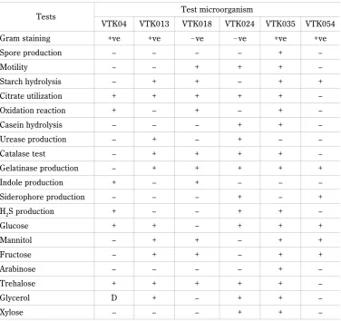
Various staining methods (gram staining and spore staining) were applied to observe the morphological cha-racteristics of the bacterial isolates. The VTK013, VTK04, VTK054, and VTK035 isolates were Gram posi-tive, whereas the remaining two strains (VTK018 and VTK024) were Gram negative and rod-shaped. The re-sults of the microscopic and biochemical assay (such as motility, starch hydrolysis, citrate utilization, oxidation potential, casein hydrolysis, urease production, catalase activity, gelatinase production, indole production, sidero-phore production, H2S production, and various sugar utilization) of all isolates are listed in Table 2.
Identification of bacterial species
Molecular characterizations of all the six isolates were performed using 16S rDNA sequencing (Karnwal, 2017b). As described previously, universal 16S rDNA bac-terial primers 534r (5N!ATTACCGCGGCTGCTG G!3N)
and U1517R (5N!ACGGCTACCTTGTTACGACTT!3N)
Table 1. Colony characteristics of selected bacterial isolates
Bacterial isolate Shape Color Margin Elevation Gram stain
VTK04 cocci light orange entire raised +
VTK013 cocci whitish irregular convex +
VTK018 rod whitish unbonate convex –
VTK024 rod pale yellow regular raised –
VTK035 rod pale yellow unbonate convex +
VTK054 cocci whitish entire convex +
Table 2. Microscopic and biochemical properties of bacterial isolates
Tests Test microorganism
VTK04 VTK013 VTK018 VTK024 VTK035 VTK054
Gram staining +ve +ve !ve !ve +ve +ve
Spore production – – – – + –
Motility – – + + + –
Starch hydrolysis – + + – + +
Citrate utilization + + + + + –
Oxidation reaction + – + – + –
Casein hydrolysis – – – + + –
Urease production – + – + – –
Catalase test – + + + + –
Gelatinase production – + + + + +
Indole production + – + – – –
Siderophore production – – – + – +
H2S production + – – + + –
Glucose + + – + + +
Mannitol – + + – + +
Fructose – + + – + +
Arabinose – – – – + –
Trehalose + + + + + –
Glycerol D + – + + –
Xylose – – – + + –
The ability to produce biofilm of tested isolates using adherence assay
A biofilm is an assemblage of microbial cells that are irreversibly associated (not removed by gentle rinsing) with a surface and enclosed in a matrix of primarily poly-saccharide material (Belouhova et al., 2014). Microor-ganisms attach to surfaces and develop biofilms, and biofilm-associated cells can be distinguished from their
A
B
D
E
F
Fig. 1. A) Phylogenetic tree showing close neighboring of VTK04 with Enterococcus faecalis strain LMG 7937; B) VTK013 with
Staphylococcus aureus strain S33 R; C) VTK18 with Pseudomonas aeruginosa DSM 50071; D) VTK024 with Proteus mirabilis
120 100 80 60 40 20 0 D ye de co lo ur iz a tio n [ % ]
1 2 3 4 5 6 7 8
— — — — — — — — VTK013 VTK018 Time [days] of the most appropriate method for textile azo dye
de-colorization is based on advantages in terms of economy, practicality, and efficiency. Biofilm processes degrade pollutants (such as dye, oil, or waste water) to a higher extent compared to active sludge treatment. They have better resistance to fluctuations in loads, starvation pe-riods, and washouts (Chandran and Das, 2011). In the previous investigation (Belouhova et al., 2014), the bac-teria from genus Pseudomonas played an important role in the adaptation of biofilms for the biodegradation of azo dyes. This study focused on increasing the potential of tested isolates to generate stable bonding with the abiotic surface in the form of a biofilm (Rajendran et al., 2015). A bacterial adherence study on the abiotic surface was investigated using 96-well microtitre polystyrene plates, as reported by Chaieb et al. (2016). The results confirmed that VTK013, VTK054, and VTK024 have the potential to form stable biofilms on abiotic surfaces (OD570 = 1.37, 2.38 and 1.52 respectively). The VTK035 and VTK018 isolated showed a lower adherence potential (OD570 = 0.41 and 0.38, respectively) compared to VTK04 (OD570 = 0.87). Anjaneya et al. (2013) reported that bacterial biofilms are more efficient in docolorizing Ama-ranth dye at three different dye concentrations (200, 400, and 600 mg/l). Wong and Yuen (1996) reported that
the strains of Klebsiella pneumoniae RS-13 and
Aceto-bacter liquefaciens S-1 have the ability to decolorize the azo dye methyl red as well as its possible suitability for treating azo dye-containing textile effluents. The aerobic-anaerobic decolorization and degradation of red HE7B in
the textile effluent was achieved using Pseudomonas
desmolyticum (Cariell et al., 1995; Ogawa and Yatome, 1990). Zissi et al. (1997) reported that Bacillus subtilis could be used to degrade a specific azo dye, i.e., p-aminobenzene. Coughlin et al. (1999) reported that the Sphingomonas sp., strain 1CX had the ability to decolo-rize 20 mg/l orange II, acid orange 8 and 10, acid red 4, and acid red dyes.
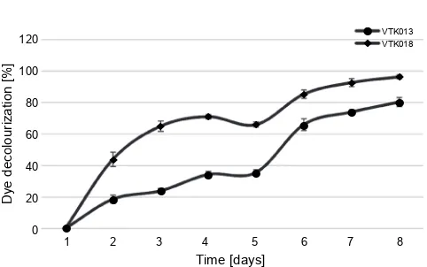
Dye decolorization assay
The wastewater from the textile industry contains a large amount of coloring chemicals (Yuan et al., 2016). In this study, bacterial strains were examined for dye decolorization potential using three textile azo dyes (congo red, methyl orange, and reactive red 198). Se-lected bacterial isolates showed a variable dye color re-moval potential at 100 mg/l concentration of the
respec-Fig. 2. Decolorization of congo red by bacterial isolates
120 100 80 60 40 20 0 D ye d e co lour iz at ion [% ]
1 2 3 4 5 6 7 8
Time [days] – – – – – – – – – — — — — — — — — VTK035 VTK054 VTK04 VTK024
1 2 3 4 5 6 7 8
Time [days] — — — — — — — — • • • • • • • • • 100 90 80 70 60 50 40 30 20 10 0 Dye de co lo ur iz at io n [ % ]
Fig. 3. Decolorization of methyl orange by bacterial isolates
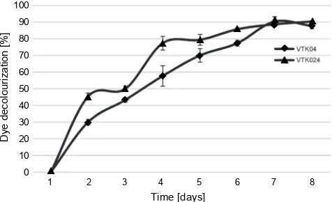
Fig. 4. Decolorization of reactive red 198 by bacterial isolates
2015). Many studies have confirmed that glucose and yeast extract can work as electron donors for dye degra-ding enzymes and enhance the dye degradation process (Bilal and Asgher, 2015). The decolorization of congo red at a concentration of 100 mg/l by VTK013 and VTK018 isolates was recorded at 80% and 96%, respecti-vely, after 8 days of incubation (Fig. 2). Furthermore, the dye decolorization by the VTK013 isolate reached 34% after three days of incubation and stabilized at the 4th day of incubation. On the 5th day, an increase in the
decolorization potential of VTK013 was observed from 37% to 67%. The VTK018 results show a variation in dye decolorization after the first to seventh day of incuba-tion. Furthermore, after one day of incubation, the de-colorization was recorded with an efficiency of 44%. Moreover, we observed efficient decolourization on the 4th, 5th, and 6th day. Finally, on the 8th day, maximum
de-colorization (with 96% efficiency) was noted for congo red, as shown in Figure 2. Figure 3 shows the decolori-zation of methyl orange at a concentration of 100 mg/l by selected bacterial isolates. On the first day, no
de-colorization by the isolates VTK054 and VTK035 was observed. However, with the increase in the incubation time, a significant decolorization of methyl orange was noted for both bacterial isolates. After five days of in-cubation, the bacterial isolate VTK054 showed the highest dye decolorization with 100% efficiency. Initially, on the 2nd day and 5th day, the decolorization efficiency
for VTK054 increased from 76% to 84% and reached 100% on the 6th day under controlled conditions. The
isolate VTK035 showed the maximum dye decolorization of 75% after seven days of incubation. Moreover, the dye decolorization after one day of incubation for methyl orange with VTK053 was recorded as 33% and reached 61% on the 6th day of the experiment. A small drop in the
decolorization efficiency was observed on the 7th day
with 59% with VTK035 isolate, as shown in Figure 3. In this study, maximum decolourization (90%) for reactive red 198 was observed for the VTK04 bacterial isolate on the 7th day of incubation. As shown in Figure 4, a sharp
increase from 53% to 77% was observed for the VTK04 dye decolorization efficiency after the 2nd day of
incuba-tion. A small decreased in dye decolorization efficiency was recorded for VTK04 on the 8th day from 90% to 88%.
However, as shown in Figure 4, the isolate VTK024 pre-sented 90% dye decolorization on the 8th day of
incuba-tion. Finally, on the 3rd day, decolorization was recorded
with 50% efficiency, which then changed to 86% on the 6th day of experiment for VTK024. The divergence in the
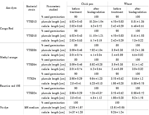
Table 3. Phytotoxicity study of azo dyes and their degradation product
Azo dyes Bacterialstrain Parametersstudied
Chick pea Wheat
before
treatment biodegradationafter treatmentbefore biodegradationafter
Congo Red
VTK013
% seed germination 90 100 90 100
plumule length [cm] 5.02 ± 0.43 10.25 ± 1.04 4.78 ± 0.83 8.81 ± 1.35 radicle length [cm] 2.02 ± 0.53 5.2 ± 0.72 2.42 ± 0.29 5.46 ± 0.51
VTK018
% seed germination 90 100 90 100
plumule length [cm] 5.02 ± 0.43 11.10 ± 1.21 4.78 ± 0.83 8.51 ± 1.03 radicle length [cm] 2.02 ± 0.53 5.7 ± 0.19 2.42 ± 0.29 7.3 ± 0.22
Methyl orange
VTK035
% seed germination 80 100 80 100
plumule length [cm] 3.09 ± 0.46 7.92 ± 1.04 3.9 ± 0.38 10.2 ± 1.36 radicle length [cm] 3.01 ± 0.74 4.1 ± 0.34 2.4 ± 0.89 5.5 ± 0.82
VTK054
% seed germination 80 100 80 100
plumule length [cm] 3.09 ± 0.46 8.92 ± 0.28 3.9 ± 0.38 8.1 ± 1.42 radicle length [cm] 3.01 ± 0.74 5.2 ± 0.54 2.4 ± 0.89 3.9 ± 0.32
Reactive red 198
VTK04
% seed germination 90 100 90 100
plumule length [cm] 3.98 ± 0.29 9.64 ± 1.23 3.78 ± 0.52 8.55 ± 1.2 radicle length [cm] 2.8 ± 0.41 5.23 ± 0.13 1.98 ± 0.28 5.88 ± 0.78
VTK024
% seed germination 90 100 90 100
plumule length [cm] 3.98 ± 0.29 7.25 ± 0.37 3.78 ± 0.52 8.98 ± 0.72 radicle length [cm] 2.8 ± 0.41 4.8 ± 1.12 1.98 ± 0.28 9.2 ± 1.78
No dye BH medium
% seed germination 100 100
plumule length [cm] 12.85 ± 1.67 13.61 ± 0.65 radicle length [cm] 14.37 ± 1.28 9.25 ± 1.24
Values represent the mean of three replicates ±standard error of the mean
Bacillus sp., Alkaligenes sp., and Aeromonas sp., in va-rious soil and wastewater samples obtained from the neighboring locations of fabric-coloring companies and waste materials management dumping sites (Mnif et al., 2016; Ng et al., 2014). However, bacterial isolates, i.e.,
Staphylococcus aureus, Pseudomonas aeruginosa, and
Enterococcus faecalis, are considered as pathogens for humans, and their application for treating textile efflu-ents might create major medical issues, particularly if these isolates are not correctly maintained.
Phytotoxicity study results
For environment sustainability and reusability in agri-cultural practice, it is necessary to study the toxic nature of decolorized solutions (Chen et al., 2018). In this study, after seven days of dye decolorization
experi-ments, a toxicity study for all six decolorized dye solu-tions before and after dye decolorization was performed, as described by Chaieb et al. (2016). The results of the phytotoxicity study are shown in Table 3. Fresh BH me-dium without any dye (positive control) did not show any negative effect on the germination of seeds (chick pea and wheat) and both seeds reached 100% germination in
the in vitro study. In the phytotoxic studies, a similar
chick-pea seeds was observed with the decolorized dye solu-tion of VTK035 (60%) and VTK054 (65%) over non-de-colorized dye medium (Table 3). In this study, a positive impact of a dye-decolorized medium on wheat shoot growth was recorded. Note that, after seven days of incubation with a bacterial solution, a decolorized me-dium promoted/induced better wheat shoot growth, which ranged from 43% to 62%, compared to when using a non-decolorized medium, as shown in Table 3. An ear-lier study (Telke et al. 2010) demonstrated that Su-EBT solutions containing azo dyes biodegraded by Pseudo-monas sp. decreased the dye toxicity on the growth of great millet (Sorghum bicolor), mung bean (Vigna
ra-diata), lentil (Lens culinaris), and rice (Oryza sativa)
plants. In this study, the effect of non-decolorized and decolorized dye media on chickpea and wheat seeds was tested. The VTK035 isolate increased chick pea root growth to 36% over the non-treated dye medium. While VTK054, VTK04, and VTK024 showed improved root growth from 41% to 49%, compared to the VTK035-de-colorized and non-treated dye medium. The maximum root growth (28%) for wheat was observed with the VTK024 bacterial isolate. A similar observation was per-formed for VTK04- and VTK018-based dye-decolorized medium, in which the root growth was recorded as 66% and 62%, respectively. Moreover, there was an increase in the root growth compared with that observed for a non-decolorized medium, as shown in Table 3. Saratale et al. (2015) reported the non-toxic nature of a decolori-zed dye solution on sorghum and black gram. Further-more, the results of Wilcoxon signed-rank test demon-strated a statistically considerable variation in the plu-mule and radicle growth between the treated crop seeds (P < 0.05) and control seeds (Table 3).
Conclusions
Pollution in any form is highly dangerous for the su-stainability of the environment. Effluents from textile dyeing industries have created an alarming situation for both aquatic and terrestrial life. Environmental sustaina-bility depends on the remediation of textile effluents through physical, chemical, or biological processes. In this study, the screening of dye-decolorizing bacteria isolates from textile effluents and their dye decoloriza-tion efficiency on three azo dyes was investigated. The results demonstrated the support of the biological
re-mediation of azo dyes and made it a cost-effective pro-cess for the decolorization of textile dye effluents. The results of our study clearly demonstrate that bacterial isolates did not use azo dyes as carbon sources, whereas glucose and yeast extract supplemented into media were used by bacterial isolates. The treated dye effluents were tested for phytotoxic properties to determine the possi-bility of reusing them for agricultural irrigation pur-poses. The results of the phytotoxic study demonstrated that the remediated effluent showed a non-toxic effect on chickpea and wheat seeds as well as their germination. However, the detoxification mechanism must be impro-ved further to enhance the use of the remediated efflu-ents with other plants in field.
Acknowledgement
I am very thankful to the Bhojia Institute of Life Sciences, Budh, Baddi, H.P., India, for the technical support that al-lowed me to complete this study, as well as for the help exten-ded to me in all steps of my research.
References
Anjaneya O., Shrishailnath S.S., Guruprasad K., Nayak A.S., Mashetty S.B., Karegoudar T.B. (2013) Decolourization of Amaranth dye by bacterial biofilm in batch and continuous packed bed bioreactor. Int. Biodeterior. Biodegradation. 79: 64–72.
Belouhova M., Schneider I., Chakarov S., Ivanova I., Topalova Y. (2014) Microbial community development of biofilm in Amaranth decolourization technology analysed by FISH.
Biotechnol. Biotechnol. Equip. 28(4): 635–642.
Bilal M., Asgher M. (2015) Dye decolourization and detoxifica-tion potential of ca-alginate beads immobilized manganese peroxidase. BMC Biotechnol. 15: 111.
Cariell C.M., Barclay S.J., Naidoo N., Buckley C.A., Mulhol-land D.A., Senior E. (1995) Microbial decolourisation of a reactive azo dye under anaerobic conditions. Water S.A. 21: 61–69.
Celebi M., Kaya M.A., Altikatoglu M., Yildirim H. (2013) Enzy-matic decolourization of anthraquinone and diazo dyes using horseradish peroxidase enzyme immobilized onto various polysulfone supports. Appl. Biochem. Biotechnol. 171(3): 716–730.
Cerron L.M., Romero-Su arez D., Vera N., Ludena Y., Gretty V.K., Gutierrez-Correa M. (2015) Decolourization of textile Reactive dyes and effluents by biofilms of Trametes poly-zona LMB-TM5 and Ceriporia sp. LMB-TM1 isolated from the Peruvian Rainforest. Water. Air. Soil. Pollut. 226: 1–13.
Chandran P., Das N. (2011) Degradation of diesel oil by im-mobilized Candida tropicalis and biofilm formed on gra-vels. Biodegradation, 22(6): 1181–1189.
Chauhan P.S., Goradia B., Saxena A. (2017) Bacterial laccase: Recent update on production, properties and industrial ap-plications. 3 Biotech. 7(5): 323.
Chen Y., Feng L., Li H., Wang Y., Chen G., Zhang Q. (2018)
Biodegradation and detoxification of direct black g textile dye by a newly isolated thermophilic microflora. Bioresour. Technol. 250: 650–657.
Coughlin M.F., Kinkle B.K., Bishop P.L. (1999) Degradation of azo dyes containing aminonaphthol by Sphingomonas sp. strain 1CX. J. Ind. Microbiol. Biotechnol. 23: 341–346. Dereeper A., Guignon V., Blanc G., Audic S., Buffet S., Cheve-net F., Dufayard J.F., Guindon S., Lefort V., Lescot M. et al. (2008) Phylogeny. Fr: Robust phylogenetic analysis for the non-specialist. Nucleic. Acids. Res. 36(suppl. 2): W465–W469.
Haq I., Raj A., Markandeya (2018) Biodegradation of azure-b dye by serratia liquefaciens and its validation by phytotoxi-city, genotoxicity and cytotoxicity studies. Chemosphere 196: 58–68.
Jha P., Jobby R., Desai N.S. (2016) Remediation of textile azo dye acid red 114 by hairy roots of ipomoea carnea jacq. and assessment of degraded dye toxicity with human kera-tinocyte cell line. J. Hazard. Mater. 311: 158–167. Karim M.E., Dhar K., Hossain M.T. (2018) Decolourization of
textile reactive dyes by bacterial monoculture and consor-tium screened from textile dyeing effluent. Genet. Eng. Biotechnol. J. 16(2): 375–380.
Karnwal A. (2017a) Biosurfactant production by kocuria rosea and arthrobacter luteolus using sugar cane waste as sub-strate. II Ponte. 73(2): 20–29.
Karnwal A. (2017b) Isolation and identifi cation of plant growth promoting rhizobacteria from maize (zea mays l.) rhizosphere and their plant growth promoting eff ect on rice (oryza sativa l.). J. Plant. Prot. Res. 57(2): 144–151. Khandare R.V., Govindwar S.P. (2015) Phytoremediation of
textile dyes and effluents: current scenario and future prospects. Biotechnol Adv. 33(8): 1697–1714.
Krieg N.R., Holt J.G. (1984) Bergey's manual of systematic bacteriology. Baltimore, London: Williams & Wilkins. Liu G., Zhou J., Meng X., Fu S.Q., Wang J., Jin R., Lv H.
(2013) Decolourization of azo dyes by marine shewanella strains under saline conditions. Appl. Microbiol. Bio-technol. 97(9): 4187–4197.
Ma X., Liu L., Li Q., Liu Y., Yi L., Ma L., Zhai C. (2017) High-level expression of a bacterial laccase, cueo from esche-richia coli k12 in pichia pastoris gs115 and its application on the decolourization of synthetic dyes. Enzyme. Microb. Technol. 103: 34–41.
Miranda R.C., Gomes E.B., Pereira N. Jr., Marin-Morales M.A., Machado K.M., Gusmao N.B. (2013) Biotreatment of textile effluent in static bioreactor by curvularia lunata urm 6179 and phanerochaete chrysosporium urm 6181.
Bioresour. Technol. 142: 361–367.
Mnif I., Fendri R., Ghribi D. (2015) Biosorption of congo red from aqueous solution by bacillus weihenstephanensis ri12; effect of spb1 biosurfactant addition on biodecolouri-zation potency. Water. Sci. Technol. 72(6): 865–874. Mnif I., Maktouf S., Fendri R., Kriaa M., Ellouze S., Ghribi D.
(2016) Improvement of methyl orange dye biotreatment by a novel isolated strain, aeromonas veronii gri, by spb1 bio-surfactant addition. Environ. Sci. Pollut. Res. Int. 23(2): 1742–1754.
Muhammad Nasir Iqbal H., Asgher M. (2013) Decolourization applicability of sol-gel matrix immobilized manganese per-oxidase produced from an indigenous white rot fungal strain ganoderma lucidum. BMC Biotechnol. 13: 56. Neifar M., Chouchane H., Mahjoubi M., Jaouani A., Cherif A.
(2016) Pseudomonas extremorientalis bu118: A new salt-tolerant laccase-secreting bacterium with biotechnological potential in textile azo dye decolourization. 3 Biotech. 6(1): 107.
Neoh C.H., Lam C.Y., Lim C.K., Yahya A., Bay H.H., Ibrahim Z., Noor Z.Z. (2015) Biodecolourization of recalcitrant dye as the sole sourceof nutrition using curvularia clavata nz2 and decolourization ability of its crude enzymes. Environ. Sci. Pollut. Res. Int. 22(15): 11669–11678.
Ng I.S., Chen T., Lin R., Zhang X., Ni C., Sun D. (2014) De-colourization of textile azo dye and congo red by an isola-ted strain of the dissimilatory manganese-reducing bac-terium shewanella xiamenensis bc01. Appl. Microbiol. Biotechnol. 98(5): 2297–2308.
Nisar N., Aleem A., Saleem F., Aslam F., Shahid A., Chaudhry H., Malik K., Albaser A., Iqbal A., Qadri R. et al. (2017) Re-duction of reactive red 241 by oxygen insensitive azoreduc-tase purified from a novel strain staphylococcus ku898286. PLoS One. 12(5): e0175551.
Ogawa T., Yatome C. (1990) Biodegradation of azo dyes in multistage rotating biological contactor immobilized by assimilating bacteria. Bull. Environ. Contam. Toxicol. 44: 561–566.
Paz A., Carballo J., Perez M.J., Dominguez J.M. (2017) Bio-logical treatment of model dyes and textile wastewaters.
Chemosphere. 181: 168–177.
Rajendran R., Prabhavathi P., Karthiksundaram S., Pattab S., Kumar S.D., Santhanam P. (2015) Biodecolourization and bioremediation of denim industrial wastewater by adapted bacterial consortium immobilized on inert polyurethane foam (puf) matrix: A first approach with biobarrier model.
Pol. J. Microbiol. 64(4): 329–338.
Saratale R.G., Saratale G.D., Chang J.S., Govindwar S.P. (2011) Decolourization and degradation of reactive azo dyes by fixed bed bioreactors containing immobilized cells of proteus vulgaris ncim-2027. Biotechnol. Bioprocess. Eng. 16(4): 830–842.
Saratale R.G., Saratale G.D., Govindwar S.P., Kim D.S. (2015)
Skariyachan S., Prasanna A., Manjunath S.P., Karanth S.S., Nazre A. (2016) Environmental assessment of the degrada-tion potential of mushroom fruit bodies of pleurotus os-treatus (jacq.: Fr.) p. Kumm. Towards synthetic azo dyes and contaminating effluents collected from textile indus-tries in karnataka, india. Environ. Monit. Assess. 188(2): 121.
Srinivasan R., Karaoz U., Volegova M., MacKichan J., Kato-Maeda M. (2015) Use of 16s rrna gene for identification of a broad range of clinically relevant bacterial pathogens. PLOS One. 10(2).
Telke A.A., Joshi S.M., Jadhav S.U., Tamboli D.P., Govindwar S.P. (2010) Decolourization and detoxification of congo red and textile industry effluent by an isolated bacterium pseudomonas sp. Su-ebt. Biodegradation 21(2): 283–296. Vats A., Mishra S. (2017) Decolourization of complex dyes and textile effluent by extracellular enzymes of cyathus bulleri cultivated on agro-residues/domestic wastes and proposed pathway of degradation of kiton blue a and reactive orange 16. Environ. Sci. Pollut. Res. Int. 24(12): 11650–11662.
Wan J., Sun X., Liu C., Tang M., Li L., Ni H. (2017) Decolouri-zation of textile dye rb19 using volcanic rock matrix immo-bilized bacillus thuringiensis cells with surface displayed laccase. World. J. Microbiol. Biotechnol. 33(6): 123. Wang D., Zou J., Cai H., Huang Y., Li F., Cheng Q. (2018)
Effective degradation of orange g and rhodamine b by al-kali-activated hydrogen peroxide: Roles of HO2G and O2G.
Environ. Sci. Pollut. Res. Int. 26(2): 1445–1454.
Wong P.K., Yuen P.Y. (1996) Decolourization and biodegrada-tion of methyl red by Klebsiella pneumoniae RS-13. Water Res. 30: 1736–1744.
Yuan X., Tian G., Zhao Y., Zhao L., Wang H., Ng T.B. (2016)
Degradation of dyes using crude extract and a thermo-stable and ph-thermo-stable laccase isolated from pleurotus nebro-densis. Biosci. Rep. 36(4).