Mahsa Zare1, Fariborz Faeghi1*, Ashrafsadat Hosseini2, Mohammad Sobhan Ardekani2, Mohammad Hossein Heidari3, Ehsan Zarei4
Research Paper:
Comparison Between
Three-Dimen-sional Diffusion-Weighted PSIF Technique and Routine
Imaging Sequences in Evaluation of Peripheral Nerves in
Healthy People
Introduction: The present study aims to evaluate the Three-Dimensional Diffusion-Weighted reversed fast imaging with steady state free precession (3D DW-PSIF) sequence with respect to imaging of the peripheral nerves; the tibial, medial, and lateral plantar nerves in the lower extremity, ulnar and median nerve in the upper extremity, sciatic nerve, brachial plexus, and lumbosacral plexus, and also to compare its usefulness with the current two-dimensional sequences on a 1.5 T MR scanner.
Methods: A total of 25 healthy subjects underwent MR imaging of peripheral nerves, 5 subjects in each area. In each imaging sequence, including T2W SPAIR and 3D DW-PSIF, images were evaluated for ability to identify the nerves in the related area using a 3-score scale (0-2). Then, by summing up the conspicuity scores, a total certainty score was recorded for each sequence. Results: With combining the results of all studies, the conspicuity mean (SD) score was 1.57(0.67) on the 3D DW-PSIF images, and 0.74(0.76) on the T2-weighted images (P<0.001). Regarding the lumbosacral plexus, the corresponding certainty mean (SD) scores were 1.80(0.40) and 1.07(0.74) (P<0.001) and with regard to the brachial plexus, they were 1.23(0.83) and 0.75(0.84), (P<0.001). Regarding the ankle/hind foot they were 1.87(0.35) and 0.40(0.50) (P<0.001) and in the wrist/ proximal hand, 1.70(0.48) and 0.50(0.52) (P<0.001). Regarding the sciatic nerve, they were 1.80(0.44) and 0.20(0.44) (P=0.003).
Conclusion: 3D DW PSIF provides better manifestation of nerves compared to routine imaging sequences particularly fat saturated T2W images. This novel imaging technique can be used in MR neurography examination protocol for exact localization of the nerve and evaluation of the nerve pathology.
A B S T R A C T
Key Words: MR neurography, 3D DW PSIF, Peripheral nerves, Lumbosacral plexus, Brachial plexus Article info:
Received: 06 October 2016
First Revision: 21 October 2016
Accepted: 13 May 2017
1. Department of Radiology Technology, School of Allied Medical Sciences, Shahid Beheshti University of Medical Sciences, Tehran, Iran. 2. Department of Radiology, Shahid Sadoughi Hospital, Shahid Sadoughi University of Medical Sciences, Yazd, Iran.
3. Department of Basic Sciences, School of Allied Medical Sciences, Shahid Beheshti University of Medical Sciences, Tehran, Iran. 4. Department of Physical Education, School of Education & Psychology, Shiraz University, Shiraz, Iran.
* Corresponding Author:
Fariborz Faeghi, PhD
Address: Department of Radiology Technology, School of Allied Medical Sciences, Shahid Beheshti University of Medical Sciences, Tehran, Iran. Tel:+98 (21) 22711131
E-mail: [email protected]
Citation:Zare, M., Faeghi, F., Hosseini, A., Ardekani, M. S., Heidari, M. H., & Zarei, E. (2018). Comparison Between Three-Dimensional Diffusion-Weighted PSIF Technique and Routine Imaging Sequences in Evaluation of Peripheral Nerves in Healthy People. Basic and Clinical Neuroscience, 9(1), 65-71. https://doi.org/10.29252/NIRP.BCN.9.1.65
:
: https://doi.org/10.29252/NIRP.BCN.9.1.65
1. Introduction
here are various methods for the evalu-ation of peripheral nerves. Clinical ex-aminations and electrodiagnostic tests are traditional methods for evaluating the pe-ripheral nerves. In most cases, these meth-ods provide incomplete information about the anatomic details and the nerve intensity (Chhabra et al., 2011a;
Chhabra et al., 2013). Sonography is a complementary
method used to evaluate the peripheral nerves injury. However, this method has also certain restrictions, for example its dependency on a well-experienced and knowledgeable operator with the pretentious sonogra-phy of the soft tissues structures (Khachi, Skirgaudes,
Lee, & Wollstein, 2007).
MR imaging of the nerves, called Magnetic Reso-nance Neurography, is an advanced technique for the evaluation of normal and abnormal peripheral nerves
(Chhabra et al., 2011c). It mostly benefits from the
tech-niques of two-dimensional MR imaging (Zhang et al.,
2008a). Two-dimensional images have advantages such
as high SNR, fairly short acquisition time, and radiolo-gist’s familiarity with it, however, it barely distinguishes between small peripheral nerves and adjacent vascular
structures due to their similar size and signal intensity and also performs poorly during saturation of subcutane-ous and fascial edema, particularly in elbow and knee
(Chhabra, et al., 2011c;Chhabra, Subhawong, Bizzell,
Flammang, & Soldatos, 2011d).
Three-dimensional diffusion-weighted PSIF (3D-DW-PSIF) sequence can help cope with such restrictions and obtain images which represent detailed anatomy and ac-curate localization of the nerves. The steady state nature of this sequence, and use of diffusion gradient, leads to saturation of water signal and vascular structure. There-fore, small nerves can be distinguished from the adjacent vessels more effectively (Chhabra, et al., 2011c; Chhabra
et al., 2011d;Chhabra, Lee, Bizzell, & Soldatos, 2011b).
The current study aims to evaluate the 3D DW-PSIF se-quence in imaging peripheral nervous system including the nerves in the distal lower and upper extremities, sci-atic nerve, brachial plexus, and lumbosacral plexus, and also to compare its results with the current two-dimen-sional sequences which utilize 1.5 Tesla magnet.
2. Methods
A total of 25 healthy subjects (15 males and 10 females, mean (SD) age; 32.5(35) years; age range; 20–55 years)
T
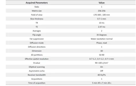
Table 1. The acquired parameters of the 3D DW-PSIF sequence
Value Acquired Parameters
1 Slabs
256-256 Matrix size
170-300 ; 100 mm Field of view
0.7-1 mm Slice thickness
10 ms TR
2.47 ms TE
2 Averages
35 Degrees Flip angle
Water excitation normal Fat suppression
Phase, read Diffusion mode
1 Diffusion directions
3D Dimension
50-90 3D partitions
0.7-1.2 ; 0.7-1.2 ; 0.7-1 mm Effective spatial resolution
90–100 s/mm2
B-value
On Elliptical scanning
Off Asymmetric echo
40 Hz/Px Receiver bandwidth
1 Acquisitions
participated voluntarily in the study. We took written consent forms from all study participants. MR imaging of each part of peripheral nerves was done in 5 subjects. Totally, 100 peripheral nerves were analyzed as follows: C1 to C8 nerves of brachial plexus; L1 to S1 nerves of lumbosacral plexus; Median and ulnar nerves in wrist and palm; Tibial, medial, and lateral plantar nerves in the ankle and hind foot; and Sciatic nerve and it bifurcation.
Images were taken by a 1.5 T MR scanner (Avanto, Siemens Medical Solutions, Erlangen, Germany) using body matrix coil, neck matrix coil, head matrix coil, and CP flexible coil. This study used T2-weighted Spectrally Adiabatic Inversion Recovery (SPAIR) sequence (TR/ TE=2500-6700/36-100 ms, Slice Thickness=2.5-4), as well as high-resolution 3D-DWI-PSIF sequence with dominant T2 contrast. The 3D DW-PSIF sequence was primarily optimized for peripheral nerve imaging in each part of the body with the change in sequence parameters according to Table 1.
In the 3D DW-PSIF sequence, water excitation method was used for fat suppression. In this sequence, low dif-fusion moment (80-90 s/mm2) was applied in the
ap-propriate direction (phase encoding, frequency encod-ing, or slice selection) depending on the related area for imaging. Furthermore, flow compensated gradient was applied in readout direction, in order to reduce the flow artifact. In lumbosacral and brachial plexus, the phase direction was in feet to head. In extremities and sciatic nerve, the phase direction was in anteroposterior direc-tion. The diffusion moment was applied along the phase encoding direction in extremities, sciatic nerve, and bra-chial plexus, but in lumbosacral plexus, applied along the readout direction.
Imaging was done on coronal or sagittal planes to keep the acquisition time low in all studies. Therefore, with us-ing these parameters, we selected images of the nerves with characteristics such as uniform fat, resulting in good nerve contrast to the background, and effective suppression of the vessels (Chhabra et al., 2011c). Then, 3D DW-PSIF images were reformatted with appropriate thickness using post-processing techniques such as MPR and MIP. Since this se-quence was isotropic, the nerve could be reformatted in any arbitrary plane (Chhabra et al., 2011c). The thickness of the reconstructed slab, varies between 3 to 20 mm depending on the nerve size in the related area.
2.1. Image analysis and statistical approaches
Two experienced radiologists evaluated the images on a Picture Archiving and Communication System
(PACS) workstation. In each study, the T2W and 3D DW-PSIF images were analyzed separately. In each imaging sequence, images were evaluated for the abil-ity to represent the nerves in the related area, uniformly fat saturation, and suppress signal from the vessels. Each radiologist independently performed primary evaluation of the images, but final conclusion about the scores given to each sequence was done with consultation.
The conspicuity of each nerve was determined using a semi-quantitative scale (0-2) as follows: 2: Nerve iden-tified with certainty; 1: Nerve probably ideniden-tified; 0: Nerve not identified. Then, by summing up the conspi-cuity scores of all evaluated nerves in the related images, a total conspicuity score was recorded for each imaging sequence. In order to evaluate differences in conspicuity scores between sequences in each study, paired samples t test was performed. A probability level of 0.05 was con-sidered as statistically significant.
3. Results
Table 2 presents the study results. In the current study,
3D DW-PSIF sequence was investigated in most pe-ripheral nerves including lumbosacral plexus, brachial plexus, median and ulnar nerves in the upper limb, tibial, medial and lateral plantar nerves in the lower limb, and sciatic nerve. Furthermore, routine sequences such as T1W-axial images, and T2W-axial and coronal images with fat saturation via SPAIR method were also used. In the 3D DW-PSIF sequence, small nerves and adjacent vessels were better distinguished compared with T2W sequence, and fat saturation was done more effectively than T2W SPAIR method.
Of 100 analyzed peripheral nerves, 67% of the nerves on 3D-DW-PSIF images and 19% of the nerves on T2W images were recognized with certainty. About 45% of the peripheral nerves on T2W images were not identified. In all study MR examinations, the conspi-cuity scores corresponding to 3D DW-PSIF images were higher than those corresponding to T2W images
(Figure 1). In conclusion, the 3D-DW-PSIF sequence
In the brachial plexus imaging, due to large FOV and accordingly suboptimal or no fat saturation in off-center areas of the neck, 3D DW-PSIF is only able to represent nerve roots ganglia and proximal part of brachial plexus (Figure 1). Therefore to obtain high quality images of brachial plexus with uniform fat sup-pression, other three-dimensional sequences, such as 3D-STIR-SPACE can be used.
4. Discussion
With recent advances in MR imaging of peripheral nerves, high quality images of the nerves can be obtained. So nerve anatomy and pathology is effectively depicted in these images (Bendszus et al., 2003;Howe, Filler, Bell,
& Griffiths, 1992). These images can also be used in
pre-operative assessments of nerve injury and entrapments
(Chhabra et al., 2011c). The routine imaging techniques
commonly used in the assessment of peripheral nerves include fat-saturated T2-weighted fast spin-echo, short
inversion-recovery with fat saturation (STIR), and T2W spectral adiabatic inversion recovery turbo spin echo (T2 SPAIR TSE) sequences. Although these methods produce high-quality images, they have limitations in represent-ing small peripheral nerves, because the peripheral nerves and vessels have similar diameter and T2 signal inten-sity resulting in poor discrimination (Zhang et al., 2008a;
Chhabra et al., 2011c; Chhabra et al., 2011d).
Further-more, high-resolution T1-weighted Spin Echo Sequence can also be used to evaluate the fascicular structure of normal nerves characterized with abundant peri- and in-tra-mural fat that is dispersed between fascicles and also distinguish them from the adjacent vessels (Chhabra et
al., 2010). However, since nerve injuries and entrapments
lead to loss of this perineural fat in involved areas, T1W images are not usually helpful (Chhabra et al., 2011c;
Chhabra et al., 2010).
3D-DW-SSFP sequence, used in this study, is a high resolution three dimensional diffusion weighted steady
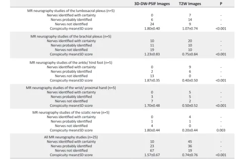
Table 2. Summary of the conspicuity scores obtained from the imaging sequences
P T2W Images 3D-DW-PSIF Images -<0.001 7 14 9 1.07±0.74 0 6 24 1.80±0.40 MR neurography studies of the lumbosacral plexus (n=5)
Nerves identified with certainty Nerves probably identified
Nerves not identified Conspicuity mean±SD score
-<0.001 20 10 10 0.75±0.84 10 11 19 1.23±0.83 MR neurography studies of the brachial plexus (n=5)
Nerves identified with certainty Nerves probably identified
Nerves not identified Conspicuity mean±SD score
-<0.001 9 6 0 0.40±0.50 0 2 13 1.87±0.35 MR neurography studies of the ankle/ hind foot (n=5)
Nerves identified with certainty Nerves probably identified
Nerves not identified Conspicuity mean±SD score
-<0.001 5 5 2 0.50±0.52 0 3 7 1.70±0.48 MR neurography studies of the wrist/ proximal hand (n=5)
Nerves identified with certainty Nerves probably identified
Nerves not identified Conspicuity mean±SD score
-0.003 4 1 0 0.20±0.44 0 1 4 1.80±0.44 MR neurography studies of the sciatic nerve (n=5)
Nerves identified with certainty Nerves probably identified
Nerves not identified Conspicuity mean±SD score
-<0.001 45 36 19 0.74±0.76 10 23 67 1.57±0.67 All MR neurography studies (n=25)
Nerves identified with certainty Nerves probably identified
Nerves not identified Conspicuity mean±SD score
MR: Magnetic Resonance; 3D DW-PSIF: 3-Dimensional Diffusion-Weighted MR based on reversed fast imaging with steady state precession; T2W: T2-weighted
state free precession imaging in combination with fat saturation and flow compensation techniques (Zhang et
al., 2008a). This new sequence has several advantages.
The signal intensity is formed by spin echo mechanism with characteristics similar to a spin echo sequence, and therefore, has T2 dominant contrast. Hence, the effect of magnetic field inhomogeneities is reduced due to the spin relaxation (Hoffmann et al., 2000). Using continu-ous fine sections in this sequence, as well as
applica-tion of high isotropic resoluapplica-tion, allows representaapplica-tion of small structures and injuries and makes it possible to reconstructive the images in any desired plane with good quality using maximum intensity projections technique
(Zhang et al., 2008a).
Since peripheral nerves pass over various tortuous paths, an attempt to suppress vascular signal using satura-tion bands will lead to failure, particularly in extremities
Figure 1. MR neurography of the distal lower extremity in a 26-year-old male
The identification of the medial plantar nerve (arrows) is poor on the sagittal fat-suppressed T2-weighted image due to adja
-cent T2 hyperintense vessels (a), and the conspicuity score is 1. In the 3D DW-PSIF image (b), the identification of the medial plantar nerves (arrows) is definite, and the conspicuity scores is 2. In the isotropic reconstructed oblique sagittal (c) image, the medial planter nerve (arrows) can be easily depicted.
Figure 2. Thin section MIP reconstruction MR images, obtained with a 3D DW-SSFP sequence
(Chhabra et al., 2011a;Chhabra et al., 2011c;Chhabra et
al., 2010). The steady state nature of 3D-DW-PSIF
se-quence, effectively eliminates the signal of vascular flow
(Taupitz et al., 1995;Zhang et al., 2008b). Furthermore,
application of low diffusion moment (b 80–90 s/mm2) in
this sequence is helpful in suppression of signals from the vessels (Chhabra et al., 2011a;Chhabra et al., 2011b;
Chavhan, Babyn, Jankharia, Cheng, & Shroff, 2008). By
using this parameters, we will be able to distinguish the high T2 signal intensity of the nerves from the nulled signal of adjacent vessels. Therefore in comparison with T2W sequence, 3D-DW-PSIF sequence provides bet-ter differentiation between small peripheral nerves and adjacent vessels (Chhabra et al., 2011c;Chhabra et al.,
2011d). In addition, with using diffusion gradients, the
subcutaneous and fascial edema that limits the evalu-ation of small peripheral nerves (in particular distal to elbow and knee), selectively suppresses and accordingly increases the relative nerve conspicuity (Chhabra et al.,
2010;Baur & Reiser, 2000). Since water excitation
tech-nique is used in this sequence, the fat signal intensity is fully suppressed. Also chemical shift effects and motion dependent phase errors are reduced (Zhang et al., 2008a).
An important factor on 3D DW-PSIF imaging is its good shimming. The large FOV and off-center areas may lead to ghosting artifacts and non-uniform fat suppression such as in brachial plexus, which makes the assessment of these neural pathways problematic. The most signifi-cant restriction in this study is high sensitivity of 3D DW-PSIF to motion artifact, breathing artifact and magnetic field inhomogeneity (Chhabra et al., 2011d; Chhabra,
Flammang, Padua, Carrino, & Andreisek, 2014), which
limits the image quality. Another restriction is low Sig-nal to Noise Ratio (SNR) of this sequence in comparison with T2W sequence; as a result, fascicular structure of the nerves is always better represented in fat suppressed T2W. Therefore, 3D DW-PSIF may not replace the T2-weighted imaging which is commonly used in MR Neu-rography (Chhabra et al., 2011c;Chhabra et al., 2011d).
3D-DW-PSIF imaging was first successfully used for the evaluation of cranial nerves in healthy people by Zhang et al. in 2008. (Zhang et al., 2008b). The stud-ies conducted in this respect have emphasized on the usefulness of this imaging method in the assessment of central and peripheral nerves in human body. In a study in 2008, the 3D DW-PSIF sequence used to determine structures of the human Lumbosacral Plexus (LSP). This study showed that this technique can clearly reveal de-tailed anatomy of the LSP and its branches (Zhang et al.,
2008a). In 2010, this imaging method was used for
visu-alization of the intraparotid facial nerve and parotid duct
in 10 patients with suspected parotid gland disease. Ac-cording to the results, the excellent fat suppression, high spatial resolution, vascular signal suppression, and suf-ficient T2-weighting of 3D-PSIF allowed for simultane-ous visualization (Reimer, Parizel, Meaney, & Stichnoth,
2010). In another study in 2011, the researchers
investi-gated the 3D-PSIF sequence in the evaluation of upper and lower extremities and compared its results with rou-tine 2D sequence. They concluded that the 3D-PSIF im-ages provide better identification of the nerves compared to the T2-weighted images (Chhabra et al., 2011c).
In this study, we used this new sequence in peripheral nerves imaging of distal lower and upper extremities, sciatic nerve, brachial plexus, and lumbosacral plexus. We found that the conspicuity scores in 3D DW-PSIF images were higher compared to corresponding scores in T2W images. In addition, 3D PSIF sequence provides better manifestation of nerves compared to fat-saturated T2W sequence. Our data suggest that this novel imaging sequence can be used in MR neurography examination protocol for exact localization of the nerve and evalua-tion of the nerve pathology.
Acknowledgements
This study was the result of MSc. thesis of Mahsa Zare in the Department of Radiology Technology, School of Allied Medical Sciences, Shahid Beheshti University of Medical Sciences, Tehran.
Conflict of Interest
The authors declared no conflict of interest.
References
Baur, A., & Reiser, M. F. (2000). Diffusion-weighted imaging
of the musculoskeletal system in humans. Skeletal Radiology, 29(10), 555–62. doi: 10.1007/s002560000243
Bendszus, M., Wessig, C., Reiners, K., Bartsch, A. J., Solymosi, L., & Koltzenberg, M. (2003). MR imaging in the differential
diagnosis of neurogenic foot drop. American Journal of
Neuro-radiology, 24(7), 1283-9. PMID: 12917113
Chavhan, G. B., Babyn, P. S., Jankharia, B. G., Cheng, H. L. M., & Shroff, M. M. (2008). Steady-state MR imaging sequences: Physics, classification, and clinical applications. RadioGraphics, 28(4), 1147–60. doi: 10.1148/rg.284075031
Chhabra, A., Soldatos, T., Flammang, A., Gilson, W., Padua, A., & Carrino, J. A. (2010). 3T MR imaging of peripheral nerves using
clinical-mri.com/wp-content/uploads/new_technologies/ Carrino.pdf
Chhabra, A., Andreisek, G., Soldatos, T., Wang, K. C., Flam
-mang, A. J., Belzberg, A. J., et al. (2011a). MR neurography: Past, present, and future. American Journal of Roentgenology, 197(3), 583–91. doi: 10.2214/ajr.10.6012
Chhabra, A., Lee, P. P., Bizzell, C., & Soldatos, T. (2011b). 3 Te
-sla MR neurography—technique, interpretation, and pitfalls.
Skeletal Radiology, 40(10), 1249–60. doi:
10.1007/s00256-011-1183-6
Chhabra, A., Soldatos, T., Subhawong, T. K., Machado, A. J., Thawait, S. K., Wang, K. C., et al. (2011c). The application of
three-dimensional diffusion-weighted PSIF technique in pe-ripheral nerve imaging of the distal extremities. Journal of
Mag-netic Resonance Imaging, 34(4), 962–7. doi: 10.1002/jmri.22684
Chhabra, A., Subhawong, T. K., Bizzell, C., Flammang, A., & Soldatos, T. (2011d). 3T MR neurography using three-dimen -sional diffusion-weighted PSIF: Technical issues and advan-tages. Skeletal Radiology, 40(10), 1355–60. doi: 10.1007/s00256-011-1162-y
Chhabra, A., Zhao, L., Carrino, J. A., Trueblood, E., Koceski, S., Shteriev, F., et al. (2013). MR neurography: Advances.
Radiol-ogy Research and Practice, 1–14. doi: 10.1155/2013/809568
Chhabra, A., Flammang, A., Padua, A., Carrino, J. A., & Andre
-isek, G. (2014). Magnetic resonance neurography: technical
considerations. Neuroimaging Clinics, 24(1), 67-78.
Hoffmann, K. T., Hosten, N., Meyer, B. U., Röricht, S., Sprung, C., Oellinger, J., et al. (2000). CSF flow studies of intracranial
cysts and cyst-like lesions achieved using reversed fast imag-ing with steady-state precession MR sequences. American
Jour-nal of Neuroradiology, 21(3), 493-502. PMID: 10730641
Howe, F. A., Filler, A. G., Bell, B. A., & Griffiths, J. R. (1992). Mag -netic resonance neurography. Magnetic Resonance in Medicine, 28(2), 328–38. doi: 10.1002/mrm.1910280215
Khachi, G., Skirgaudes, M., Lee, W. P. A., & Wollstein, R. (2007).
The clinical applications of peripheral nerve imaging in the upper extremity. The Journal of Hand Surgery, 32(10), 1600–4. doi: 10.1016/j.jhsa.2007.09.017
Reimer, P., Parizel, P. M., Meaney, J. F. M., & Stichnoth, F. A. (2010). Clinical MR imaging. New York: Springer. doi:
10.1007/978-3-540-74504-4
Taupitz, M., Speidel, A., Hamm, B., Deimling, M., Reichel, M., Bock, A., et al. (1995). T2-weighted breath-hold MR imaging of the liver at 1.5 T: Results with a three-dimensional steady-state free precession sequence in 87 patients. Radiology, 194(2), 439–46. doi: 10.1148/radiology.194.2.7824724
Zhang, Z. W., Song, L. J., Meng, Q. F., Li, Z. P., Luo, B. N., Yang, Y. H., et al. (2008a). High-resolution diffusion-weighted MR
imaging of the human lumbosacral plexus and its branches based on a steady-state free precession imaging technique at 3T. American Journal of Neuroradiology, 29(6), 1092–4. doi: 10.3174/ajnr.a0994
Zhang, Z., Meng, Q., Chen, Y., Li, Z., Luo, B., Yang, Z., et al. (2008b). 3-T imaging of the cranial nerves using three-dimen -sional reversed FISP with diffusion-weighted MR sequence.