Open Access
Research
Modulation of expression and cellular distribution of p21 by
macrophage migration inhibitory factor
Elliott Taranto, Jin R Xue, Eric F Morand and Michelle Leech*
Address: Centre for Inflammatory Diseases, Monash University Department of Medicine, Monash Medical Centre, Clayton, Melbourne, Australia
Email: Elliott Taranto - [email protected]; Jin R Xue - [email protected]; Eric F Morand - [email protected]; Michelle Leech* - [email protected]
* Corresponding author
Abstract
Background: The pleiotropic protein MIF, (macrophage migration inhibitory factor), has been demonstrated to modulate several key proteins governing cell cycle control and is considered to contribute to cell growth and differentiation. In this study we investigated the effect of MIF on the expression and cellular distribution of the CDK inhibitor p21.
Methods: The effect of endogenous MIF on p21 expression and distribution was examined by comparing murine dermal fibroblasts derived from wt and MIF -/- mice. The effect of MIF on cell growth and apoptotic rates was compared using 3H-Thymidine incorporation assays and annexin V/PI assays respectively. Total p21 protein levels were compared using flow cytometry and western blotting. p21 mRNA was assessed by RT-PCR. Intracellular p21 staining was performed to assess cellular distribution of total protein. To further confirm observations siRNA was used to knockdown MIF protein in wt cells. Cell cycle analysis was performed using PI incorporation assays.
Results: MIF-/- murine dermal fibroblasts exhibited reduced proliferative responses and were more susceptible to apoptosis. This was associated with reduced p21 expression and nuclear distribution. Treatment with recombinant MIF protein was demonstrated to reduce both basal and induced apoptosis and increase nuclear p21 expression. Reduced nuclear p21 expression was also observed in MIF siRNA treated wt cells.
Conclusion: The results demonstrate that in the absence of MIF p21 expression and nuclear distribution is reduced which is associated with a reduction in cell growth and increased apoptosis. MIF may therefore play a role in maintaining homeostatic control of p21.
Background
Phase transition of the cell cycle is dependent upon the ordered activation and subsequent assembly of cyclin-dependent kinases (CDKs) with their regulatory subunit proteins, cyclins. Quiescent cells are stimulated to pro-duce cyclins in response to specific mitogenic signals. The pro-proliferative activity of cyclin-CDK complexes are
tightly regulated by CDK- inhibitors (CKIs) that block the catalytic activity of the holoenzymes. P21 is a member of the Cip/Kip family of CKIs which also consists of p27 and p57. p21 is therefore traditionally identified as a cell cycle arrest protein. Its function in the p53 tumour suppressor pathway as an inhibitor of G1 CDK's resulting in cell cycle arrest in response to various cellular stresses is well
Published: 24 August 2009
Journal of Inflammation 2009, 6:24 doi:10.1186/1476-9255-6-24
Received: 1 April 2009 Accepted: 24 August 2009
This article is available from: http://www.journal-inflammation.com/content/6/1/24 © 2009 Taranto et al; licensee BioMed Central Ltd.
described in the literature [1,2]. However, more recent investigations have revealed new roles for p21 in normal cell cycle progression [3-6] and in active proliferation [7,8].
Recent studies have shown that the proinflammatory cytokine macrophage migration inhibitory factor (MIF) is an important contributor to normal cell division as well as oncogene-induced malignant transformation and tumor-igenesis [9]. One mechanism through which MIF may exert these effects is via escape from p53 mediated cell cycle control. This concept is supported by studies from our lab wherein recombinant MIF (rMIF) treatment leads to decreased p53 protein expression as measured by West-ern blotting [10]. MIF mediated escape from p53 effect is also demonstrated in studies by Hudson et al where the addition of either purified recombinant MIF or expression of an ectopically expressed MIF could over-come p53-induced growth arrest, senescence and apopto-sis [11]. Similarly, MIF has shown to sustain the survival of macrophages through suppression of p53 [12].
In this study we find that the absence of MIF impacts sig-nificantly on the expression and cellular distribution of p21. This observation was associated with a marked reduction in cell growth and increased apoptosis, suggest-ing a role for MIF in maintainsuggest-ing normal homeostatic control of p21 expression and distribution.
Methods
AnimalsMIF-/- mice were generated via homologous recombina-tion in J1 embryonic stem cells as described previously [13]. They were maintained on a mixed background of 129/Sv C57BL/6. MIF+/+ wild-type (wt) mice of the same background were bred from MIF-/- littermates and used as controls. Mice were housed in a conventional housing facility. All animal experiments were performed in accord-ance with the regulations of Monash University Animal Ethics Committee.
Isolation and culture of murine dermal fibroblasts
Murine dermal fibroblasts (MDF) were obtained from MIF-/- and wt mice. Dermal fibroblasts were isolated by dissection of the dermal layer. A single cell suspension was obtained by digesting minced dermal tissue with 2.4 mg/ml Dispase (grade II, 5 U/mg; Boehringer Mannheim, Melbourne, Australia), 1 mg/ml collagenase (type II; Sigma, Melbourne, Australia) and DNase (type I; Boe-hringer Mannheim). MDF were propagated at a cell den-sity of 15 × 106 cells per 10 cm culture plates in RPMI (ICN
Biomedicals, Cincinatti OH)/10% FCS (ICN Pty Ltd, Mel-bourne, Australia) at 37°C in a 5% CO2 humidified incu-bator. Cells were used between passages 4 and 14.
Comparisons between MIF-/- and wt cells were made using cells of the same passage.
Fibroblast proliferation by 3H-Thymidine assay
MDF were seeded onto a 24 well plate at a density of 5 × 10⁴ cells/well (70% confluence) in RMPI (10%FCS) and
incubated for 24 hours at 37°C. Cells were then washed twice with Hanks buffered saline solution (HBSS) before starving in 500 ul RPMI (0.1%BSA) at 37C for 24 hours and were then cultured for 30 hours in 10% FCS. Cells were pulsed with 1 uCi/ml 3H/Thymidine in 50 ul RPMI
for 18 hours at 37°C. Radioactivity content was deter-mined by scintillation counting using a Wallac 1409 Liq-uid Scintillation Counter (3H/Thy, 60 seconds/tube). DNA synthesis was estimated by measurement of 3H-Thy
incorporation in fibroblasts. Results were expressed as the average CPM count with the standard error of the mean.
Fibroblast proliferation by cell count
Wt and MIF-/- MDF were seeded onto a 24 well plate at a density of 5 × 10⁴ cells/well (70% confluence) in RMPI
(10%FCS) and incubated for 72 hours at 37°C. At 24 hour time points, cells were washed with Hanks buffered saline solution (HBSS) and removed by trypsinisation. A single cell suspension was prepared 1:1 ratio with trypan blue (Sigma) and counted using a haemocytometer. Results were expressed as the total number of viable cells (cell count).
Fibroblast apoptosis
Murine Dermal Fibroblasts (MDF) seeded onto a 6 well plate at 2 × 10⁵ cells per well in RPMI (10% FCS) and
per-centage of annexin V/PI and annexin V positive cells rela-tive to total number of cells gated.
Detection of P21 in FLS by Western blotting
P21 expression in wt and MIF-/- MDF were compared using Western blotting with a monoclonal antibody spe-cific for p21 (F-5 Santa Cruz, CA). Briefly, cells taken from semi confluent (70%) culture were washed with cold PBS and then lysed with 2× SDS sample buffer. The protein samples were boiled for 10 minutes and stored at -20°C. Samples were subjected to 10% Tris Glycine iGel SDS/ PAGE (Gradipore, Sydney Australia), transferred to a Hybond C membrane and detected using ECL detection system (Amersham).
RNA extraction and real-time polymerase chain reaction (RT-PCR) analysis
Total RNA was extracted from MDF using TRIzol reagent (Gibco BRL, Grand Island, NY), according to the manu-facturer's protocol. Two micrograms of total RNA was reverse transcribed using Superscript II reverse tran-scriptase and oligo (dT)18 (Gibco BRL). PCR amplifica-tion was performed on a LightCycler (Roche Diagnostics, Castle Hill, Australia) by using SYBR Green I as a double-stranded DNA-specific binding dye and continuous fluo-rescence monitoring. MIF, p21 and β-actin PCR products were purified on agarose gel electrophoresis using the QIAEX II gel extraction system (Qiagen, Clifton Hill, Aus-tralia). The level of expression of messenger RNA (mRNA) of MIF and or p21 was determined relative to the standard preparation using the LightCycler computer software.
For PCR, 5 μl each of the standard and the sample com-plementary DNA dilutions were added to individual cap-illary tubes. Amplification was carried out in a total volume of 10 μl containing primer concentrations of 3 pmoles and 1 μl of dNTP mix, Taq, reaction buffer, and SYBR Green I dye as supplied in the LightCycler DNA Mas-ter SYBR Green I kit (Roche). Forty cycles of PCR were pro-grammed. Relative quantification of target mRNA expression was calculated and normalized to the expres-sion of β-actin. The results, based on the ratio of target mRNA to β-actin amplification, are presented as the fold induction in mRNA expression relative to the amount present in control samples.
Detection of p21 in murine fibroblasts by immunofluorescence
MDF were seeded on mini-slides placed within a 24 well plate at a semi confluent density (70%) of 2.5 × 10⁴ cells
per slide for 24 hours. Adherent cells were then washed with HBSS, and fixed in 2% paraformaldehyde. Cells were again washed and then permeabilised in 0.1% Triton X (Sigma), or 0.1% Saponin (Sigma) for extranuclear stain-ing, before being washed again in PBS/0.1% Na azide/
0.1% BSA and incubated with specific monoclonal anti-body to p21 (F-5) or isotype-matched negative control antibodies (Santa Cruz, CA) for 30 minutes. Unbound antibody was removed by washing in PBS/0.1% Na azide/ 0.1% BSA. Cells were then incubated for 30 minutes with FITC conjugated-secondary antibody (Silenus, Mel-bourne, Australia) before being washed and analysed under UV microscope.
Detection of p21 in murine fibroblasts by flow cytometry
MDF taken from semi confluent culture were washed with HBSS, and fixed in 2% paraformaldehyde. Cells were again washed and then permeabilised in 0.1% Triton X (Sigma) for 30 minutes before being washed again in PBS/ 0.1% Na azide/0.1% BSA and incubated with specific monoclonal antibody to p21 (F-5) or isotype-matched negative control antibodies (Santa Cruz, CA) for 30 min-utes. Unbound antibody was removed by washing in PBS/ 0.1% Na azide/0.1% BSA. Cells were then incubated for 30 minutes with FITC conjugated-secondary antibody (Silenus, Melbourne, Australia) before being washed and analysed by flow cytometry. Results are expressed as delta mean fluorescence intensity (ΔMFI)
For intracellular analysis of p21 protein by flow cytometry 0.05% Saponin was used to permeabilise the cell wall leaving intracellular membranes intact. As above cells were incubated with p21 antibody or negative control antibody and analysed by flow cytometry. Nuclear p21 was estimated by subtracting ΔMFI of cytoplasmic p21 from ΔMFI of total p21.
SiRNA
RNA interference was used to knockdown MIF protein expression by introducing a homologous double-stranded RNA. The nucleotide sequences of dsRNA and complimentary dsRNA for mouse MIF mRNA were 5'-CCGCAACUACAGUAAGCUGdTdT-3' and 5'-CAGCU-UACUGUAGUUGCGGdTdT-3', respectively. As a control RNA duplex 5'-GCGCGCUUUGUAGGAUUCGdTdT-3' and 5'-CGAAUCCUACAAAGCGCGCdTdT-3' were used. MDF grown in RPMI 1640 with FCS (10%) were trans-fected with either the MIF siRNA or the control RNA duplex using Amaxa nucleofection electroporation (Amaxa AG, Germany) according to the manufacturer's protocol [14]. After 24 hours, cells were transfected with either MIF siRNA or the control siRNA a second time. 24 hours after the second transfection the culture medium was removed and cells were examined for p21 immun-ofluorescence.
Cell cycle analysis
suspension was then centrifuged and ethanol thoroughly decanted. The pellet was resuspended in a staining solu-tion of 0.1% triton-X, DNase free RNase and Propidium iodide (PI) for 30 minutes at room temperature. Samples were then analysed by flow cytometry.
Data Analysis
ΔMFI was calculated by subtracting the mean fluorescence of the samples stained with an isotype matched negative control antibody from that of the samples stained with specific antibodies. Results are expressed as the mean +SEM. Statistical analysis was performed using the Stu-dents t test. P < 0.05 was considered statistically signifi-cant.
Results
MIF-/- cells exhibit reduced proliferative responses
To determine the role of endogenous MIF in cell growth we examined proliferation in MIF-/- and wt MDF using a
3H-thymidine incorporation assay and cell count assay.
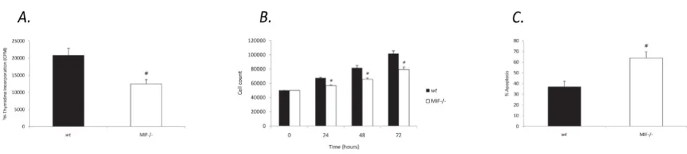
After a 42 hour analysis of 6 independent experiments we found that cells lacking endogenous MIF displayed signif-icantly reduced cell growth, (12437.9 +/- 1253.3), com-pared to wt MDF (20841.7 +/- 2040.6) (Figure 1a). This was confirmed by cell count assays which revealed signif-icantly reduced numbers of viable MIF-/- cells compared to wt over 24, 48 and 72 hours (Figure 1b).
MIF-/- cells are more susceptible to apoptosis
To further characterise the effects of endogenous MIF on cell cycle profile, we examined apoptotic events in MIF-/-and wt MDF by annexin V-FITC and PI dual labeling. Compared with wt MDF, MIF-/- MDF were more prone to undergo cell death exhibiting increased SNP induced apoptosis, (63.8 +/- 5.5), compared to wt MDF, (36.7 +/-5.2) (Figure 1c).
rMIF rescues cells from basal and SNP induced apoptosis
To explore the potential for exogenous MIF to alter cell cycle events we examined both basal and induced apop-totic events in MIF-/- and wt MDF with and without rMIF treatment. Compared with control, treatment with rMIF protein was demonstrated to significantly increase basal survival and to significantly reduce SNP-induced apopto-sis in MIF-/- MDF (Figure 2).
p21 expression is reduced in MIF-/- MDF
To examine the role of endogenous MIF in the regulation of p21 levels, MIF-/- and wt MDF expression of p21 was analysed using Western blotting and flow cytometry anal-ysis. MIF-/- cells expressed lower levels of p21 protein compared to wt MDF (Figure 3a, 3b). Accordingly P21 mRNA was reduced in MIF-/- MDF as determined by RT-PCR (Figure 3c).
Subcellular localisation of p21 in MIF-/- MDF
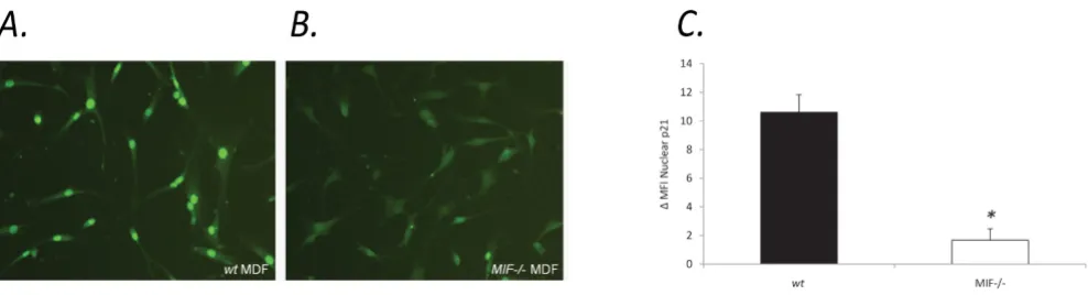
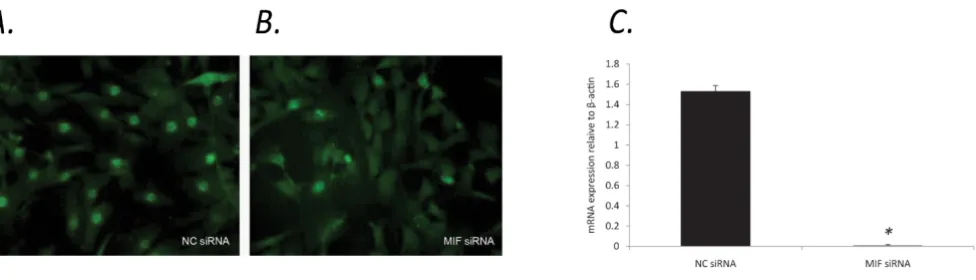
To further explore the potential for MIF to influence p21, we examined the subcellular localisation of p21 in MIF-/-and wt MDF. Interestingly, MIF-/- MDF exhibited diffuse, predominantly cytoplasmic p21 staining (Figure 4b) com-pared to wt MDF, which displayed strong nuclear staining for p21 (Figure 4a). Flow cytometric analysis of nuclear p21 protein revealed a significant reduction in MIF-/-compared to wt MDF (Figure 4c). Similar results were seen in wt MDF spiked with MIF siRNA which exhibited a sig-nificant reduction in nuclear p21 (figure 5b) compared to control siRNA (Figure 5a). The efficiency of MIF knock-down in wt MDF was quantified using RT-PCR. After 40 cycles of amplification MIF mRNA was not detectable in samples transfected with MIF siRNA (figure 5c).
MIF-/- cells exhibit reduced proliferative responses and increased apoptosis
Figure 1
MIF-/- cells exhibit reduced proliferative responses and increased apoptosis. a. Quantification of basal MIF-/- and wt
rMIF induces p21 expression & nuclear localisation in MIF-/- MDF
To further confirm the involvement of MIF in p21 redistri-bution, MIF-/- and wt MDF were treated with rMIF and examined for p21 by immunofluorescence. Randomly cycling wt MDF from semi-confluent culture exhibited predominantly nuclear localisation of p21 and rMIF treat-ment of these cells did not yield any further increase in nuclear p21 expression. However compared to vehicle, rMIF induced a shift from predominantly cytoplasmic to nuclear localisation of p21 protein in MIF-/- MDF demon-strating MIF involvement in induction and nuclear trans-location of p21 protein (Figure 6).
P21 redistribution is not the result of cell cycle phase differences
Cell cycle phase is well described to alter expression of key cell cycle proteins including p21. To address this issue, we examined the cell cycle phase profile of MIF-/- and wt
MDF taken from semi-confluent culture using flow cyto-metric analysis of PI incorporation. No significant differ-ence in the percentage of cells in each phase of the cell cycle was seen. Therefore the difference in p21 expression and subcellular localisation observed between MIF-/- and
wt MDF could not be explained by cell cycle phase differ-ences between the cells in these experiments (Figure 7).
Discussion
MIF is a pro-inflammatory cytokine which is expressed constitutively by almost all cell types and is significantly
upregulated in the setting of inflammatory stimulation [15,16]. The findings in this study suggest that MIF is involved in the promotion of normal cell cycle and prolif-erative responses in murine dermal fibroblasts. Support-ing this, recent studies have demonstrated a functional role for MIF in normal cell division as well as cell survival and oncogene-induced malignant transformation making this cytokine an important new focus in tumour biology research [17-19]. Evidence suggests that MIF may also indirectly support tumour growth via promotion of ang-iogenic responses [20,21]. Other studies have shown that rMIF treatment or forced over expression of MIF can stim-ulate cell growth [22]. A number of cell types have also been shown to secrete MIF in response to growth factor stimulation [23,24]. We have previously shown dose-dependent regulation of human fibroblast proliferation by rMIF [25].
In the current study, reduced proliferative responses in MIF-/- MDF were associated with increased baseline and SNP induced apoptosis. Moreover, treatment with recom-binant MIF was associated with a reduction in SNP induced apoptosis. This promotion of cell survival may have relevance to inflammatory disease in terms of per-sistence of inflammatory cells or growth of intrinsic cells in an inflammatory lesion. For example endogenous MIF has been shown to be important in promoting macro-phage viability in the setting of inflammatory stimuli and rMIF treatment or its overexpression in a variety of cell types including fibroblasts, macrophages and endothelial rMIF rescues cells from basal and SNP induced apoptosis
Figure 2
cells rendered them resistant to apoptosis induced by a variety of stimuli [20,26]. Previous studies in our own lab-oratory have shown that rMIF treatment of human fibrob-lasts leads to rescue from SNP-induced apoptosis [10]. Consistent with this data, others have reported impaired DNA damage response in cells lacking MIF [27].
Our finding in this study that genetic or siRNA mediated deficiency of MIF is associated with reduced expression and/or nuclear localisation of p21 and that rMIF treat-ment induces nuclear localisation of p21 must be inter-preted in the setting of emerging new literature regarding this protein. p21 is traditionally conceptualised as a cell cycle arrest protein. p53-induced nuclear accumulation of high level p21 expression, resulting in G1 cell cycle arrest,
in response to various stressful stimuli, is well described in the literature [1,2,28]. Evidence for an unexpected requirement for basal expression of p21 in normal cell cycle progression is however found in several recent stud-ies [3-6]. p21-/- cells exhibit a failure of normal cell cycle progression which has been related to a role for p21 as an assembly factor which promotes cyclin-D1-CDK4 binding thus contributing to cellular proliferation [29]. Moreover, LaBaer et al 1997 demonstrated that p21 provides the localisation signal for cyclin-D1-CDK4 nuclear import [29]. Other studies have shown that p21 promotes nuclear accumulation of cyclin-D1-CDK4 via its ability to inhibit cyclin D1 nuclear export [4]. These studies suggest some duality in p21 function with evidence for an unex-Reduced p21 expression in MIF-/- cells compared with wt
Figure 3
Reduced p21 expression in MIF-/- cells compared with wt. a. Levels of p21 expression in MIF-/- and wt MDF assessed by Western blotting using a monoclonal antibody to p21. The equivalence of protein loading was measured by detection of β -tubulin. Each lane represents a single MIF-/- or wt MDF mouse. b. Flow cytometric detection of p21 protein in untreated MDF taken from semi-confluent culture. Results are representative of 4 independent experiments, each measuring at least 5000 events. Expressed as delta mean fluorescence intensity (ΔMFI) which is the difference between the MFI of tested cells and the MFI of background staining (N = 4, * P < 0.05). c. Quantification of p21 mRNA to β-actin in MIF-/- and wt MDF by reverse tran-scription polymerase chain reaction (RT-PCR).
Decreased nuclear p21 in MIF-/- MDF compared with wt MDF
Figure 4
Cells spiked with MIF siRNA display reduced nuclear p21
Figure 5
Cells spiked with MIF siRNA display reduced nuclear p21. In four separate experiments wt MDF nucleofected with MIF siRNA exhibited a significant decrease in nuclear p21 expression (b), while MDF nucleofected with control siRNA remained largely unchanged (a). The efficiency of MIF knockdown in wt MDF was quantified using RT-PCR. Compared with control siRNA, nucleofection of MIF siRNA leads to undetectable MIF mRNA in MDF as determined by RT-PCR (c) (N = 4, * P < 0.05).
rMIF treatment of MIF-/- MDF induces p21 expression and nuclear localisation
Figure 6
pected permissive role for p21 in normal cell cycle pro-gression.
In addition to these findings, evidence also exists for p21 as a direct inhibitor of apoptosis, separate from its func-tion in arrest. Studies examining targeted overexpression of P21 have shown a significant reduction in induced apoptosis by interaction with proapoptotic molecules including procaspase-3, caspase-8 and the kinase apopto-sis signal-regulating kinase 1 (ASK1) [30-34]. In p21 defi-cient cells a significant decrease in death receptor mediated apoptosis, including FAS-dependent apoptosis, was reported [35]. Furthermore animal studies examining p21 deficient mice have demonstrated an increased sus-ceptibility to and aggressiveness of various tumors [36-39].
The mechanism by which MIF influences the expression and nuclear localisation of the cell cycle protein p21 requires further elucidation. Several possibilities are sup-ported in the current literature. The potential for MIF to impact upon cell cycle via its well described down-regula-tion of the cell-cycle protein p53 is the subject of many previous studies [10,11,40,41]. If however the impact of MIF on p21 is entrained by p53, MIF deficiency with its attendant increase in p53 protein expression would be expected to result in increased basal p21 expression. Accordingly rMIF treatment of cells leading to decreased p53 expression would be expected to lead to decreased p21 expression and nuclear localisation. Our results are in contrast to this and suggest a p53-independent effect of
MIF on p21. Clearly p53 has a powerful impact on both p21 and cell cycle but this may be more relevant in the set-ting of immune or oxidative stress rather than during nor-mal proliferative responses.
We and others have shown MIF-dependant activation of ERK MAP kinase pathways and reduced ERK and other MAPK phosphorylation in cells derived from MIF-/- cells [25,42,43]. Sustained activation of MAPK is known to be important in cell cycle progression in response to particu-lar stimuli especially growth factors. For example ERK pathway activation is required for p21 induction in vascu-lar smooth muscle cells in response to PDGF [44] and in keratinocytes in response to TGFb [45]. Further evidence of MAPK dependant expression of p21 comes from stud-ies using specific ERK inhibitors [46]. There is also evi-dence to suggest that MAPK activation leads to phosphorylation of specific sites on p21 [47].
Studies have shown that MIF is involved in adhesion dependant signaling and facilitates cell cycle progression via transcriptional regulation of cyclin D levels [48]. Addi-tionally it has been demonstrated that MIF-mediated induction of ERK leads to cyclin D1 transcription [49]. It is therefore conceivable that p21 expression and nuclear localisation are influenced by the reduction in cyclin D, specifically the reduction in available cyclin D to complex with p21 and CDK2 which would promote nuclear reten-tion of all three molecules.
The cytokine MIF is increasingly appreciated to play a role in oncogenic transformation and the promotion of tumor growth in addition to a broad spectrum of pro-inflamma-tory actions in a range of tissues and inflammapro-inflamma-tory dis-eases. Its validity as a target for therapeutic blockade in a spectrum of immune-mediated disease in humans is now well established [50]. Given the broad spectrum of effects MIF has on the cell cycle, it cannot be assumed that our observation of reduced proliferation and increased cell death in the absence of MIF are attributable to modula-tion of p21 alone. It is however feasible that it may be a contributing factor to both. The findings in this study sug-gest that endogenous MIF may be important in the facili-tation of normal cell growth and that MIF promotes growth responses and cell viability both basally and in the setting of oxidative stress, such as SNP. This may be of rel-evance in the assessment and application of MIF blockade strategies. The finding of reduced p21 expression and nuclear localisation in MIF-/- mice and the induction of p21 nuclear localisation by MIF suggest a novel mecha-nism, namely the maintenance of basal nuclear p21 lev-els, by which MIF mediates its permissive effects on cell growth and cell cycle progression.
p21 localisation is not caused by cycle phase differences in MIF-/- compared with wt
Figure 7
Abbreviations
CDK: Cyclin-Dependent Kinase; ERK: Extracellular Signal-Regulated Kinase; MAPK: Mitogen-Activated Protein Kinase; MIF: Macrophage migration Inhibition Factor; PDGF: Platelet-Derived Growth Factor; TgF: Transforming Growth Factor; SNP: Sodium Nitroprusside; PI: Propid-ium Iodide.
Competing interests
The authors declare that they have no competing interests.
Authors' contributions
JRX carried out the RT PCR and western blotting studies. ET carried out all other studies and drafted the manu-script. ML conceived of the study and helped draft the manuscript. Both EFM and ML participated in the design and coordination of the study. All authors read and approved the final manuscript.
Author information
Elliott Taranto BSc (Hons)
Jin Rong Xue, MD
Eric F. Morand MBBS (Hons) FRACP PhD
Michelle Leech MBBS (Hons) FRACP PhD
Acknowledgements
The authors wish to acknowledge the funding support for this research provided by the National Health & Medical Research Council of Australia and the Arthritis Australia.
References
1. Waldman T, Kinzler KW, Vogelstein B: p21 is necessary for the p53-mediated G1 arrest in human cancer cells. Cancer Res
1995, 55:5187-5190.
2. el-Deiry WS, Harper JW, O'Connor PM, Velculescu VE, Canman CE, Jackman J, Pietenpol JA, Burrell M, Hill DE, Wang Y, et al.: WAF1/ CIP1 is induced in p53-mediated G1 arrest and apoptosis.
Cancer Res 1994, 54:1169-1174.
3. Cheng M, Olivier P, Diehl JA, Fero M, Roussel MF, Roberts JM, Sherr CJ: The p21(Cip1) and p27(Kip1) CDK 'inhibitors' are essen-tial activators of cyclin D-dependent kinases in murine fibroblasts. Embo J 1999, 18:1571-1583.
4. Alt JR, Gladden AB, Diehl JA: p21(Cip1) Promotes cyclin D1 nuclear accumulation via direct inhibition of nuclear export.
J Biol Chem 2002, 277:8517-8523.
5. Atanasoski S, Boller D, De Ventura L, Koegel H, Boentert M, Young P, Werner S, Suter U: Cell cycle inhibitors p21 and p16 are required for the regulation of Schwann cell proliferation. Glia
2006, 53:147-157.
6. Scatizzi JC, Hutcheson J, Bickel E, Woods JM, Klosowska K, Moore TL, Haines GK 3rd, Perlman H: p21Cip1 is required for the development of monocytes and their response to serum transfer-induced arthritis. Am J Pathol 2006, 168:1531-1541. 7. Dong Y, Chi SL, Borowsky AD, Fan Y, Weiss RH: Cytosolic
p21Waf1/Cip1 increases cell cycle transit in vascular smooth muscle cells. Cell Signal 2004, 16:263-269.
8. Dupont J, Karas M, LeRoith D: The cyclin-dependent kinase inhibitor p21CIP/WAF is a positive regulator of insulin-like growth factor I-induced cell proliferation in MCF-7 human breast cancer cells. J Biol Chem 2003, 278:37256-37264.
9. Sun B, Nishihira J, Yoshiki T, Kondo M, Sato Y, Sasaki F, Todo S: Mac-rophage migration inhibitory factor promotes tumor inva-sion and metastasis via the Rho-dependent pathway. Clin Cancer Res 2005, 11:1050-1058.
10. Leech M, Lacey D, Xue JR, Santos L, Hutchinson P, Wolvetang E, David JR, Bucala R, Morand EF: Regulation of p53 by macrophage migration inhibitory factor in inflammatory arthritis. Arthritis Rheum 2003, 48:1881-1889.
11. Hudson JD, Shoaibi MA, Maestro R, Carnero A, Hannon GJ, Beach DH: A proinflammatory cytokine inhibits p53 tumor sup-pressor activity. J Exp Med 1999, 190:1375-1382.
12. Mitchell RA, Liao H, Chesney J, Fingerle-Rowson G, Baugh J, David J, Bucala R: Macrophage migration inhibitory factor (MIF) sus-tains macrophage proinflammatory function by inhibiting p53: regulatory role in the innate immune response. Proc Natl Acad Sci USA 2002, 99:345-350.
13. Bozza M, Satoskar AR, Lin G, Lu B, Humbles AA, Gerard C, David JR:
Targeted disruption of migration inhibitory factor gene reveals its critical role in sepsis. J Exp Med 1999, 189:341-346. 14. Lonza [http://www.lonzabio.com/fileadmin/groups/marketing/
Downloads/Protocols/Primary_cells/
amaxa_OP_Basic_Mammal_Fibro_VPI-1002.pdf]
15. Morand EF, Leech M, Bernhagen J: MIF: a new cytokine link between rheumatoid arthritis and atherosclerosis. Nat Rev Drug Discov 2006, 5:399-410.
16. Santos LL, Morand EF: The role of macrophage migration inhib-itory factor in the inflammatory immune response and rheu-matoid arthritis. Wien Med Wochenschr 2006, 156:11-18. 17. Mitchell RA: Mechanisms and effectors of MIF-dependent
pro-motion of tumourigenesis. Cell Signal 2004, 16:13-19.
18. Nishihira J, Ishibashi T, Fukushima T, Sun B, Sato Y, Todo S: Macro-phage migration inhibitory factor (MIF): Its potential role in tumor growth and tumor-associated angiogenesis. Ann N Y Acad Sci 2003, 995:171-182.
19. Mitchell RA, Bucala R: Tumor growth-promoting properties of macrophage migration inhibitory factor (MIF). Semin Cancer Biol 2000, 10:359-366.
20. Amin MA, Volpert OV, Woods JM, Kumar P, Harlow LA, Koch AE:
Migration inhibitory factor mediates angiogenesis via mitogen-activated protein kinase and phosphatidylinositol kinase. Circ Res 2003, 93:321-329.
21. Chesney J, Metz C, Bacher M, Peng T, Meinhardt A, Bucala R: An essential role for macrophage migration inhibitory factor (MIF) in angiogenesis and the growth of a murine lymphoma.
Mol Med 1999, 5:181-191.
22. Hagemann T, Robinson SC, Thompson RG, Charles K, Kulbe H, Balk-will FR: Ovarian cancer cell-derived migration inhibitory fac-tor enhances tumor growth, progression, and angiogenesis.
Mol Cancer Ther 2007, 6:1993-2002.
23. Onodera S, Suzuki K, Kaneda K, Fujinaga M, Nishihira J: Growth fac-tor-induced expression of macrophage migration inhibitory factor in osteoblasts: relevance to the plasminogen activator system. Semin Thromb Hemost 1999, 25:563-568.
24. Pelosi L, Giacinti C, Nardis C, Borsellino G, Rizzuto E, Nicoletti C, Wannenes F, Battistini L, Rosenthal N, Molinaro M, Musaro A: Local expression of IGF-1 accelerates muscle regeneration by rap-idly modulating inflammatory cytokines and chemokines.
Faseb J 2007, 21:1393-1402.
25. Lacey D, Sampey A, Mitchell R, Bucala R, Santos L, Leech M, Morand E: Control of fibroblast-like synoviocyte proliferation by mac-rophage migration inhibitory factor. Arthritis Rheum 2003,
48:103-109.
26. Nguyen MT, Lue H, Kleemann R, Thiele M, Tolle G, Finkelmeier D, Wagner E, Braun A, Bernhagen J: The cytokine macrophage migration inhibitory factor reduces pro-oxidative stress-induced apoptosis. J Immunol 2003, 170:3337-3347.
27. Nemajerova A, Mena P, Fingerle-Rowson G, Moll UM, Petrenko O:
Impaired DNA damage checkpoint response in MIF-defi-cient mice. Embo J 2007, 26:987-997.
28. Boulaire J, Fotedar A, Fotedar R: The functions of the cdk-cyclin kinase inhibitor p21WAF1. Pathol Biol (Paris) 2000, 48:190-202. 29. LaBaer J, Garrett MD, Stevenson LF, Slingerland JM, Sandhu C, Chou
Publish with BioMed Central and every scientist can read your work free of charge
"BioMed Central will be the most significant development for disseminating the results of biomedical researc h in our lifetime."
Sir Paul Nurse, Cancer Research UK
Your research papers will be:
available free of charge to the entire biomedical community
peer reviewed and published immediately upon acceptance
cited in PubMed and archived on PubMed Central
yours — you keep the copyright
Submit your manuscript here:
http://www.biomedcentral.com/info/publishing_adv.asp
BioMedcentral
30. Xu SQ, El-Deiry WS: p21(WAF1/CIP1) inhibits initiator cas-pase cleavage by TRAIL death receptor DR4. Biochem Biophys Res Commun 2000, 269:179-190.
31. Suzuki A, Tsutomi Y, Akahane K, Araki T, Miura M: Resistance to Fas-mediated apoptosis: activation of caspase 3 is regulated by cell cycle regulator p21WAF1 and IAP gene family ILP.
Oncogene 1998, 17:931-939.
32. Steinman RA, Johnson DE: p21WAF1 prevents down modula-tion of the apoptotic inhibitor protein c-IAP1 and inhibits leukemic apoptosis. Mol Med 2000, 6:736-749.
33. Suzuki A, Tsutomi Y, Miura M, Akahane K: Caspase 3 inactivation to suppress Fas-mediated apoptosis: identification of binding domain with p21 and ILP and inactivation machinery by p21.
Oncogene 1999, 18:1239-1244.
34. Asada M, Yamada T, Ichijo H, Delia D, Miyazono K, Fukumuro K, Mizutani S: Apoptosis inhibitory activity of cytoplasmic p21(Cip1/WAF1) in monocytic differentiation. EMBO J 1999,
18:1223-1234.
35. Hingorani R, Bi B, Dao T, Bae Y, Matsuzawa A, Crispe IN: CD95/Fas signaling in T lymphocytes induces the cell cycle control pro-tein p21cip-1/WAF-1, which promotes apoptosis. J Immunol
2000, 164:4032-4036.
36. Martin-Caballero J, Flores JM, Garcia-Palencia P, Serrano M: Tumor susceptibility of p21(Waf1/Cip1)-deficient mice. Cancer Res
2001, 61:6234-6238.
37. Franklin DS, Godfrey VL, O'Brien DA, Deng C, Xiong Y: Functional collaboration between different cyclin-dependent kinase inhibitors suppresses tumor growth with distinct tissue spe-cificity. Mol Cell Biol 2000, 20:6147-6158.
38. Philipp J, Vo K, Gurley KE, Seidel K, Kemp CJ: Tumor suppression by p27Kip1 and p21Cip1 during chemically induced skin car-cinogenesis. Oncogene 1999, 18:4689-4698.
39. Adnane J, Jackson RJ, Nicosia SV, Cantor AB, Pledger WJ, Sebti SM:
Loss of p21WAF1/CIP1 accelerates Ras oncogenesis in a transgenic/knockout mammary cancer model. Oncogene
2000, 19:5338-5347.
40. Yasuda Y, Kasuya K, Nishihira J, Sasaki Y, Tsuchida A, Aoki T, Koyan-agi Y: Induction of cell arrest by transfection of macrophage migration inhibitory factor antisense plasmid. Int J Mol Med
2002, 10:463-467.
41. Mitchell RA, Liao H, Chesney J, Fingerle-Rowson G, Baugh J, David J, Bucala R: Macrophage migration inhibitory factor (MIF) sus-tains macrophage proinflammatory function by inhibiting p53: regulatory role in the innate immune response. Proc Natl Acad Sci USA 2002, 99:345-350.
42. Santos LL, Lacey D, Yang Y, Leech M, Morand EF: Activation of syn-ovial cell p38 MAP kinase by macrophage migration inhibi-tory factor. J Rheumatol 2004, 31:1038-1043.
43. Lue H, Kapurniotu A, Fingerle-Rowson G, Roger T, Leng L, Thiele M, Calandra T, Bucala R, Bernhagen J: Rapid and transient activation of the ERK MAPK signalling pathway by macrophage migra-tion inhibitory factor (MIF) and dependence on JAB1/CSN5 and Src kinase activity. Cell Signal 2006, 18:688-703.
44. Lee B, Moon SK: Ras/ERK signaling pathway mediates activa-tion of the p21WAF1 gene promoter in vascular smooth muscle cells by platelet-derived growth factor. Arch Biochem Biophys 2005, 443:113-119.
45. Kim YK, Bae GU, Kang JK, Park JW, Lee EK, Lee HY, Choi WS, Lee HW, Han JW: Cooperation of H2O2-mediated ERK activation with Smad pathway in TGF-beta1 induction of p21WAF1/ Cip1. Cell Signal 2006, 18:236-243.
46. Das D, Pintucci G, Stern A: MAPK-dependent expression of p21(WAF) and p27(kip1) in PMA-induced differentiation of HL60 cells. FEBS Lett 2000, 472:50-52.
47. Kim GY, Mercer SE, Ewton DZ, Yan Z, Jin K, Friedman E: The stress-activated protein kinases p38 alpha and JNK1 stabilize p21(Cip1) by phosphorylation. J Biol Chem 2002,
277:29792-29802.
48. Liao H, Bucala R, Mitchell RA: Adhesion-dependent signaling by macrophage migration inhibitory factor (MIF). J Biol Chem
2003, 278:76-81.
49. Swant JD, Rendon BE, Symons M, Mitchell RA: Rho GTPase-dependent signaling is required for macrophage migration inhibitory factor-mediated expression of cyclin D1. J Biol Chem
2005, 280:23066-23072.
50. Calandra T, Roger T: Macrophage migration inhibitory factor: a regulator of innate immunity. Nat Rev Immunol 2003,