Open Access
Research
Structural genomics of human proteins – target selection and
generation of a public catalogue of expression clones
Konrad Büssow*
1,2, Christoph Scheich
1,2, Volker Sievert
1,2, Ulrich Harttig
1,4,5,
Jörg Schultz
3,8, Bernd Simon
3, Peer Bork
3, Hans Lehrach
1,2and
Udo Heinemann
1,6,7Address: 1Protein Structure Factory, Heubnerweg 6, 14059 Berlin, Germany, 2Max-Planck-Institut für Molekulare Genetik, Ihnestr. 73, 14195 Berlin, Germany, 3EMBL Heidelberg, Meyerhofstr. 1, 69117 Heidelberg, Germany, 4RZPD German Resource Center for Genome Research GmbH, Heubnerweg 6, 14059 Berlin, Germany, 5DIFE, Arthur-Scheunert-Allee 114–116, 14558 Nuthetal, Germany, 6Max-Delbrück-Centrum für Molekulare Medizin, Robert-Rössle-Str. 10, 13092 Berlin, Germany, 7Institut für Chemie/Kristallographie, Freie Universität, Takustr. 6, 14195 Berlin, Germany and 8Department of Bioinformatics, University of Würzburg, Biocenter, Am Hubland, 97074 Würzburg, Germany
Email: Konrad Büssow* - [email protected]; Christoph Scheich - [email protected]; Volker Sievert - [email protected]; Ulrich Harttig - [email protected]; Jörg Schultz - [email protected]; Bernd Simon - [email protected]; Peer Bork - [email protected]; Hans Lehrach - [email protected]; Udo Heinemann - [email protected]
* Corresponding author
Abstract
Background: The availability of suitable recombinant protein is still a major bottleneck in protein structure analysis. The Protein Structure Factory, part of the international structural genomics initiative, targets human proteins for structure determination. It has implemented high throughput procedures for all steps from cloning to structure calculation. This article describes the selection of human target proteins for structure analysis, our high throughput cloning strategy, and the expression of human proteins in Escherichia coli host cells.
Results and Conclusion: Protein expression and sequence data of 1414 E. coli expression clones representing 537 different proteins are presented. 139 human proteins (18%) could be expressed and purified in soluble form and with the expected size. All E. coli expression clones are publicly available to facilitate further functional characterisation of this set of human proteins.
Background
The Protein Structure Factory
The Protein Structure Factory (PSF) is a joint endeavour of universities, research institutes and companies from the Berlin area [1,2]. It takes part in the international struc-tural genomics initiative [3,4] and aims at the determina-tion of human protein structures by X-ray diffracdetermina-tion methods and NMR spectroscopy using standardised high-throughput procedures. A complete pipeline has been established for this purpose that comprises cloning,
tein expression in small and large scale, biophysical pro-tein characterisation, crystallisation, X-ray diffraction and structure calculation.
It is known that eukaryotic proteins are often difficult to express in Escherichia coli [5]. Only a certain fraction of these proteins can be overproduced in E. coli in sufficient yield without formation of inclusion body aggregates or proteolytic degradation. Alternative expression systems include cell cultures of various eukaryotic organisms and
Published: 05 July 2005
Microbial Cell Factories 2005, 4:21 doi:10.1186/1475-2859-4-21
Received: 23 May 2005 Accepted: 05 July 2005
This article is available from: http://www.microbialcellfactories.com/content/4/1/21
© 2005 Büssow et al; licensee BioMed Central Ltd.
cell-free, in vitro protein expression. These systems have been greatly improved since 1999, when the PSF project was initiated. In the meantime, E. coli [5-7] and wheat germ [8]in vitro protein synthesis is routinely used by structural genomics projects. At the PSF, yeast expression hosts, Saccharomyces cerevisiae and Pichia pastoris, were suc-cessfully established as alternative systems to E. coli, as described in detail previously [9-11]. We will focus here on the results obtained with the E. coli expression system.
E. coli strains and vectors
The T7 RNA polymerase-dependent E. coli expression vec-tor system (pET-vecvec-tors) is a universal system to generate recombinant protein for structural analysis [12]. pET vec-tors are usually combined with the E. coli B strain BL21 and derivatives that are engineered to carry the T7 RNA polymerase gene. These strains, however, have limitations in cloning and stable propagation of the expression con-structs. Expression vectors which are regulated by the lac
operator are independent of the host strain. Recombina-tion-deficient E. coli K-12 strains are suitable for cloning because of their high transformation rates and because they allow for stable propagation of recombinant con-structs. The strain SCS1 (Stratagene; hsdR17(rK- m
K+) recA1
endA1 gyrA96 thi-1 relA1 supE44) was found to perform well at the PSF in cloning experiments. It grows relatively fast and allows for robust protein expression.
Affinity tags allow for standardised protein purification procedures. The first vector that was used routinely in the PSF, pQStrep2 (GenBank AY028642, Figure 1), is based on pQE-30 (Qiagen) and adds an N-terminal His-tag [13] for metal chelate affinity chromatography (IMAC) and a C-terminal Strep-tag II [14,15] to the expression product. pQStrep2 allows for an efficient two-step affinity purifica-tion of the encoded protein, as demonstrated in a study of an SH3 domain [16]. The eluate of the initial IMAC is directly loaded onto a Streptactin column. Thereby, only full-length expression products are purified and degrada-tion products are removed. However, the two tags, which are flexible unfolded peptides, remain on the protein and may interfere with protein crystallisation, although we could show that crystal growth may be possible in their presence even for small proteins [16]. To exclude any neg-ative influence by the affinity tags, another vector, pQTEV (GenBank AY243506, Figure 1), was constructed. pQTEV allows for expression of N-terminal His-tag fusion pro-teins that contain a recognition site of the tobacco etch virus (TEV) protease for proteolytic removal of the tag.
Codon usage has a major influence on protein expression levels in E. coli [17], and eukaryotic sequences often con-tain codons that are rare in E. coli. Especially the arginine codons AGA and AGG lead to low protein yield [18]. This can be alleviated by introducing genes for overexpression
Vector maps
Figure 1
Vector maps. Vector maps of pQStrep2, pQTEV and pSE111 beta-lactamase (2434-3292) BglI (2627) XhoI (1) XbaI (1201) pMB1/ColE1 ori (1673) P (1-87) O (30-48) (60-87) EcoRI (88) RBS (101-107) MRGSH (115-144) BamHI (145) SalI (173) BglII (184) NotI (191) Strep tag (199-222) HindIII (224)
t terminator (245-330) t5 lac lac 6 O O KpnI (167) - II λ
rrnB T1 terminator (1102-1199)
pQStrep2
3499 bp
TAACTATGAGAGGATCGCATCACCATCACCATCACGGATCCGCATGCGAGCTCGGTACCC
| | | | | |
110 120 130 140 150 160
M R G S H H H H H H G S A C E L G T P
|
CGGGTCGACCTGCAAGATCTGCGGCCGCTTGGAGCCACCCGCAGTTCGAAAAATAAGCTT
| | | | | |
170 180 190 200 210 220
G R P A R S A A A W S H P Q F E K *
Bam Kpn
Sma
Sal Bgl Not Hin
HI I
I
I II I dIII
Strep-tag II His6-tag beta-lactamase (3738-4596) lacI (1301-2258) Q BglI (3931) XhoI (1) XbaI (1215) XbaI (2505) pMB1/ColE1 ori (2977) P (1-87) O (30-48) (60-87) EcoRI (88) RBS (101-107) Met-Arg-His (115-141) TEV-site (166-189) BamHI (184) SalI (192) BglII (205) KpnI (216) NotI (219) HindIII (239)
t terminator (259-354) t5 lac lac 7 O O λ
rrnB T1 terminator (1116-1213) pQTEV 4803 bp EcoRI cleavage site GAATTCATTAAAGAGGAGAAATTAACTATGAAACATCACCATCACCATCACCATAGCGATTACGACATCCCCACTACT | | | | | | | |
90 100 110 120 130 140 150 160
M K H H H H H H H S D Y D I P T T
GAGAATCTTTATTTTCAGGGATCCGGGTCGACTGTTGATAGATCTCGGTACCGCGGCCGCTCGACCTGCAGCCAA
| | | | | | | |
170 180 190 200 210 220 230 240
E N L Y F Q G S G S T V D R S R Y R G R S T C S Q His7-tag
HI I II I I dIII
TEV protease
Bam Sal Bgl Kpn Not Hin
of the corresponding tRNAs into the E. coli host cells. We have used the plasmid pSE111 (Figure 1) carrying the
argU gene for this purpose. pSE111 is compatible with pQTEV and other common expression vectors. It carries the lacIQ gene for overexpression of the Lac repressor,
which is required when using promoters regulated by lac
operators. pSE111 was used at the PSF in combination with the expression vectors pQStrep2 and pGEX-6P-1. Strains for overexpression of rare tRNAs are available from Invitrogen (BL21 Codon Plus) and Novagen (Rosetta). The Rosetta strain contains the chloramphenicol-resistant pRARE plasmid that supplies tRNAs for the codons AUA, AGG, AGA, CUA, CCC, GGA [19]. This plasmid is used at the PSF in combination with pQTEV and pGEX-6P-2.
The Additional file 1 psfClones.xml lists the vector and helper plasmid for overexpression of rare tRNAs that was used for each individual clone.
Selection of target proteins
We selected target proteins with higher-than-average chances of successful expression in E. coli and crystallisa-tion [1]. Proteins were excluded for which sequence anal-ysis predicted that structure determination would be difficult. Starting from the complete set of known human proteins, potentially difficult target proteins and proteins of known structure were excluded according to the follow-ing criteria:
• Membrane proteins are known to be complicated targets for structure determination and were excluded. Mem-brane proteins were identified with the program TMHMM [20,21].
• Since very large proteins are often difficult to express, the maximal length of target proteins was set to 500 amino acids.
• Protein regions that are unstructured or only partially structured [22] may lead to difficulties during protein expression and purification. Unstructured regions are sus-ceptible to proteolyic attack, and represent an obstacle to protein crystallisation. A large proportion of intrinsically unstructured protein sequences are characterised by sequence stretches of low complexity and tandem repeats [23]. Proteins with low complexity regions of more than 20 amino acids length, detected by the SEG program, or with more than one region were excluded [24,25].
• Coiled-coil proteins were excluded from our target list, since this fold is not novel, and structural analysis of coiled coils requires special attention. Many coiled coil proteins form hetero-complexes with other coiled-coil proteins and cannot be studied without their binding partner. Coiled-coils domains are long, extended
structures which can usually only be crystallised as domains, i.e. expression constructs lacking other domains have to be prepared. To identify coiled-coil proteins, the program COILS was used [26-28].
• The cellular localisation of target proteins was assigned with the Meta_A(nnotator) [29,30]. Target proteins anno-tated to be localised in the extracellular space, endoplas-mic reticulum, Golgi stack, peroxisome or mitochondria were excluded from expression in E. coli. Many of these proteins require formation of disulphide bonds for correct folding, but these are generally not formed in the reducing environment of the E. coli cytoplasm. Therefore, these proteins were allocated only for extracellular expression by yeast host cells. Proteins with predicted intracellular localisation or which were not assigned with a localisa-tion by Meta_A(nnotator) were expressed in the cytosol of
E. coli.
• Potential target proteins were matched to the sequences of proteins with known structure at the Protein Data Bank (PDB) [31]. PSI-BLAST [32] was used to detect even very distinct homologies to PDB entries to rule out proteins with known folds. This filter was later replaced by a less stringent one, which considers the sequence identity and 'coverage' of matches to PDB sequences. The coverage is the length of the sequence match divided by the protein length. According to the less stringent filter, proteins with 60% or more sequence identity over 90% or more of the sequence length were excluded. Thereby target proteins could be included of which only a part, e.g. a single domain, has a known structure.
Results
Target listsThe first target list was generated in 1999, when the PSF project started, from the set of all human proteins known at that time. This set was filtered as described above. PSI-BLAST was used to match potential target proteins to the PDB and to include only proteins of presumably novel folds. To enable high-throughput cloning, we selected tar-get proteins for which full-length cDNA clones were avail-able. In 1999, cDNA clones of the IMAGE consortium represented the main public source of sequenced cDNA clones [33]. However, only partial sequence information existed for these clones – the EST sequences of the dbEST database [34] – and only a small proportion contained complete open reading frames (full-ORF clones). 490 pro-teins were selected which met the selection criteria, had no match to PDB detected by PSI-BLAST and for which full-ORF IMAGE cDNA clones were available.
list were applied, except that cellular localisation was not taken into account. Proteins with a PDB match of 60% or more sequence identity and 90% sequence coverage were excluded, resulting in a target list of 259 proteins.
A third set of target proteins was selected from a human cDNA expression library (hEx1), which was cloned in a bacterial expression vector. This library was screened for expression clones on high density arrays and by high-throughput protein expression and purification experi-ments [37-40]. This identified 2,700 clones expressing soluble His-tag fusion proteins that could be purified by affinity chromatography [39]. The cDNA inserts of these clones were sequenced and assigned to sequences of the Ensembl database [41]. 141 proteins represented by clones of the expression library where selected as targets for structural analysis [39]. These clones express soluble, full-length proteins, of which the three-dimensional struc-ture was unknown.
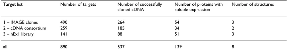
The numbers of targets and success rates grouped by the type of target cDNA clone are summarised in Table 1.
Generation and characterisation of expression clones We established a common cloning strategy that allows for easy shuttling of cDNA fragments between different E. coli
and yeast vectors. We adopted a cloning system that adds only a minimal number of extra amino acids to the pro-tein of interest and therefore decided to clone with restric-tion enzymes instead of using alternative systems, such as Invitrogen's Gateway system or ligation independent cloning [42].
The PSF has been working with more than a thousand tar-get proteins to date. Suitable cDNA clones were selected and subcloned into the E. coli expression vectors pQTEV GenBank AY243506 and pQStrep2 AY028642[16]. These vectors provide for an N-terminal His-tag; pQStrep2 also encodes an C-terminal Strep-tag-II. Some proteins have also been expressed as GST fusion proteins using the vec-tors pGEX-4T2 or pGEX-6P1 (Amersham Biosciences).
Expression of protein coding genes from multiple trans-formants per target was tested under multiple conditions. Standardisation and automation was introduced to achieve this throughput. Expression clones were charac-terised by small scale protein synthesis at different tem-peratures, 37°C, 30°C and 25°C, in 1 ml volumes in deep-96-well microplates. Proteins were purified in paral-lel by a pipetting robot, as described previously [43]. 10% of the purified protein eluate from a 1 ml culture was ana-lysed by SDS-PAGE. For each protein expression experi-ment, the size of the expression product was recorded and the amount of protein was classified into four categories: none, weak, moderate and strong expression. This classifi-cation is arbitrary to a certain degree, however, we found it sufficient to select suitable clones for protein produc-tion scale-up.
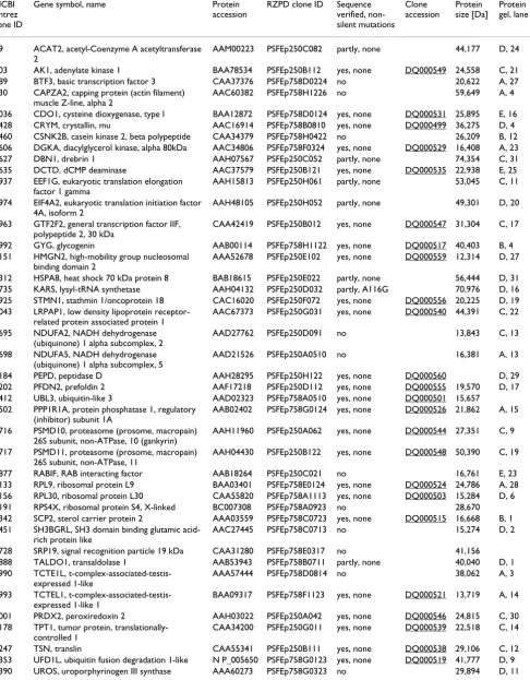
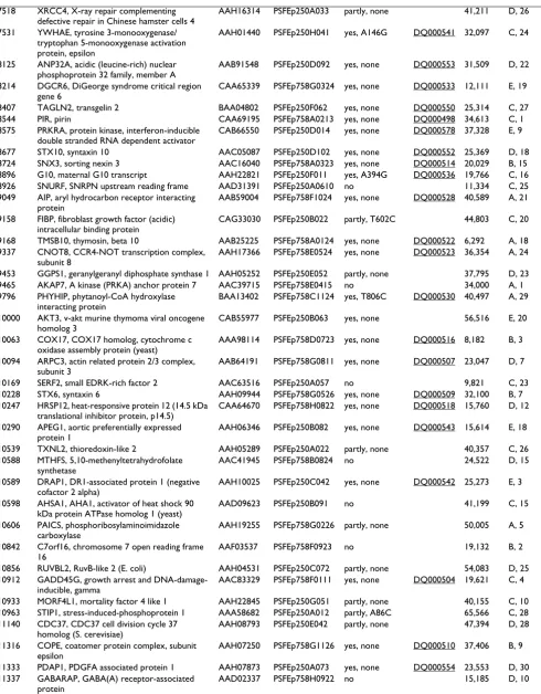
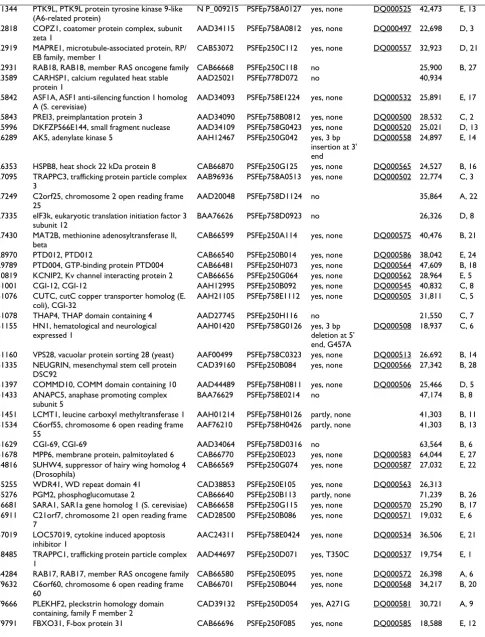
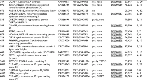
1414 clones for 537 target proteins were successfully cloned in E. coli expression vectors, with 473, 191 and 94 target proteins corresponding to target lists one (IMAGE clones), two (DKFZ clones) and three (hEx1 clones), respectively. Clones for 139 different target proteins were found to be expressed in soluble form by E. coli. Figure 2 and Table 2 show the result of small scale expression and purification of these proteins. The yield varied signifi-cantly among different target proteins. The Additional file 1 psfClones.xml contains further details on the expression clones, such as vector, strain and helper plasmid for over-expression of rare tRNAs.
Biophysical properties of proteins which could be expressed in soluble form in E. coli were compared against all tested proteins. We found no significant correlation between expression success and either protein length or mean net charge (data not shown). However, when ana-lysing the mean hydrophobicity, we found that hydro-phobic proteins are less likely to be expressed in soluble form. Only one of 139 well expressed proteins has a mean hydrophobicity of more than 0.2, while 8% of the other proteins are above this value. This group of proteins does not contain transmembrane helices according to
Table 1: Origin of template cDNA clones Numbers of targets grouped by type of template cDNA clone
Target list Number of targets Number of successfully cloned cDNA
Number of proteins with soluble expression
Number of structures
1 – IMAGE clones 490 264 54 3
2 – cDNA consortium 259 185 34 2
3 – hEx1 library 141 88 51 3
SDS-PAGE of purified human proteins
Figure 2
SDS-PAGE of purified human proteins. 15% SDS-PAGE (Coomassie-stained) of proteins expressed in small scale in E. coli and purified by automated immobilised metal chelate affinity chromatography as described in [43]. The identities of the purified proteins are indicated in Table 2. Protein expression was induced at the temperature that is optimal for the individual clone. These temperatures are listed in the supplementary file psfClones.xml. M: Molecular weight marker.
A
B
C
E
D
M 1 2 3 4 5 M 6 7 8 9 10 11 12 13 M14 15 16 17 18 19 20 21 22 23 24 25 26 27 28 29
M 1 2 3 4 5 M 6 7 8 9 10 11 12 13 14 15 M16 17 18 19 20 21 22 23 24 25 26 27 28 29
M 1 2 3 4 5 6 7 M 8 9 10 11 12 13 14 15 M16 17 18 19 20 21 22 23 24 25 26 27 28 29 30 31
M 1 2 3 4 5 6 M 7 8 9 10 11 12 13 14 15 M16 17 18 19 20 21 22 23 24 25 26 27 28 29 30 31
M 1 2 3 4 5 M 6 7 8 9 10 M11 12 13 14 15 M16 17 18 19 20 21 22 23 24 25 26 27
Table 2: PSF E. coli expression clones with soluble expression products. The table corresponds to the proteins shown in Figure 2. More detailed information is available in the supplementary XML file, Additional file 1.
NCBI Entrez gene ID
Gene symbol, name Protein accession
RZPD clone ID Sequence verified, non-silent mutations
Clone accession
Protein size [Da]
Protein gel, lane
39 ACAT2, acetyl-Coenzyme A acetyltransferase 2
AAM00223 PSFEp250C082 partly, none 44,177 D, 24
203 AK1, adenylate kinase 1 BAA78534 PSFEp250B112 yes, none DQ000549 24,558 C, 21 689 BTF3, basic transcription factor 3 CAA37376 PSFEp758D0224 no 20,622 A, 27 830 CAPZA2, capping protein (actin filament)
muscle Z-line, alpha 2
AAC60382 PSFEp758H1226 no 59,649 A, 4
1036 CDO1, cysteine dioxygenase, type I BAA12872 PSFEp758D0124 yes, none DQ000531 25,895 E, 16 1428 CRYM, crystallin, mu AAC16914 PSFEp758B0810 yes, none DQ000499 36,275 D, 4 1460 CSNK2B, casein kinase 2, beta polypeptide CAA34379 PSFEp758H0422 no 26,209 B, 12 1606 DGKA, diacylglycerol kinase, alpha 80kDa AAC34806 PSFEp758F0324 yes, none DQ000529 16,408 A, 23 1627 DBN1, drebrin 1 AAH07567 PSFEp250C052 partly, none 74,354 C, 31 1635 DCTD, dCMP deaminase AAC37579 PSFEp250B121 yes, none DQ000535 22,938 E, 25 1937 EEF1G, eukaryotic translation elongation
factor 1 gamma
AAH15813 PSFEp250H061 partly, none 53,045 C, 11
1974 EIF4A2, eukaryotic translation initiation factor 4A, isoform 2
AAH48105 PSFEp250H052 partly, none 49,301 D, 20
2963 GTF2F2, general transcription factor IIF, polypeptide 2, 30 kDa
CAA42419 PSFEp250B012 yes, none DQ000547 31,304 C, 17
2992 GYG, glycogenin AAB00114 PSFEp758H1122 yes, none DQ000517 40,403 B, 4 3151 HMGN2, high-mobility group nucleosomal
binding domain 2
AAA52678 PSFEp250E102 yes, none DQ000559 12,314 D, 27
3312 HSPA8, heat shock 70 kDa protein 8 BAB18615 PSFEp250E022 partly, none 56,444 D, 31 3735 KARS, lysyl-tRNA synthetase AAH04132 PSFEp250D032 partly, A116G 70,976 D, 16 3925 STMN1, stathmin 1/oncoprotein 18 CAC16020 PSFEp250F072 yes, none DQ000556 20,225 D, 19 4043 LRPAP1, low density lipoprotein
receptor-related protein associated protein 1
AAC67373 PSFEp250G031 yes, none DQ000540 44,391 C, 22
4695 NDUFA2, NADH dehydrogenase (ubiquinone) 1 alpha subcomplex, 2
AAD27762 PSFEp250D091 no 13,843 C, 13
4698 NDUFA5, NADH dehydrogenase (ubiquinone) 1 alpha subcomplex, 5
AAD21526 PSFEp250A0510 no 16,381 A, 13
5184 PEPD, peptidase D AAH28295 PSFEp250H122 yes, none DQ000560 D, 29 5202 PFDN2, prefoldin 2 AAF17218 PSFEp250D112 yes, none DQ000555 19,570 D, 17 5412 UBL3, ubiquitin-like 3 AAD02323 PSFEp758A0510 yes, none DQ000501 15,657
5502 PPP1R1A, protein phosphatase 1, regulatory (inhibitor) subunit 1A
AAB02402 PSFEp758G0124 yes, none DQ000526 21,862 A, 15
5716 PSMD10, proteasome (prosome, macropain) 26S subunit, non-ATPase, 10 (gankyrin)
AAH11960 PSFEp250A062 yes, none DQ000544 27,351 C, 9
5717 PSMD11, proteasome (prosome, macropain) 26S subunit, non-ATPase, 11
AAH04430 PSFEp250B122 yes, none DQ000548 50,390 C, 19
5877 RABIF, RAB interacting factor AAB18264 PSFEp250C021 no 16,761 E, 23 6133 RPL9, ribosomal protein L9 BAA03401 PSFEp758E0124 yes, none DQ000524 24,786 A, 28 6156 RPL30, ribosomal protein L30 CAA55820 PSFEp758A1113 yes, none DQ000503 15,284 D, 6 6191 RPS4X, ribosomal protein S4, X-linked BC007308 PSFEp758A0923 no 28,670 6342 SCP2, sterol carrier protein 2 AAA03559 PSFEp758C0723 yes, none DQ000515 16,668 B, 1 6451 SH3BGRL, SH3 domain binding glutamic
acid-rich protein like
AAC27445 PSFEp758C0713 no 15,274 D, 2
6728 SRP19, signal recognition particle 19 kDa CAA31280 PSFEp758E0317 no 41,156 6888 TALDO1, transaldolase 1 AAB53943 PSFEp758B0711 partly, none 40,040 D, 1 6990 TCTE1L,
t-complex-associated-testis-expressed 1-like
AAA57444 PSFEp758D0814 no 38,062 A, 3
6993 TCTEL1, t-complex-associated-testis-expressed 1-like 1
BAA09317 PSFEp758F1123 yes, none DQ000521 13,719 A, 14
7001 PRDX2, peroxiredoxin 2 AAH03022 PSFEp250A042 yes, none DQ000546 24,815 C, 30 7178 TPT1, tumor protein,
translationally-controlled 1
CAA34200 PSFEp250G011 yes, none DQ000539 22,518 C, 14
7518 XRCC4, X-ray repair complementing defective repair in Chinese hamster cells 4
AAH16314 PSFEp250A033 partly, none 41,211 D, 26
7531 YWHAE, tyrosine 3-monooxygenase/ tryptophan 5-monooxygenase activation protein, epsilon
AAH01440 PSFEp250H041 yes, A146G DQ000541 32,097 C, 24
8125 ANP32A, acidic (leucine-rich) nuclear phosphoprotein 32 family, member A
AAB91548 PSFEp250D092 yes, none DQ000553 31,509 D, 22
8214 DGCR6, DiGeorge syndrome critical region gene 6
CAA65339 PSFEp758G0324 yes, none DQ000533 12,111 E, 19
8407 TAGLN2, transgelin 2 BAA04802 PSFEp250F062 yes, none DQ000550 25,314 C, 27 8544 PIR, pirin CAA69195 PSFEp758A0213 yes, none DQ000498 34,613 C, 1 8575 PRKRA, protein kinase, interferon-inducible
double stranded RNA dependent activator
CAB66550 PSFEp250D014 yes, none DQ000578 37,328 E, 9
8677 STX10, syntaxin 10 AAC05087 PSFEp250D102 yes, none DQ000552 25,369 D, 18 8724 SNX3, sorting nexin 3 AAC16040 PSFEp758A0323 yes, none DQ000514 20,029 B, 15 8896 G10, maternal G10 transcript AAH22821 PSFEp250F011 yes, A394G DQ000536 19,766 C, 16 8926 SNURF, SNRPN upstream reading frame AAD31391 PSFEp250A0610 no 11,334 C, 25 9049 AIP, aryl hydrocarbon receptor interacting
protein
AAB59004 PSFEp758F1024 yes, none DQ000528 40,589 A, 21
9158 FIBP, fibroblast growth factor (acidic) intracellular binding protein
CAG33030 PSFEp250B022 partly, T602C 44,803 C, 20
9168 TMSB10, thymosin, beta 10 AAB25225 PSFEp758A0124 yes, none DQ000522 6,292 A, 18 9337 CNOT8, CCR4-NOT transcription complex,
subunit 8
AAH17366 PSFEp758E0524 yes, none DQ000523 36,354 A, 24
9453 GGPS1, geranylgeranyl diphosphate synthase 1 AAH05252 PSFEp250E052 partly, none 37,795 D, 23 9465 AKAP7, A kinase (PRKA) anchor protein 7 AAC39715 PSFEp758E0415 no 34,000 A, 1 9796 PHYHIP, phytanoyl-CoA hydroxylase
interacting protein
BAA13402 PSFEp758C1124 yes, T806C DQ000530 40,497 A, 29
10000 AKT3, v-akt murine thymoma viral oncogene homolog 3
CAB55977 PSFEp250B063 yes, none 56,516 E, 20
10063 COX17, COX17 homolog, cytochrome c oxidase assembly protein (yeast)
AAA98114 PSFEp758D0723 yes, none DQ000516 8,182 B, 3
10094 ARPC3, actin related protein 2/3 complex, subunit 3
AAB64191 PSFEp758G0811 yes, none DQ000507 23,047 D, 7
10169 SERF2, small EDRK-rich factor 2 AAC63516 PSFEp250A057 no 9,821 C, 23 10228 STX6, syntaxin 6 AAH09944 PSFEp758G0526 yes, none DQ000509 32,100 B, 7 10247 HRSP12, heat-responsive protein 12 (14.5 kDa
translational inhibitor protein, p14.5)
CAA64670 PSFEp758H0822 yes, none DQ000518 15,760 D, 12
10290 APEG1, aortic preferentially expressed protein 1
AAH06346 PSFEp250B082 yes, none DQ000543 15,614 E, 18
10539 TXNL2, thioredoxin-like 2 AAH05289 PSFEp250A022 partly, none 40,357 C, 26 10588 MTHFS, 5,10-methenyltetrahydrofolate
synthetase
AAC41945 PSFEp758B0824 no 24,522 D, 15
10589 DRAP1, DR1-associated protein 1 (negative cofactor 2 alpha)
AAH10025 PSFEp250C042 yes, none DQ000542 25,273 E, 3
10598 AHSA1, AHA1, activator of heat shock 90 kDa protein ATPase homolog 1 (yeast)
AAD09623 PSFEp250B091 no 41,199 C, 15
10606 PAICS, phosphoribosylaminoimidazole carboxylase
AAH19255 PSFEp758G0226 partly, none 50,005 A, 5
10842 C7orf16, chromosome 7 open reading frame 16
AAF03537 PSFEp758F0923 no 19,132 B, 2
10856 RUVBL2, RuvB-like 2 (E. coli) AAH04531 PSFEp250C072 partly, none 54,083 D, 25 10912 GADD45G, growth arrest and
DNA-damage-inducible, gamma
AAC83329 PSFEp758F0111 yes, none DQ000504 19,621 C, 4
10933 MORF4L1, mortality factor 4 like 1 AAH22845 PSFEp250G051 partly, none 40,155 C, 10 10963 STIP1, stress-induced-phosphoprotein 1 AAA58682 PSFEp250A012 partly, A86C 65,566 C, 28 11140 CDC37, CDC37 cell division cycle 37
homolog (S. cerevisiae)
AAH08793 PSFEp250E042 partly, none 47,394 D, 28
11316 COPE, coatomer protein complex, subunit epsilon
AAH07250 PSFEp758G1126 yes, none DQ000510 37,406 B, 9
11333 PDAP1, PDGFA associated protein 1 AAH07873 PSFEp250A073 yes, none DQ000554 23,553 D, 30 11337 GABARAP, GABA(A) receptor-associated
protein
11344 PTK9L, PTK9L protein tyrosine kinase 9-like (A6-related protein)
N P_009215 PSFEp758A0127 yes, none DQ000525 42,473 E, 13
22818 COPZ1, coatomer protein complex, subunit zeta 1
AAD34115 PSFEp758A0812 yes, none DQ000497 22,698 D, 3
22919 MAPRE1, microtubule-associated protein, RP/ EB family, member 1
CAB53072 PSFEp250C112 yes, none DQ000557 32,923 D, 21
22931 RAB18, RAB18, member RAS oncogene family CAB66668 PSFEp250C118 no 25,900 B, 27 23589 CARHSP1, calcium regulated heat stable
protein 1
AAD25021 PSFEp778D072 no 40,934
25842 ASF1A, ASF1 anti-silencing function 1 homolog A (S. cerevisiae)
AAD34093 PSFEp758E1224 yes, none DQ000532 25,891 E, 17
25843 PREI3, preimplantation protein 3 AAD34090 PSFEp758B0812 yes, none DQ000500 28,532 C, 2 25996 DKFZP566E144, small fragment nuclease AAD34109 PSFEp758G0423 yes, none DQ000520 25,021 D, 13 26289 AK5, adenylate kinase 5 AAH12467 PSFEp250G042 yes, 3 bp
insertion at 3' end
DQ000558 24,897 E, 14
26353 HSPB8, heat shock 22 kDa protein 8 CAB66870 PSFEp250G125 yes, none DQ000565 24,527 B, 16 27095 TRAPPC3, trafficking protein particle complex
3
AAB96936 PSFEp758A0513 yes, none DQ000502 22,774 C, 3
27249 C2orf25, chromosome 2 open reading frame 25
AAD20048 PSFEp758D1124 no 35,864 A, 22
27335 eIF3k, eukaryotic translation initiation factor 3 subunit 12
BAA76626 PSFEp758D0923 no 26,326 D, 8
27430 MAT2B, methionine adenosyltransferase II, beta
CAB66599 PSFEp250A114 yes, none DQ000575 40,476 B, 21
28970 PTD012, PTD012 CAB66540 PSFEp250B014 yes, none DQ000586 38,042 E, 24 29789 PTD004, GTP-binding protein PTD004 CAB66481 PSFEp250H073 yes, none DQ000564 47,609 B, 18 30819 KCNIP2, Kv channel interacting protein 2 CAB66656 PSFEp250G064 yes, none DQ000562 28,964 E, 5 51001 CGI-12, CGI-12 AAH12995 PSFEp250B092 yes, none DQ000545 40,832 C, 8 51076 CUTC, cutC copper transporter homolog (E.
coli), CGI-32
AAH21105 PSFEp758E1112 yes, none DQ000505 31,811 C, 5
51078 THAP4, THAP domain containing 4 AAD27745 PSFEp250H116 no 21,550 C, 7 51155 HN1, hematological and neurological
expressed 1
AAH01420 PSFEp758G0126 yes, 3 bp deletion at 5' end, G457A
DQ000508 18,937 C, 6
51160 VPS28, vacuolar protein sorting 28 (yeast) AAF00499 PSFEp758C0323 yes, none DQ000513 26,692 B, 14 51335 NEUGRIN, mesenchymal stem cell protein
DSC92
CAD39160 PSFEp250B084 yes, none DQ000566 27,342 B, 28
51397 COMMD10, COMM domain containing 10 AAD44489 PSFEp758H0811 yes, none DQ000506 25,466 D, 5 51433 ANAPC5, anaphase promoting complex
subunit 5
BAA76629 PSFEp758E0214 no 47,174 B, 8
51451 LCMT1, leucine carboxyl methyltransferase 1 AAH01214 PSFEp758H0126 partly, none 41,303 B, 11 51534 C6orf55, chromosome 6 open reading frame
55
AAF76210 PSFEp758H0426 partly, none 41,303 B, 13
51629 CGI-69, CGI-69 AAD34064 PSFEp758D0316 no 63,564 B, 6
51678 MPP6, membrane protein, palmitoylated 6 CAB66770 PSFEp250E023 yes, none DQ000583 64,044 E, 27 54816 SUHW4, suppressor of hairy wing homolog 4
(Drosophila)
CAB66569 PSFEp250G074 yes, none DQ000587 27,032 E, 22
55255 WDR41, WD repeat domain 41 CAD38853 PSFEp250E105 yes, none DQ000563 26,313
55276 PGM2, phosphoglucomutase 2 CAB66640 PSFEp250B113 partly, none 71,239 B, 26 56681 SARA1, SAR1a gene homolog 1 (S. cerevisiae) CAB66658 PSFEp250G115 yes, none DQ000570 25,290 B, 17 56911 C21orf7, chromosome 21 open reading frame
7
CAD28500 PSFEp250B086 yes, none DQ000571 19,032 E, 6
57019 LOC57019, cytokine induced apoptosis inhibitor 1
AAC24311 PSFEp758E0424 yes, none DQ000534 36,506 E, 21
58485 TRAPPC1, trafficking protein particle complex 1
AAD44697 PSFEp250D071 yes, T350C DQ000537 19,754 E, 1
64284 RAB17, RAB17, member RAS oncogene family CAB66580 PSFEp250E095 yes, none DQ000572 26,398 A, 6 79632 C6orf60, chromosome 6 open reading frame
60
CAB66701 PSFEp250B044 yes, none DQ000568 34,217 B, 20
79666 PLEKHF2, pleckstrin homology domain containing, family F member 2
CAD39132 PSFEp250D054 yes, A271G DQ000581 30,721 A, 9
TMHMM, and therefore may represent peripheral mem-brane proteins with hydrophobic surface regions.
The E. coli expression clones of the PSF are publicly avail-able from the RZPD German Resource Center [44]. The Additional file 1 is an XML list of these clones. It can be viewed in a web browser (Figure 3). The Additional file contains, for each clone:
• Gene ID and name,
• Accession number,
• Cloning details,
• Strain and vector,
• Expected sequence,
• Protein expression results,
• Sequence verification.
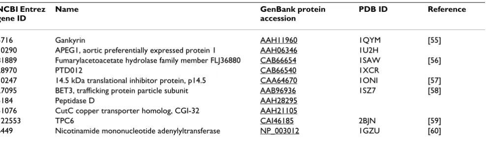
Solved structures
As a result of the target selection and cloning described in this paper, ten novel X-ray structures of human proteins were determined (Table 3). The structure of one protein, TRAPPC3/BET-3, was determined after protein expression in S. cerevisae, while the other proteins were produced in
E. coli.
Discussion
We describe here the strategies and experiments of our structural genomics project on human proteins. In addi-tion to the expression of full length proteins, the Protein Structure Factory has also studied protein domains by NMR spectroscopy, which has been described elsewhere [45-47]. Our selection of full length target proteins was mainly determined by the availability of full length cDNA clones. In addition, biophysical and bioinformatical crite-ria were applied, leading to a biased selection of target proteins from the human proteome. Therefore, we expect that the percentage of proteins that we could express and purify in soluble form, 18%, is higher than it would be in a randomly selected set. The low proportion of success-fully expressed proteins indicates that E. coli is not the appropriate expression host for many full length human proteins. High throughput protein expression in
alterna-80347 COASY, Coenzyme A synthase AAF87955 PSFEp250A053 yes, none DQ000551 33,147 C, 29 80895 ILKAP, integrin-linked kinase-associated
serine/threonine phosphatase 2C
CAB66784 PSFEp250D083 yes, none DQ000569 45,832 B, 19
81876 RAB1B, RAB1B, member RAS oncogene family CAB66570 PSFEp250C128 no 25,094 A, 12 81889 DKFZP566J2046, fumarylacetoacetate
hydrolase domain containing 1
CAB66654 PSFEp250B074 yes, none DQ000567 27,766 B, 24
83538 DKFZP434H0115, hypothetical protein DKFZp434H0115
CAB66694 PSFEp250G093 partly, none 79,584 E, 11
83543 C9orf58, chromosome 9 open reading frame 58
CAB66501 PSFEp250B085 yes, none DQ000573 19,990 B, 25
83667 SESN2, sestrin 2 CAB66486 PSFEp250F043 yes, none DQ000576 57,420 E, 7 84072 NOHMA, HORMA domain containing protein CAB66689 PSFEp250F083 yes, none DQ000561 47,343 E, 4 84324 CIP29, cytokine induced protein 29 kDa CAC37950 PSFEp758H1026 yes, none DQ000512 26,594 A, 2 84457 PHYHIPL, phytanoyl-CoA hydroxylase
interacting protein-like
CAD39006 PSFEp250A084 yes, none DQ000579 45,425 E, 10
84557 MAP1LC3A, microtubule-associated protein 1 light chain 3 alpha
CAD38714 PSFEp250E106 yes, none DQ000584 17,194 E, 26
91603 MGC20398, hypothetical protein MGC20398 BAB70992 PSFEp758H0526 yes, none DQ000511 44,915 B, 10 94240 EPSTI1, epithelial stromal interaction 1
(breast)
CAD38599 PSFEp250C015 yes, none DQ000580 25,527 A, 11
112611 RWDD2, RWD domain containing 2 CAB52345 PSFEp758A1024 partly, T709C 35,159 B, 5 118812 C10orf83, chromosome 10 open reading
frame 83
CAD38849 PSFEp250H085 yes, none DQ000574 19,158 B, 23
122060 FLJ30046, hypothetical protein FLJ30046 CAD38891 PSFEp250E015 yes, none DQ000577 23,468 E, 8 136319 MTPN, myotrophin CAD38909 PSFEp250D016 yes, none DQ000582 15,817 A, 7 140856 C20orf79, chromosome 20 open reading
frame 79
tive system such as yeast [9-11] or insect cells/baculovirus [48] has been established and will lead to better success rates in future projects.
Generally, clones that did express a soluble protein were verified by DNA sequencing, while clones that did not express or expressed an insoluble product were usually not sequence verified. It cannot be ruled out that some of the unsuccessful clones contain sequence errors intro-duced during cloning. Since template cDNA clones of the IMAGE consortium with only partial sequence informa-tion were used for most cloning experiments, expression clones that were not sequence verified might represent splice variants or isoforms of the original target. The dis-tribution of mean net charge and length was similar among successfully expressed and all proteins, while very hydrophobic proteins were generally not expressed well in our E. coli expression system.
Future efforts in structural genomics of mammalian pro-teins will benefit from a much better supply of full length cDNA clones. Clones prepared for protein expression by resource centres and commercial suppliers are becoming available now. With such resources, alternative target selection strategies will become feasible that will not be
restricted by the availability of cDNA clones. Instead, all potential target proteins, including splice variants, could be clustered by similarity and the most suitable members of each cluster could be selected by appropriate criteria as outlined in the Background section.
In our approach, we have excluded certain types of pro-teins such as membrane propro-teins and very large propro-teins. A structural genomics approach that includes membrane proteins would require standard protocols to optimise expression conditions and detergents [49]. The best strat-egy to study large proteins is to divide them into domains and smaller regions. However, such smaller constructs usually have to be designed manually.
All clones listed in the supplementary file (Additional file 1) and Table 2 are available to the research community. Thereby we hope to facilitate further functional character-isation of this set of human proteins.
Materials and Methods
Cloning with restriction enzymescDNA inserts were amplified by PCR primers carrying tails with BamHI and NotI sites and cloned into the respective sites of one of the expression vectors pQTEV, pQStrep2 or pGEX-6p1. This had the drawback that the restriction sites chosen for cloning might occur in the insert. In such cases, compatible overhangs were produced by alternative enzymes or by the hetero-stagger cloning method [50]. Alternative enzymes are BglII for BamHI and the type IIs enzymes BpiI, Eco31I, Esp3I, which can replace both
BamHI and NotI. Type IIs enzymes cut outside their recog-nition sequence and can produce arbitrary overhangs.
PCR Primer design
PCR primers with tails carrying restriction enzyme cleav-age sites were designed automatically by a Perl program. The primer design program adjusts the length of the prim-ers to achieve a melting temperature close to a common default. Then, restriction enzymes that do not cut within the respective cDNA sequence are selected by the program and restriction enzyme sites are attached to the primer sequences. Finally, since restriction enzymes do not cut well at the very end of a DNA molecule, an additional short nucleotide tail is automatically attached to the prim-ers. The sequence of this tail is optimised to minimise for-mation of secondary structure, hairpins or dimerisation. A Java version of the primer design software, 'ORFprimer', is publicly available [51].
Automated high-throughput cloning, protein expression and purification
PCR primers and cDNA clones were delivered in 96-well microplate format. Upon delivery, plates with PCR prim-ers and template clones were reformatted to obtain
corre-The supplementary XML Additional file 1 file displayed in a web browser
Figure 3
sponding plate positions by a Zinsser Speedy pipetting robot. The PCR master mix (Roche Expand) and cDNA primers (10 µM stocks) were pipetted into a PCR micro-plate with a multichannel pipet. Temmicro-plate clone bacteria were added with a 96-pin steel replicating device from overnight cultures in microtitre plates. PCR product size and yield was determined by agarose gel electrophoresis and the software Phoretix 1D Quantifier (Nonlinear dynamics). PCR products were purified with magnetic beads on the pipetting robot with a system that has been developed at the Max Planck Institute of Molecular Genet-ics in collaboration with Bruker DaltonGenet-ics (Bruker genop-ure kit). The correct restriction enzyme master mixes were automatically added and the digested fragments were purified again, analysed by agarose gel electrophoresis and quantified. The robot then adjusted DNA concentrations to a common default by dilution. Liga-tions were set up manually with a multichannel pipet, and SCS1 E. coli cells carrying pRARE were transformed in a PCR microplate by chemical transformation on a PCR machine [52]. Transformed cells were manually plated on individual agar plates. Four clones were picked per trans-formation and were checked by PCR using vector primers.
E. coli expression clones were ready for protein expression at this stage.
Primer sequences and template clones for cloning of the target cDNAs are listed in the supplementary XML file, Additional file 1.
The characterisation of expression clones by parallel expression and protein purification is described in refer-ence [43].
Sequence analysis
Sequence analysis software was run with default settings unless indicated otherwise. The mean charge of a protein was calculated as the difference of the number of positive
and negatively charged amino acids (Lys, Arg and Glu, Asp, respectively) divided by the protein length. The mean hydrophobicity was calculated with the Kyte and Doolit-tle hydropathy index [53], obtained from the EMBOSS package [54]. The index values were added up for a given protein and divided by the protein length.
Authors' contributions
• Study design and coordination: KB, HL, UHe
• Bioinformatics: UHa, BS, JS, PB, VS, KB
• E. coli cloning: VS, KB
• E. coli protein work: CS, KB
• Manuscript, figures, XML file preparation: KB
Additional material
Acknowledgements
We wish to thank Ulf Lenski for help with sequence analysis and Janett Tischer for technical assistance. This work was funded by the German Fed-eral Ministry of Education and Research (BMBF) through the Leitprojek-tverbund Proteinstrukturfabrik and the National Genome Network NGFN Table 3: Novel human protein structures The structures of full length proteins solved by the Protein Structure Factory.
NCBI Entrez gene ID
Name GenBank protein
accession
PDB ID Reference
5716 Gankyrin AAH11960 1QYM [55]
10290 APEG1, aortic preferentially expressed protein 1 AAH06346 1U2H
81889 Fumarylacetoacetate hydrolase family member FLJ36880 CAB66654 1SAW [56]
28970 PTD012 CAB66540 1XCR
10247 14.5 kDa translational inhibitor protein, p14.5 CAA64670 1ONI [57] 27095 BET3, trafficking protein particle subunit AAB96936 1SZ7 [58]
5184 Peptidase D AAH28295
51076 CutC copper transporter homolog, CGI-32 AAH21105
122553 TPC6 CAI46185 2BJN [59]
6449 Nicotinamide mononucleotide adenylyltransferase NP_003012 1GZU [60]
Additional File 1
A ZIP archive that contains the XML list psfClones.xml of PSF clones available at the RZPD and the XSL stylesheet psfToHtml.xsl to display the XML file in a web browser. The two files should be extracted from the ZIP archive into a local directory. Then psfClones.xml can be opened with a current web browser like Mozilla or Internet Explorer. 1414 Clones for 537 target proteins are described in the XML file. It lists the gene, cloning details, expected sequence, protein expression results and the degree of sequence verification for each clone.
Click here for file
(FKZ 01GR0471, 01GR0472) and with support by the Fonds der Che-mischen Industrie to U.H. Support by the Berlin Senate and the European Fund for Regional Development (EFRE) is also gratefully acknowledged.
References
1. Heinemann U, Büssow K, Mueller U, Umbach P: Facilities and methods for the high-throughput crystal structure analysis of human proteins. Accounts of Chemical Research 2003, 36(3):157-163.
2. Protein Structure Factory [http://www.proteinstrukturfab rik.de]
3. Zhang C, Kim SH: Overview of structural genomics: from structure to function. Current Opinion in Chemical Biology 2003, 7(1):28-32.
4. Todd AE, Marsden RL, Thornton JM, Orengo CA: Progress of structural genomics initiatives: an analysis of solved target structures. Journal of Molecular Biology 2005, 348(5):1235-1260. 5. Gilbert M, Albala Joanna S: Accelerating code to function: Sizing
up the protein production line. Current Opinion in Chemical Biology 2002, 6(1):102-105.
6. Yokoyama S: Protein expression systems for structural genomics and proteomics. Current Opinion in Chemical Biology 2003, 7(1):39-43.
7. Kigawa T, Yabuki T, Yoshida Y, Tsutsui M, Ito Y, Shibata T, Yokoyama S: Cell-free production and stable-isotope labeling of milli-gram quantities of proteins. FEBS Letters 1999, 442(1):15-19. 8. Tyler RC, Aceti DJ, Bingman CA, Cornilescu CC, Fox BG, Frederick
RO, Jeon WB, Lee MS, Newman CS, Peterson FC, Phillips GNJ, Sha-han MN, Singh S, Song J, Sreenath HK, Tyler EM, Ulrich EL, Vinarov DA, Vojtik FC, Volkman BF, Wrobel RL, Zhao Q, Markley JL: Com-parison of cell-based and cell-free protocols for producing target proteins from the Arabidopsis thaliana genome for structural studies. Proteins 2005, 59:633-643.
9. Holz C, Hesse O, Bolotina N, Stahl U, Lang C: A micro-scale proc-ess for high-throughput exprproc-ession of cDNAs in the yeast Saccharomyces cerevisiae. Protein Expression & Purification 2002, 25(3):372-378.
10. Boettner M, Prinz B, Holz C, Stahl U, Lang C: High-throughput screening for expression of heterologous proteins in the yeast Pichia pastoris. Journal of Biotechnology 2002, 99(1):51-62. 11. Holz C, Prinz B, Bolotina N, Sievert V, Büssow K, Simon B, Stahl U,
Lang C: Establishing the yeast Saccharomyces cerevisiae as a system for expression of human proteins on a proteome-scale. Journal of Structural and Functional Genomics 2003, 4(2/ 3):97-108.
12. Studier FW, Rosenberg AH, Dunn JJ, Dubendorff JW: Use of T7 RNA polymerase to direct expression of cloned genes. Meth-ods in Enzymology 1990, 185:60-89.
13. Hochuli E, Dobeli H, Schacher A: New metal chelate adsorbent selective for proteins and peptides containing neighboring histidine residues. Journal of Chromatography 1987, 411:177-184. 14. Schmidt TG, Skerra A: The random peptide library-assisted
engineering of a C-terminal affinity peptide, useful for the detection and purification of a functional Ig Fv fragment. Pro-tein Engineering 1993, 6(1):109-122.
15. Schmidt TGM, Koepke J, Frank R, Skerra A: Molecular interaction between the Strep-tag affinity peptide and its cognate tar-get, streptavidin. Journal of Molecular Biology 1996, 255(5):753-766.
16. Mueller U, Büssow K, Diehl A, Bartl FJ, Niesen FH, Nyarsik L, Heine-mann U: Rapid purification and crystal structure analysis of a small protein carrying two terminal affinity tags. Journal of Structural and Functional Genomics 2004, 4:217-225.
17. Kane JF: Effects of rare codon clusters on high-level expres-sion of heterologous proteins in Escherichia coli. Current Opin-ion in Biotechnology 1995, 6(5):494-500.
18. Brinkmann U, Mattes RE, Buckel P: High-level expression of recombinant genes in Escherichia coli is dependent on the availability of the DNAY gene product. Gene 1989, 85(1):109-114.
19. Novy R, Drott D, Yaeger K, Mierendorf R: Overcoming the codon bias of E.coli for enhanced protein expression. inNovations 2001, 12:1-3.
20. Krogh A, Larsson B, von Heijne G, Sonnhammer ELL: Predicting transmembrane protein topology with a hidden Markov
model: Application to complete genomes. Journal of Molecular Biology 2001, 305(3):567-580.
21. TMHMM [http://www.cbs.dtu.dk/services/TMHMM/]
22. Dyson HJ, Wright PE: Coupling of folding and binding for unstructured proteins. Current Opinion in Structural Biology 2002, 12(1):54-60.
23. Wootton JC, Federhen S: Analysis of compositionally biased regions in sequence databases. Methods in Enzymology 1996, 266:554-571.
24. Wootton JC, Federhen S: Statistics of local complexity in amino acid sequences and sequence databases. Computers & Chemistry 1993, 17:149-163.
25. Wootton JC: Non-globular domains in protein sequences: automated segmentation using complexity measures. Com-puters & Chemistry 1994, 18(3):269-285.
26. COILS [http://www.russell.embl.de/cgi-bin/coils-svr.pl]
27. Lupas A, Van Dyke M, Stock J: Predicting coiled coils from pro-tein sequences. Science 1991, 252(5010):1162-1164.
28. Lupas A: Prediction and analysis of coiled-coil structures. Methods in Enzymology 1996, 266:513-525.
29. Eisenhaber F, Bork P: Wanted: Subcellular localization of pro-teins based on sequence. Trends in Cell Biology 1998, 8(4):169-170. 30. Meta_Annotator [http://mendel.imp.univie.ac.at/aat/
CellLoc_MetaA]
31. Berman HM, Battistuz T, Bhat TN, Bluhm WF, Bourne PE, Burkhardt K, Feng Z, Gilliland GL, Iype L, Jain S, Fagan P, Marvin J, Padilla D, Rav-ichandran V, Schneider B, Thanki N, Weissig H, Westbrook JD, Zard-ecki C: The protein data bank. Acta Crystallographica Section D 2002, 58(Pt 6 Pt 1):899-907.
32. Altschul SF, Madden TL, Schaeffer AA, Zhang J, Zhang Z, Miller W, Lipman DJ: Gapped BLAST and PSI-BLAST: A new genera-tion of protein database search programs. Nucleic Acids Research 1997, 25(17):3389-3402.
33. Lennon G, Auffray C, Polymeropoulos M, Soares MB: The I.M.A.G.E. consortium: an integrated molecular analysis of genomes and their expression. Genomics 1996, 33:151-152. 34. dbEST [http://www.ncbi.nlm.nih.gov/dbEST]
35. Bannasch D, Mehrle A, Glatting KH, Pepperkok R, Poustka A, Wie-mann S: LIFEdb: a database for functional genomics experi-ments integrating information from external sources, and serving as a sample tracking system. Nucleic Acids Research 2004, 32(Special Issue SI):D505-D508.
36. Wiemann S, Weil B, Wellenreuther R, Gassenhuber J, Glassl S, Ansorge W, Bocher M, Blocker H, Bauersachs S, Blum H, Lauber J, Dusterhoft A, Beyer A, Kohrer K, Strack N, Mewes HW, Ottenwal-der B, Obermaier B, Tampe J, Heubner D, Wambutt R, Korn B, Klein M, Poustka A: Toward a catalog of human genes and proteins: sequencing and analysis of 500 novel complete protein cod-ing human cDNAs. Genome Research 2001, 11(3):422-435. 37. Büssow K, Nordhoff E, Lübbert C, Lehrach H, Walter G: A human
cDNA library for high-throughput protein expression screening. Genomics 2000, 65(1):1-8.
38. Büssow K, Cahill D, Nietfeld W, Bancroft D, Scherzinger E, Lehrach H, Walter G: A method for global protein expression and anti-body screening on high-density filters of an arrayed cDNA library. Nucleic Acids Research 1998, 26(21):5007-5008.
39. Büssow K, Quedenau C, Sievert V, Tischer J, Scheich C, Seitz H, Hieke B, Niesen FH, Götz F, Harttig U, Lehrach H: A catalogue of human cDNA expression clones and its application to struc-tural genomics. Genome Biology 2004, 5:R71.
40. The hEx1 Database [http://www.proteinstrukturfabrik.de/hex1] 41. Hubbard T, Barker D, Birney E, Cameron G, Chen Y, Clark L, Cox T,
Cuff J, Curwen V, Down T, Durbin R, Eyras E, Gilbert J, Hammond M, Huminiecki L, Kasprzyk A, Lehvaslaiho H, Lijnzaad P, Melsopp C, Mongin E, Pettett R, Pocock M, Potter S, Rust A, Schmidt E, Searle S, Slater G, Smith J, Spooner W, Stabenau A, Stalker J, Stupka E, Ureta-Vidal A, Vastrik I, Clamp M: The Ensembl genome database project. Nucleic Acids Research 2002, 30(1):38-41.
42. Aslanidis C, de Jong PJ: Ligation-independent cloning of PCR products (LIC-PCR). Nucleic Acids Research 1990, 18(20):6069-6074.
43. Scheich C, Sievert V, Büssow K: An automated method for high-throughput protein purification applied to a comparison of His-tag and GST-tag affinity chromatography. BMC Biotechnology 2003, 3:12.
Publish with BioMed Central and every scientist can read your work free of charge "BioMed Central will be the most significant development for disseminating the results of biomedical researc h in our lifetime."
Sir Paul Nurse, Cancer Research UK
Your research papers will be:
available free of charge to the entire biomedical community
peer reviewed and published immediately upon acceptance
cited in PubMed and archived on PubMed Central
yours — you keep the copyright
Submit your manuscript here:
http://www.biomedcentral.com/info/publishing_adv.asp
BioMedcentral 45. Scheich C, Leitner D, Sievert V, Leidert M, Schlegel B, Simon B,
Letu-nic I, Büssow K, Diehl A: Fast identification of folded human protein domains expressed in E. coli suitable for structural analysis. BMC Structural Biology 2004, 4:4.
46. Brockmann C, Leitner D, Labudde D, Diehl A, Sievert V, Büssow K, Kühne R, Oschkinat H: The solution structure of the SODD BAG domain reveals additional electrostatic interactions in the HSP70 complexes of SODD subfamily BAG domains. FEBS Letters 2004, 558(1-3):101-106.
47. Soukenik M, Diehl A, Leidert M, Sievert V, Büssow K, Leitner D, Labudde D, Ball LJ, Lechner A, Nagler DK, Oschkinat H: The SEP domain of p47 acts as a reversible competitive inhibitor of cathepsin L. FEBS Letters 2004, 576(3):358-362.
48. McCall EJ, Danielsson A, Hardern IM, Dartsch C, Hicks R, Wahlberg JM, Abbott WM: Improvements to the throughput of recom-binant protein expression in the baculovirus/insect cell system. Protein Expr Purif 2005, 42(1):29-36.
49. Eshaghi S, Hedren M, Nasser MI, Hammarberg T, Thornell A, Nord-lund P: An efficient strategy for high-throughput expression screening of recombinant integral membrane proteins. Pro-tein Sci 2005, 14(3):676-683.
50. Li C, Evans RM: Ligation independent cloning irrespective of restriction site compatibility. Nucleic Acids Research 1997, 25(20):4165-4166.
51. ORFprimer [http://www.proteinstrukturfabrik.de/ORFprimer] 52. Inoue H, Nojima H, Okayama H: High efficiency transformation
of Escherichia coli with plasmids. Gene 1990, 96(1):23-28. 53. Kyte J, Doolittle RF: A simple method for displaying the
hydro-pathic character of a protein. Journal of Molecular Biology 1982, 157(1):105-132.
54. Rice P, Longden I, Bleasby A: EMBOSS: the European Molecular Biology Open Software Suite. Trends in Genetics 2000, 16(6):276-277.
55. Manjasetty BA, Quedenau C, Sievert V, Büssow K, Niesen F, Del-brück H, Heinemann U: X-ray structure of human gankyrin, the product of a gene linked to hepatocellular carcinome. Pro-teins: Structure, Function and Genetics 2004, 55(1):214-217.
56. Manjasetty BA, Niesen FH, Delbruck H, Gotz F, Sievert V, Bussow K, Behlke J, Heinemann U: X-ray structure of fumarylacetoacetate hydrolase family member homo sapiens FLJ36880. Biological Chemistry 2004, 385(10):935-942.
57. Manjasetty BA, Delbrück H, Pham DT, Mueller U, Fieber-Erdmann M, Scheich C, Sievert V, Büssow K, Niesen F, Weihofen W, Loll B, Saenger W, Heinemann U: Crystal structure of Homo sapiens protein hp14.5. Proteins: Structure, Function and Bioinformatics 2004, 54:797-800.
58. Turnbull AP, Kümmel D, Prinz B, Holz C, Schultchen J, Lang C, Niesen FH, Hofmann KP, Delbrück H, Behlke J, Müller EC, Jarosch E, Sommer T, Heinemann U: Structure of palmitoylated BET3: insights into TRAPP complex assembly and membrane localization. EMBO Journal 2005, 24(5):875-884.
59. Kümmel D, Müller JJ, Roske Y, Misselwitz R, Büssow K, Heinemann U: Structure of the TRAPP subunit TPC6 suggests a model for a TRAPP subcomplex. EMBO Reports 2005, 6(8):787-793. 60. Werner E, Ziegler M, Lerner F, Schweiger M, Heinemann U: Crystal