R E S E A R C H
Open Access
High performance of targeted next
generation sequencing on variance
detection in clinical tumor specimens in
comparison with current conventional
methods
Dan Su
1,2*†, Dadong Zhang
3,4†, Kaiyan Chen
1,2, Jing Lu
3, Junzhou Wu
1,2, Xinkai Cao
3, Lisha Ying
1,2, Qihuang Jin
3,4,
Yizhou Ye
3, Zhenghua Xie
3, Lei Xiong
3, Weimin Mao
1,2*and Fugen Li
3*Abstract
Background:Next generation sequencing (NGS) is being increasingly applied for assisting cancer molecular
diagnosis. However, it is still needed to validate NGS accuracy on detection of DNA alternations based on a large number of clinical samples, especially for DNA rearrangements and copy number variations (CNVs). This study is to set up basic parameters of targeted NGS for clinical diagnosis and to understand advantage of targeted NGS in comparison with the conventional methods of molecular diagnosis.
Methods:Genomic DNA from 1000 Genomes Project and DNA from cancer cell lines have been used to establish the
basic parameters for targeted NGS. The following confirmation was conducted by clinical samples. The multiple variants tested by amplification-refractory mutation system (ARMS), fluorescence in situ hybridization (FISH) and immunohistochemistry (IHC) were evaluated by targeted NGS to determine the sensitivity. Furthermore, the multiple variants detected by targeted NGS were confirmed by current conventional methods to elucidate the specificity.
Results:At sequencing depth of 500×, the maximal sensitivities on detecting single nucletic variances (SNVs) and small
insertions/deletions (Indels) can reach 99% and 98.7% respectively, and in 20% of cancer cells, CNV detection can reach to the maximal level. The following confirmation of the sensitivity and specificity was conducted by a large cohort of clinical samples. For SNV and indel detection in clinical samples, targeted NGS can identify all hotspot mutations with 100% sensitivity and specificity. OnALKfusion detection, about 86% IHC-identified cases could be identified by targeted NGS and allALKfusion detected by targeted NGS were confirmed by IHC. ForHER2-amplification, 14HER2 -amplification cases identified by target NGS were all confirmed by FISH and about 93.3% of Her-2 IHC (3+) cases were identified by targeted NGS. Finally, the targeted NGS platform developed here has accurately detectedEGFRhotspot mutations in 215 NSCLC patients.
(Continued on next page)
* Correspondence:[email protected];[email protected]; [email protected]
†Equal contributors
1Pathology Department, Zhejiang Cancer Hospital, Hangzhou 310022, China 3The Research and Development Center of Precision Medicine, 3D Medicine Inc., Shanghai 201114, China
Full list of author information is available at the end of the article
(Continued from previous page)
Conclusions:DNA from cancer cell lines is better than standard DNA as a reference to establish basic parameters for
targeted NGS. Comparison of the conventional methods using a large cohort of patient samples confirmed the high preformance of targeted NGS on detecting DNA alterations.
Keywords:Targeted next generation sequencing, Amplification-refractory mutation system, Fluorescence in situ
hybridization, Immunohistochemistry, Clinical tumor samples
Background
Cancer is considered to be caused by both inherited and acquired genomic alterations, which leads to uncon-trolled cell growth. Over the past 20 years, new-drug de-velopment has focused on the known oncogenic drivers and heralded an era of targeted therapies. Compared to traditional cytotoxic chemotherapy, targeted therapies are safer, more efficacious and less side effects. [1] For example, it has been reported the efficacy of targeted
drugs, herceptin to patients withERBB2-amplified breast
cancer and gefitinib and erlotinib to patients with
mu-tated EGFR lung cancer, is better. So far, hundreds of
targeted drugs have been developed or under develop-ment, targeting the corresponding genomic alterations,
including site mutations, insertions and deletions
(Indels), copy number variants (CNVs) and DNA rear-rangements. Identification of these alterations in cancer patients is the first step to provides the targets for ther-apy. The cancer biology is complex. For instance, the
pa-tients with EGFR mutated lung cancer are benefit from
erlotinib and gelfitinib, but if EFGR harboring T790 M
mutation, then resistant to these drugs. [2, 3] These sug-gest it is important to fully understand the DNA alter-ations in cancer patients.
Currently, the conventional technologies for identifying
the genomic alterations in patients include the
amplification-refractory mutation system (ARMS), fluor-escence in situ hybridization (FISH) and immunohisto-chemistry (IHC). All of these methods have both advantages and limitations in application. These methods are well installed and highly reliable, but they have a com-mon shortness: each genomic alteration is analyzed in a specific assay. The sensitivity of ARMS to detect site mu-tation from genomic DNA can reache to 0.10%, [4] but the technology is only used to detect the known base sub-stitutions or Indels. ARMS can also be used for gene fu-sion detection at mRNA level, but the good quality RNA could be limited from formalin fixed, paraffin-embedded (FFPE) tissues. FISH is used to detect DNA rearrange-ments and amplifications at genomic level. This method is
relatively rapid, well-standardized, rather expensive
method. However, FISH can not distinguish fusion vari-ants. IHC mainly detected the changes of gene expression at the protein level, which usually resulted in by gene amplification or DNA rearrangement.
Next generation sequencing (NGS) is the most powerful tool to accurately detect most gene alterations on a massive scale, allowing interrogation of all genes or selected genes in a single assay. This technology re-quires low amounts of DNA, and has high sensitivity and specificity. [5] Moreover, cancers are frequently caused by alterations on multiple genes, which collab-orate to promote tumor development. [6] A combin-ation of drugs targeting the multiple altercombin-ations may be an approach to achieve the best therapeutic efficacy. [7] The conventional methods are impossible to massively screen cancer-related genes in a single assay. Therefore, NGS has been increasingly used in clinical diagnosis. However, we need throughly validate the sensitivity and accuracy of NGS to detect multiple types of DNA alter-ations from a large number of clinical specimens, which have been confirmed by the current clinical methods.
In this study, we developed and validated a panel for targeted NGS which is able to detect multiple types of genomic alterations in 365 genes commonly associated with cancers. A number of clinical samples were col-lected, including 131 specimens for base substitutions
and Indels, 18 for ALK fusions, 86 for HER2
amplifica-tions. These 235 clinical tissues were used to throughly compare the results from targeted NGS and conven-tional methods, including ARMS, FISH or IHC, and ex-plore the cause of disconcordance. Finally, the capability of targeted NGS on detecting multiple types of genomic alterations was performed in 215 non-small-cell lung cancer (NSCLC) samples. The results may guide the clinician to select the right method for the diagnosis based on the characteristics of each method and clinical needs.
Methods
Standard DNA, cell lines and clinical tumor specimens
Clinical cancer specimens
Standard DNA
For base substitution validation, purified DNA from 15 lymphoblastoid cell lines from the 1000 Genomes Project were purchased from the Coriell Institute (Additional file 2: Table S2).
Cell lines
For indel validation, 28 immortalized tumor cell lines (Additional file 3: Table S3) were purchased from American Type Culture Collection (ATCC) (http://www.atcc.org/). For copy number validation, HCC1143 matched tumor and normal cell lines were purchased from ATCC as either cell pellets or DNA.
Targeted NGS
Pathological examinations of the clinical tumor specimens
4-μm paraffin sections out of the clinical tumor
speci-mens were stained with hematoxylin and eosin (HE) for pathological review to determine that a sample has a
volume of≥1 mm3and≥20% tumor cells. If the
percent-age of tumor cells was ≤20%, a macro-dissection was
used for enrichement of tumor cells.
DNA extraction
Paraffin in Formalin Fixed Paraffin Embedded (FFPE) sec-tions and cores was removed by xylenes, followed by etha-nol washing. Tissues were digested by proteinase K at 56 ° C overnight and incubated at 90 °C for 5 min to reverse DNA crosslink. Genomic DNA was then extracted with QIAamp DNA FFPE Tissue Kit (Qiagen) and quantified by PicoGreen fluorescence assay (Invitrogen).
Construction of sequencing libraries
50–200 ng of DNAs were fragmented to around ~200 bp
by sonication (Covaris), and constructed into the libraries with KAPA Hyper Prep Kit (Kapa Biosystems). [8]
Capture of the targeted DNAs and sequencing
The baits, a pool of 16,198 individually synthesized 5′
-biotinylated 120 bp DNA oligonucleotides (Integrated DNA Technology), cover 4557 exons of 365 cancer-related genes, 47 introns of 25 genes frequently re-arranged in cancer (Additional file 4: Table S4). Intronic baits were filtered for repetitive elements as defined by the UCSC Genome RepeatMasker track. [9] The tar-geted regions were captured with the baits as described previously. [10] Briefly, a pool of indexed sequencing li-braries, total 1000 ng, was lyophilized and resuspended in water, heated to denature and kept at 68 °C. Then the bait, Cot, salmon sperm and adaptor-specific blocker DNA were added in the pool. After incubation, the library-bait duplexes were captured with Dynabeads M270 Streptavidin (Invitrogen) and off-target parts in the libraries were washed off by SSC. The PCR master
mix was added to directly amplify the captured libraries, and followed by purification with 1.8 × SPRI, quantifica-tion by Qubit 3.0 (Life Technologies) and determinaquantifica-tion of the DNA size on LabChip GX (Caliper). Libraries were adjusted to 1.05 nM and seqeuneced in next gener-ation sequencing platform illumina Nextseq 500.
Analysis of DNA alterations
Sequence data processing
Sequence data were mapped to the human genome (hg19) using BWA aligner v0.7.12. PCR duplicate read removal and sequence metric collection were done using Picard 1.130 (https://github.com/broadinstitute/picard/ releases/tag/1.130) and Samtools 0.1.19. Variant calling was done only in the targeted genomic regions.
Base substitution analysis
We used a Bayesian methodology, which allows detec-tion of novel somatic mutadetec-tions at low mutadetec-tion allele frequency (MAF) and increase sensitivity for calling mu-tations at hotspots through the incorporation of tissue-specific prior expectations. The total reads in the variant position could not be less than 30, and the maximum variant frequency of normal controls was 0.03. Final calls were cut off at MAF > 1% (MAF > 0.5% at hotspots) after filtering for strand bias.
Indel analysis
To detect Indels, de novo local assembly in each targeted exon was performed using the de Bruijn approach. Pindel
version 0.2.5a7
(https://github.com/genome/pindel/leases/tag/v0.2.5a7) was used to detect indels in this re-search. Filtering of Indel candidates was carried out as described for base substitutions above (strand bias >0.9 or <0.1, MAF threshold <1% while MAF < 0.5% at hotspots), with an empirically increased requirement at repeats.
CNA analysis
DNA arrangement analysis
Genomic rearrangements were identified by analyzing the clipped reads which can be extracted by the tag in-formation of bam files mapped by bwa software. Then candidate reads which are discordant or with the same direction are performed to be filtered. Read pairs for which reads mapped to separate chromosomes, or at a distance of over 2 kb are kept for fusion detection in probe level. Output rearrangements contain transloca-tion, inversion, long deletransloca-tion, etc.
Amplification-refractory mutation system (ARMS) PCR
Mutational analyses of the EGFR, KRAS, NRAS and
BRAF in 34 FFPE samples were carried out by
ADx-ARMS Test Kits (Xiamen AmoyDx Biomedical Technol-ogy Co., Ltd.) in Zhejiang Province Cancer Hospital.
Mutational analyses of the EGFR and KRAS in 97 FFPE
samples from lung adenocarcinomas and colorectal can-cers were carried out according to the ARMS method
using Human EGFR Gene Twenty-nine Mutations
De-tection Kit and Human KRAS Gene Seven Mutations
Detection Kit (PCR fluorescence probe method) (Wuhan
YouZhiYou Medical Technology Co., Ltd.). SomeEGFR
mutations were confirmed by Applied Biosystems® 7500 Real-Time PCR Systems. After the reaction, the fluores-cent signal curves and the threshold line were used to interpret the mutation results.
Immunohistochemistry (IHC)
IHC was carried out using established methods. [11] In brief, sections were deparaffinized and incubated with the ALK work fluid (ALK IHC-5A4, Leica Biosystems) and ERBB2 work fluid (Her-2 IHC-UMAB36, ZSGB-BIO). A three-stage indirect immunoperoxidase technique was performed on a Benchmark Ventana staining module (Ventana, Tucson, AZ). Antigen retrieving was performed on the module using the cell conditioning buffer (CC1) pH 8.4 with Tris/Borate/EDTA (Ventana), for 1 h with the Amplification Kit (Ventana). The percentage of positive cells was evaluated, and staining scores were assessed as follows: 0, no staining; 1+, faint cytoplasmic staining; 2+, moderate cytoplasmic staining; and 3+, intense granular cytoplasmic staining.
Fluorescence in situ hybridization (FISH)
FISH was carried out using established methods. [11] Briefly, FFPE tissue sections were hybridized with probes toERBB2and the centromere region of chromosome 17 (CEP17) in the PathVysion ERBB2 FISH assay (Abbott-Vysis). Hybridized slides were digitally imaged and 20 no
overlapping cells were evaluated for ERBB2 and CEP17
copy numbers using the Ikoniscope ERBB2 Analysis
Software.ERBB2copy number,CEP17copy number and
ERBB2:CEP17 ratio were calculated and reported ac-cording to the package insert.
ALKrearrangement status was assessed by FISH using
the Vysis ALK Break Apart FISH Probe Kit, and tests
were performed according to the kit instruction. In brief, slides were baked for one hour at 60 °C followed by deparaffinization and rehydration. Pretreatment was per-formed at 80 °C for 20 min, followed by protease treat-ment for 22 min at 37 °C. The slides were dehydrated at 73 °C for 3 min and incubated with probes at 37 °C overnight. After washed at 75 °C for 3 min, the slides
were mounted with 4′,6-diamidino-2-phenylindole
(DAPI) (ProLong Gold Antifade Mountant with DAPI, ThermoFisher Scientific), and analyzed under a ×60-×100 oil immersion objective using an Olympus BX-61 fluorescence microscope (Center Valley, PA). A tumor
was consideredALKrearrangement positive if more than
15% of 50 (minimum) or 100 analyzed tumor cells showed split probes signals or isolated orange signals in accordance with published IASLC guidelines (IASLC
Atlas ofALKTesting in Lung Cancer).
Results
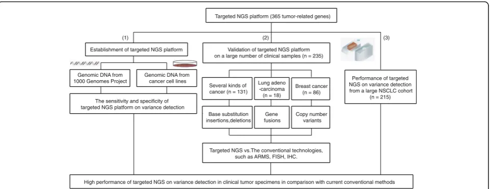
Establishment of the targeted NGS platform to detect DNA alteration using DNA samples and cancer cell lines
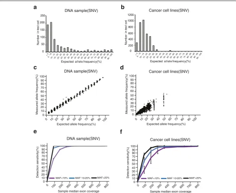
To establish a targeted NGS platform, we designed 16,198 DNA probes, targeting 4557 exons of 365 cancer-related genes, 47 introns of 25 genes frequently re-arranged in cancers. The capabality of the platform on DNA alteration detection was first tested in DNA sam-ples and cancer cell lines (Fig. 1).
In DNA samples, we created DNA pools of normal cell lines from the 1000 Genomes Project, containning thousands of single nucleotide polymorphisms (SNPs) across the targeted exons that spans a broad range of MAF
(5–100%) (Additional files 5 and 6: Table S5 and S6). On
the other hand, 10 cancer cell lines haboring known som-atic base substitutions and Indels were collected for this study. Two to ten of these 10 cell lines were randomly chosen and mixed with equal amount of DNA to form 21 pools. These 21 pools were sequenced by the targeted NGS, and the minimum sequencing coverage is 811× (Additional file 7: Table S7). 548 sites for base substitutions and 65 sites for Indels were selected for analysis across 21 pools (Additional files 8 and 9: Table S8 and S9).
In standard DNA pools, 97.5% base substitutions’
MAF in cancer cell line pools are less than 30%, while
72.3% base substitutions’MAF in DNA samples are less
genomes in cancer cells. The complexity of the cancer cell lines themselves may better represent the real world clinical samples. Therefore cancer cell lines instead of the normal cell DNA could be better for the NGS plat-form validation.
To test the correlation between sensitivity and sequen-cing depth, the sequensequen-cing reads were randomly selected to in silico form a set of fastq files with sequencing depth from 0× to 800×. As expected, detection sensitiv-ity declined with decrease of coverages, especially for those base substitutions with a MAF lower than 10% (Fig. 2e and f ). The average detection sensitivities reach plateau at the coverage of 400× for base substitutions in normal DNA samples and at 500× in cancer cell line pools (Fig. 2e and f ). For base substitutions with a MAF
of ≤10%, 10–20% and ≥20%, the sensitivities at 500×
coverage in cancer cell lines were 97.3% (1200/1233), 99.7% (1648/1653) and 100% (719/719), respectively (Fig. 2f ). These data demonstrate that the targeted NGS has high sensitivity on detection of base substitutions.
For the base substitutions with a MAF of ≤10%, 10–
20% and ≥20%, the high sensitivities of detection are
reached in both DNA samples and cancer cell lines at relative high coverage, while the speed of reaching the maximum in cancer cell lines is much slower than the one in DNA samples (Fig. 2e and f ). Furthermore, the variances at different MAF groups in caner cell lines are larger than the ones in DNA samples. In other words, when the sequence depth is low, all of the real mutations can not be detected in cancer cell lines. It is suggested that the cancer cell lines can better represent the com-plicated features of tumor heterogeneity, and may be better for NGS platform validation.
To assess the capacity of the targeted NGS on
detec-tion of Indels, the total 1365 (MAF ≥ 1%) Indels were
known in cancer cell lines and most MAF is less than 30% (Additional file 10: Fig. S1a). At 400×, the sensitivity can reach to 97.8%. Due to the complexity of the cancer cell lines, the MAFs of indels in cancer cell lines deter-mined by targeted NGS are less correlated with the ex-pected ones (Additional file 10: Fig. S1b). Overall, the targeted NGS has high sensitivity on detection of Indels with the relatively low sequencing depth compared with the base substitutions (Additional file 10: Fig. S1c).
To test the capacity of the targeted NGS on detection of CNV, DNA from the cell line HCC1143 with known
amplifications of CCND1, FGF3, FGF4, FGF19 and
AKT1, was diluted with matched normal DNA from
50% to 10%. At 50%, all known amplifications were
de-tected, but at 20%,AKT1-amplification was undetectable
(Additional file 10: Fig. S1d and S1e). It is concluded that more than 20% tumor cells in the mixed cell line pool are required to reach a high sensitivity of CNV de-tection. This is used as the guidline for a clinical test.
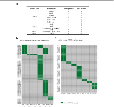
Targeted NGS to identify base substitutions and Indels from clinical specimens
To evaluate the capacity of targeted NGS to detect base substitutions and Indels from clinical specimens, we col-lected 34 FFPE resection specimens including 17 lung cancers, 13 colorectal cancers and 4 melanomas. Total
of 12 mutation sites in four oncogenes (EGFR, KRAS,
NRAS and BRAF) had been identified previously by
ARMS in the hospital. Each specimen harbors at least one DNA aberration. The DNA alterations in these sam-ples were then analyzed by targeted NGS. As was shown Targeted NGS platform (365 tumor-related genes)
Establishment of targeted NGS platform
(1) (2)
Genomic DNA from 1000 Genomes Project
Genomic DNA from cancer cell lines
The sensitivity and specificity of targeted NGS platform on variance detection
Validation of targeted NGS platform on a large number of clinical samples (n = 235)
(3)
Several kinds of cancer (n = 131)
Lung adeno -carcinoma (n = 18)
Breast cancer (n = 86)
Base substitution insertions,deletions
Gene fusions
Copy number variants
Targeted NGS vs.The conventional technologies, such as ARMS, FISH, IHC.
Performance of targeted NGS on variance detection from a large NSCLC cohort
(n = 215)
High performance of targeted NGS on variance detection in clinical tumor specimens in comparison with current conventional methods
in Fig. 3a and Additional file 11: Table S10, all DNA ab-errations were also detected by targeted NGS, indicating that the targeted NGS is as sensitive as ARMS to identify base substitutions and indels.
To further compare the detection efficiency between targeted NGS and ARMS, we gathered another batch of FFPE resection specimens, including 56 lung adenocar-cinoma and 41 colorectal cancers. These specimens had been examined by targeted NGS, and 15 hotspot mutation
sites inEGFRand KRAS(52EGFR-mutated specimens,38
KRAS-mutated specimens and 7 specimens without these
hot mutated sites) (Additional file 10: Fig. S2) were identi-fied. Because of the complexity of variant in clinical tumor samples, there are a multiple of genetic muations in one tumor sample (Additional file 10: Fig. S2). Among these hotspot mutations from 97 specimens, all samples were
confirmed by ARMS, and the concordance is 100% at the sample level (Fig. 3b, c and Additional file 12: Table S11). Overall, targeted NGS is as sensitive as ARMS to detect hotspot base substitutions and Indels from clinical FFPE samples.
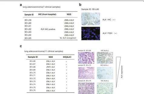
Targeted NGS to detect DNA rearrangements from clincal specimens
We collected 18 FFPE resection specimens of lung adeno-carcinoma to assess the capacity of targeted NGS to detect DNA arrangements. Among these 18 samples, 7 resection specimens had been stained by IHC and showed positive
ALK immunostaining, indicating ALK fusions (Fig. 4a).
These samples were re-examined by targeted NGS. The
data revealed that 6 samples possessedEML4-ALKfusions,
while 3D–L65 showed negative ALK fusion (Fig. 4a).
0 200 400 600 800 1000 1200 < 5 5 - 10
10 - 15 15 - 20 20 - 25 25 - 30 30 - 35 35 - 40 40 - 45 45 - 50 50 - 55 55 - 60 60 - 65 65 - 70 70 - 75 75 - 80 80 - 85 85 - 90 90 - 95 95 - 100
a
b
c
Cancer cell lines(SNV)
N u m b e r i n te st se t
Expected allele frequency(%) Expected allele frequency(%)
N u m b e r i n te st se t
d
10 20 30 40 50 60 70 90 80 0 800 400 200 600100 300 500
100
700 Sample median exon coverage
Detection sensitivity(%)
Cancer cell lines(SNV) DNA sample(SNV)
Expected allele frequency(%)
Measured allele frequency(%)
0 10 20 30 40 50 60 70 90 80 0 100 40
20 50 60
100
70 80 90 10 30
e
f
Cancer cell lines(SNV)
0 50 100 150 200 250 < 5 5 - 10
10 - 15 15 - 20 20 - 25 25 - 30 30 - 35 35 - 40 40 - 45 45 - 50 50 - 55 55 - 60 60 - 65 65 - 70 70 - 75 75 - 80 80 - 85 85 - 90 90 - 95 95 - 100
DNA sample(SNV) DNA sample(SNV) 0 10 20 30 40 50 60 70 90 80 100 Detection sensitivity(%) 0
MAF<10% MAF 10-20% MAF>20%
0
800 400
200 600
100 300 500 700
Sample median exon coverage
MAF<10% MAF 10-20% MAF>20%
Expected allele frequency(%) 0
100 40
20 50 60 70 80 90 10 30
Measured allele frequency(%) 100 20 30 40 50 60 70 90 80 100
Sample 3D–L65 was further investigated by IHC and
FISH from the third party and confirmed to be ALK
positive (Fig. 4b). The inconsistency with the targeted
NGS’s result could be contributed by the tumor
heterogen-eity or no probe coverage due to new fusion types. The remaining 11 FFPE samples were examined by targeted
NGS and were positive ALK rearrangements (Fig. 4c).
Among these variants ofALKfusions in this study,ZNF2
-ALK had not been reported previously. All these samples
were then immunostained with ALK antibody, and were
identified to be ALK positive. The specificity ofALKfusion
identification from targeted NGS is 100% (Fig. 4c).
There-fore, targeted NGS is sensitive enough to identify ALK
fusion across a large range of ALK expression level, which demonstrates high tumor heterogeneity.
Targeted NGS to identify CNVs from clinical specimens
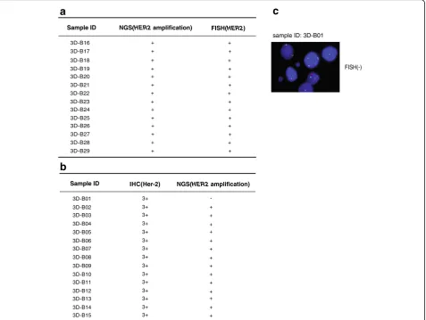
HER2 is frequently amplified in breast cancers. [12] FISH
is recognized as the“gold standard”for translocations and
HER2amplification. To investigate the detection efficiency
of targeted NGS on CNVs, FISH is used to confirm HER2-amplification detected by targeted NGS. In 14
sam-ples with the positive HER2 amplification by targeted
NGS, all samples were also positively confirmed by FISH (Fig. 5a). In comparison with the golden standerd FISH, the specificity of targeted NGS was 100% (14/14).
a
b
c
colon cancer(41 Clinical samples) lung adenocarcinoma(56 Clinical samples)
Mutated Gene Mutated Sites ARMS positive NGS positive
EGFR
3 3
L858R L861Q
2 2
T790M 1 1
Ex18 G719X 2 2
Exon 19del 5 5
Ex20 ins 4 4
KRAS
G12D 2 2
G12S 1 1
1 1
3 3
NRAS 2 2
BRAF V600E 8 8
Exon3
Exon3 G13D
T790M G719A S768I L858R 19del 20ins
3D−L01 3D−L02 3D−L03 3D−L04 3D−L05 3D−L06 3D−L07 3D−L08 3D−L09 3D−L10 3D−L11 3D−L12 3D−L13 3D−L14 3D−L15 3D−L16 3D−L17 3D−L18 3D−L19 3D−L20 3D−L21 3D−L22 3D−L23 3D−L24 3D−L25 3D−L26 3D−L27 3D−L28 3D−L29 3D−L30 3D−L31 3D−L32 3D−L33 3D−L34 3D−L35 3D−L36 3D−L37 3D−L38 3D−L39 3D−L40 3D−L41 3D−L42 3D−L43 3D−L44 3D−L45 3D−L46 3D−L47 3D−L48 3D−L49 3D−L50 3D−L51 3D−L52 3D−L53
NGSA-P NGSA-PNGSA-P NGSA-P NGS A-PNGSA-P
Detected Undeteced
G13D G12A G12C G12D G12R G12S G12V
3D−C01 3D−C02 3D−C03 3D−C04 3D−C05 3D−C06 3D−C07 3D−C08 3D−C09 3D−C10 3D−C11 3D−C12 3D−C13 3D−C14 3D−C15 3D−C16 3D−C17 3D−C18 3D−C19 3D−C20 3D−C21 3D−C22 3D−C23 3D−C24 3D−C25 3D−C26 3D−C27 3D−C28 3D−C29 3D−C30 3D−C31 3D−C32 3D−C33 3D−C34 3D−C35 3D−C36 3D−C37 3D−C38 3D−C39
NGS A-P NGSA-PNGSA-P NGSA-PNGSA-P NGSA-PNGSA-P
3D−L54 3D−L55 3D−L56
3D−C40 3D−C41
Since IHC is a widely used clinical method in China, and most breast specimens were immunostained with Her-2 antibody. The staining intensities were classified into 5 groups ranging from low to high: -,1+, 2+, 2 + ~3+ and 3+, which demonstrated that clinical breast cancer samples was too complicated to be divided into positive
and negtive groups. Among the 15 HER2-overexpressed
samples (IHC 3+), 14 were identified to beHER2
amplifi-cation by targeted NGS (Fig. 5b). The disconcordant case,
3D–B01, was further identified to be noHER2
amplifica-tion by FISH (Fig. 5c), which suggests that overexpression
of Her-2 protein in this case was not contributed byHER2
amplification. All 35 Her-2 negative (−, +) specimens were
noHER2amplification by targeted NSG (Additional file 10:
Fig. S3a). In comparison with IHC 3+ results, the sensitiv-ity and specificsensitiv-ity of targeted NGS were 93.3% (14/15) and 100% (35/35), respectively (Fig. 5b and Additional file 10: Fig. S3a). For the specimens with the IHC transient state (2+ and 2 + ~3+), the concordance between IHC and tar-geted NGS is less than 50% (Additional file 10: Fig. S3a). It is suggested that high Her-2 protein expression is not only
contributed byHER2amplification.
To further elucidate the specificity of CNV identification by NGS, eight samples from multiple types of cancers have
been identified to beHER2amplification by targeted NGS.
All of them have been stained by IHC and 7 out of 8 sam-ples were scored 3+ and one scored 2+. This suggested that
HER2 amplification detected by NGS leads to high Her-2
protein expression (Additional file 10: Fig. S3b).
Performance of targeted NGS on variance detection from a large non-small-cell lung cancer (NSCLC) cohort
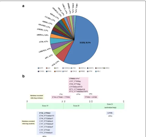
To further prove the reliability of the targeted NGS plat-form on DNA alteration detection, we analyzed the spectrum of DNA alterations identified by the platform. In 215 NSCLS cases, 17 genes were identified to have the multiple types of variants (Additional file 13: Table S12).
Most of them are known NSCLS driver genes, suchEGFR,
CDKN2A,ALKand etc. (Fig. 6a). The recurrent frequency
ofEGFRmutations was 56.3%, very close to the reported
results in NSCLC patients in China, 46.6% ~ 53.8%. [10] The other oncogenes also had similiar recurrent frequen-cies as reported in TCGA (The Cancer Genome Atlas) and other studies. [13, 14]
c
NGS IHC(ALK)
EML4-ALK +
EML4-ALK +
ZNF2-ALK
EML4-ALK
EML4-ALK
EML4-ALK
EML4-ALK
EML4-ALK +
EML4-ALK +
+
EML4-ALK +
sample ID: 3D-L66 IHC:ALK(+)
sample ID: 3D-L70 IHC:ALK(+)
sample ID: 3D-L76 IHC:ALK(+)
lung adenocarcinoma(11 cilinical samples)
3D-L66
3D-L67
3D-L68
3D-L69
3D-L70
3D-L71
3D-L72
3D-L73
3D-L74
3D-L75
3D-L76
Sample ID
+
+
+
+
+ ALK expression
a
IHC (from hospital) NGS
ALK IHC positive
EML4-ALK EML4-ALK EML4-ALK EML4-ALK EML4-ALK EML4-ALK
Sample ID
3D-L59 3D-L60 3D-L61
3D-L62 3D-L63 3D-L64
lung adenocarcinoma(7 cilinical samples)
3D-L65 NoALKarrangement
EML4-ALK
b
Sample ID: 3D-L65
In order to investigate the detection of targeted NGS
on EGFR hotspot mutations, we analyzed 121 NSCLC
samples withEGFRmutations detected by targeted NGS
(Additional file 14: Table S13). In these samples, the
EGFRmutations associated with drug sensitivity, such as
L858R and some 19 exon deletions (or insertions), were detected by targeted NGS. Moreover, the mutation rates of L858R and 19 exon deletions (or insertions) were 34% and 40% respectively (Fig. 6b), and were similar to that
from other studies. [15–17] In addition, theEGFR
muta-tions associated with drug resistance, such as T790 M
and 20 exon mutations, [18–20] were also found in these
NSCLC samples. In conclusion, the targeted NGS
plat-form developed here has accurately detectEGFRhotspot
mutations in NSCLC patients.
Discussion
We established a NGS platform targeting 365 cancer-related genes to identify genomic DNA alterations
including base substitutions, Indels, rearrangement and
CNV. In this study, we have answered to the question –
“what kind procedures need to throughly validate the new
NGS platform for clinical diagonasis”. This newly
estab-lished NGS platform has been compared with the current clinical platforms, such as ARMS, IHC and FISH in the de-tection of different types of variances. Although high con-cordance exists between the platforms, the minor difference does demonstrate the uniqueness for each plat-form. For the targeted NGS, one assay can identify mutiple types of variances. The disvantage is that the turn around time (TAT) is too long for the clinical diagnosis in compari-son with ARMS, IHC and FISH. If the clinical purpose is clear, the current clinical tool would be better in term of TAT and cost. This provides a clinician with the challenge to choose the right platform.
For the known hotspot variance detection like EGFR
L858R and 19dels, the high concordence demonstrates that targeted NGS is as efficient as ARMS does. For
c
FISH(-) sample ID: 3D-B01
IHC(Her-2) NGS(HER2 amplification)
3D-B01 3+ 3D-B02 3+
Sample ID
a
+
-FISH(HER2)
3D-B16 + + 3D-B17
Sample ID
+
b
NGS(HER2 amplification)
+
3D-B18 + +
3D-B19 +
+
3D-B20 + +
3D-B21 +
+
3D-B22 + +
3D-B23 +
+
3D-B24 + +
3D-B25 +
+
3D-B26 + +
3D-B27 +
+
3D-B28 + +
3D-B29 +
+
3D-B03 3+
3D-B04 3+ + +
3D-B05 3+
3D-B06 3+ + 3D-B07 3+
3D-B08 3+ + +
3D-B09 3+
3D-B10 3+ + 3D-B11 3+
3D-B12 3+ + +
3D-B13 3+
3D-B14 3+ + 3D-B15 3+ + + + +
those patients who do not have the hotspot mutations covered by ARMS, targeted NGS may identify the new mutations due to unbias probe design in all interesting regions, which can provide the hope for a new treat-ment. Specially for the relapse patient, he or she has de-veloped resistance to tyrosine kinase inhibitors (TKIs).
ALK rearrangement generates an oncogenic fusion
kinase leading to ALK constitutive activation. [21, 22]
ALK rearrangement occurs in around 3–6% NSCLC,
and is a promising therapeutic target. [23] An ALK in-hibitor like crizotinib has benefit the lung
adenocarcin-omas patient withALK rearrangement. [24] FISH is the
only diagnostic tool approved by Food and Drug
Admin-istration (FDA) to identify ALK rearrangement.
Al-though FISH has a high sensitivity and specificity, it can
not distinguish all ALK fusion types, which are
associ-ated with the efficacy of crizotinib in patients. [25] Sev-eral studies reported that a very high concordance between IHC and FISH exists, [26, 27] and IHC to
deter-mineALK status was also approved by China Food and
Drug Administration (CFDA). However, ALK antibody affinity to its fusion proteins may depend on specific variances. For example, ALK antibody CD246 from
Dako only has 27% sensitivity toEML4-ALKvariances 1
and 3a/b. [28] Moreover, intracellular and extracellular mucin have effect on IHC analysis, which may cause high false-negative and false-positive detection respectively. [29] Although IHC and FISH have some disvantages, they
can detect some types of ALK rearrangements that
tar-geted NGS can not identify due to the complexity of clin-ical specimens. While targeted NGS is as efficient as IHC
on ALK fusion detection and avoides IHC above
weak-ness, it can also identify newALKrearrangement.
As to HER2 amplifications, IHC is not as effective as
FISH for the detection of HER2 amplification because
IHC is mainly for protein expression. Protein expression level is highly correlated with amplification, but not one to one relationship exists. In this study, Her-2 IHC 3+ was highly concordant with FISH results, but IHC
3 +−2+ or 2+ showed a large discordance with the FISH
results. [30] FISH has a higher predictive value than IHC for response to treatment with trastuzumab which tar-gets Her-2. [31] Our study demonstrated that the
con-cordance of targeted NGS in the detection of HER2
amplification was 100%, in comparison with the golden standern FISH. In light of CNV detection, targeted NGS has advantage over IHC.
Conclusions
In conclusion, our study disclosed that DNA from can-cer cell lines is better than standard DNA as a reference to establish basic parameters for targeted NGS. In spite of the complexity of clinical specimens, comparison of the conventional methods using a large cohort of patient samples confirmed that targeted NGS has relatively high performance to identify multiple genomic alterations in a single assay. But the throughly validation of the new platform for clinical diagnosis is necessary and highly recommended.
Additional files
Additional file 1: Table S1.The information of 235 tumour specimens. (XLS 40 kb)
Additional file 2: Table S2.DNA sample information. (XLS 24 kb)
Additional file 3: Table S3.The list of immortalized tumor cell lines and pools. (XLS 26 kb)
Additional file 4: Table S4.The gene list of targeted NGS platform. (XLSX 12 kb)
Additional file 5: Table S5.The golden standard sites of SNP in DNA sample_pool1. (XLS 589 kb)
Additional file 6:The golden standard sites of SNP in DNA sample_pool2. (XLS 594 kb)
Additional file 7: Table S7.Quality control results of samples detected by NGS. (XLS 34 kb)
Additional file 8: Table S8.The golden standard sites of SNP in cancer cell line validation. (XLS 68 kb)
Additional file 9: Table S9.The golden standard sites of Indel in cancer cell line validation. (XLS 27 kb)
Additional file 10: Supplementary Figures S1-S3.Figure S1.The results of Indels and CNVs detected by the means of targeted NGS in cancer cell lines;Figure S2.Targeted NGS comparable to ARMS in Lung adenocarcinoma and Colon Cancer FFPE samples;Figure S3.The comparation between targeted NGS and IHC on the HER2 amplification in clinical FFPE samples. (DOCX 572 kb)
Additional file 11: Table S10.The comparation of ARMS and targeted NGS in 34 clinical samples. (XLS 29 kb)
Additional file 12: Table S11.The comparation of targeted NGS and ARMS in 97 clinical samples. (XLS 45 kb)
Additional file 13: Table S12.The spectrum of DNA alternations in non-small-cell lung cancer. (XLS 25 kb)
Additional file 14: Table S13.EGFR hotspot mutations identified by targeted NGS in NSCLC. (XLS 34 kb)
Abbreviations
ARMS:amplification-refractory mutation system; ATCC: American Type Culture Collection; CFDA: China Food and Drug Administration; CNV: copy number variance; FDA: Food and Drug Administration; FFPE: formalin fixed, paraffin-embedded; FISH: fluorescence in situ hybridization;
IHC: immunohistochemistry; MAF: mutation allele frequency; NGS: Next generation sequencing; NSCLC: non-small-cell lung cancer; SNP: single nucleotide polymorphisms; SNVs: single nucletic variances; TAT: turn around time; TCGA: The Cancer Genome Atlas; TKI: tyrosine kinase inhibitor
Acknowledgements
This study was partially supported by National Natural Science Foundation of China (NO. 81502549 to Dadong Zhang and NO. 81672663 to Qihuang Jin) and high-level creative and innovative health talent program of Zhejiang province (to Dan Su). We would like to thank Dr. Jingyu Li and Zisong Zhou for their help to this manuscript.
Authors’contributions
FL, DS, WM and LX designed the experiments. DZ, JW, KC, LY, QJ, YY and ZX performed the experiments. JL, XC and FL carried out data analysis. FL, DS, DZ, WM and LX wrote the manuscript. All authors read and approved the final manuscript.
Competing interests
All authors with 3D medicine affiliation are current or former employees.
Publisher’s Note
Springer Nature remains neutral with regard to jurisdictional claims in published maps and institutional affiliations.
Author details
1Pathology Department, Zhejiang Cancer Hospital, Hangzhou 310022, China. 2Key Laboratory of Diagnosis and Treatment Technology on Thoracic Oncology of Zhejiang Province, Hangzhou 310022, China.3The Research and Development Center of Precision Medicine, 3D Medicine Inc., Shanghai 201114, China.4Changhai Hospital, The Second Military Medical University, Shanghai 200433, China.
Received: 28 June 2017 Accepted: 30 August 2017
References
1. Lopez JS, Banerji U. Combine and conquer: challenges for targeted therapy combinations in early phase trials. Nature reviews. Clin Oncol. 2017;14(1):57–6. 2. Riely GJ, Pao W, Combining EGFR. Targeted therapy with chemotherapy in pancreatic cancer: is timing important? Cancer Biol Ther. 2005;4(10):1096–7. 3. Stewart EL, Tan SZ, Liu G, Tsao MS. Known and putative mechanisms of
resistance to EGFR targeted therapies in NSCLC patients with EGFR mutations-a review. Transl Lung Cancer Res. 2015;4(1):67–81.
5. Frampton GM, Fichtenholtz A, Otto GA, Wang K, Downing SR, He J, et al. Development and validation of a clinical cancer genomic profiling test based on massively parallel DNA sequencing. Nat Biotechnol. 2013;31(11):1023–31. 6. Lefebvre C, Rieckhof G, Califano A. Reverse-engineering human regulatory
networks. Wiley Interdiscip Rev Syst Biol Med. 2012;4(4):311–25. 7. Shrager J, Tenenbaum JM. Rapid learning for precision oncology. Nat Rev
Clin Oncol. 2014;11(2):109–18.
8. Fisher S, Barry A, Abreu J, Minie B, Nolan J, Delorey TM, et al. A scalable, fully automated process for construction of sequence-ready human exome targeted capture libraries. Genome Biol. 2011;12(1):R1.
9. Karolchik D, Hinrichs AS, Furey TS, Roskin KM, Sugnet CW, Haussler D, et al. The UCSC table browser data retrieval tool. Nucleic Acids Res. 2004; 32(Database issue):D493–6.
10. Shi Y, JS A, Thongprasert S, Srinivasan S, Tsai CM, Khoa MT, et al. A prospective, molecular epidemiology study of EGFR mutations in Asian patients with advanced non-small-cell lung cancer of adenocarcinoma histology (PIONEER). J Thorac Oncol. 2014;9(2):154–62.
11. McLeer-Florin A, Moro-Sibilot D, Melis A, Salameire D, Lefebvre C, Ceccaldi F, et al. Dual IHC and FISH testing for ALK gene rearrangement in lung adenocarcinomas in a routine practice: a French study. Journal of thoracic oncology : official publication of the International Association for the Study of Lung Cancer. 2012;7(2):348–54.
12. Slamon DJ, Godolphin W, Jones LA, Holt JA, Wong SG, Keith DE, et al. Studies of the HER-2/neu proto-oncogene in human breast and ovarian cancer. Science. 1989;244(4905):707–12.
13. Pao W, Girard N. New driver mutations in non-small-cell lung cancer. Lancet Oncol. 2011;12(2):175–80.
14. Swanton C, Govindan R. Clinical implications of genomic discoveries in lung cancer. N Engl J Med. 2016;374(19):1864–73.
15. Pirker R, Herth FJ, Kerr KM, Filipits M, Taron M, Gandara D, et al. Consensus for EGFR mutation testing in non-small cell lung cancer. J Thorac Oncol. 2010;5:1706–13.
16. Pao W, Chmielecki J. Rational, biologically based treatment of EGFR-mutant non-small-cell lung cancer. Nat Rev Cancer. 2010;10(11):760–74.
17. Mitsudomi T, Yatabe Y. Mutations of the epidermal growth factor receptor gene and related genes as determinants of epidermal growth factor receptor tyrosine kinase inhibitors sensitivity in lung cancer. Cancer Sci. 2007;98(12):1817–24.
18. Chen D, Song Z, Cheng G. Clinical efficacy of first-generation EGFR-TKIs in patients with advanced non-small-cell lung cancer harboring EGFR exon 20 mutations. Onco Targets Ther. 2016;9:4181.
19. Naidoo J, Sima CS, Rodriguez K, Busby N, Nafa K, Ladanyi M, et al. Epidermal growth factor receptor exon 20 insertions in advanced lung
adenocarcinomas: clinical outcomes and response to erlotinib. Cancer. 2015; 121(18):3212–20.
20. Greulich H, Chen TH, Feng W, Janne PA, Alvarez JV, Zappaterra M, et al. Oncogenic transformation by inhibitor-sensitive and -resistant EGFR mutants. PLoS Med. 2005;2(11):e313.
21. Soda M, Choi YL, Enomoto M, Takada S, Yamashita Y, Ishikawa S, et al. Identification of the transforming EML4-ALK fusion gene in non-small-cell lung cancer. Nature. 2007;448(7153):561–6.
22. Rikova K, Guo A, Zeng Q, Possemato A, Yu J, Haack H, et al. Global survey of phosphotyrosine signaling identifies oncogenic kinases in lung cancer. Cell. 2007;131(6):1190–03.
23. Shaw AT, Yeap BY, Solomon BJ, Riely GJ, Gainor J, Engelman JA, et al. Effect of crizotinib on overall survival in patients with advanced non-small-cell lung cancer harbouring ALK gene rearrangement: a retrospective analysis. Lancet Oncol. 2011;12(11):1004–12.
24. Shaw AT, Kim DW, Nakagawa K, Seto T, Crino L, Ahn MJ, et al. Crizotinib versus chemotherapy in advanced ALK-positive lung cancer. N Engl J Med. 2013;368(25):2385–94.
25. Yoshida T, Oya Y, Tanaka K, Shimizu J, Horio Y, Kuroda H, et al. Differential Crizotinib response duration among ALK fusion variants in ALK-positive non-small-cell lung cancer. Journal of clinical oncology : official journal of the American Society of Clinical Oncology. 2016;34(28):3383–9.
26. Cabillic F, Gros A, Dugay F, Begueret H, Mesturoux L, Chiforeanu DC, et al. Parallel FISH and immunohistochemical studies of ALK status in 3244 non-small-cell lung cancers reveal major discordances. J Thorac Oncol. 2014;9(3):295–6.
27. Ali G, Proietti A, Pelliccioni S, Niccoli C, Lupi C, Sensi E, et al. ALK rearrangement in a large series of consecutive non-small cell lung cancers: comparison between a new immunohistochemical approach and
fluorescence in situ hybridization for the screening of patients eligible for crizotinib treatment. Arch Pathol Lab Med. 2014;138(11):1449–58. 28. Wallander ML, Geiersbach KB, Tripp SR, Layfield LJ. Comparison of reverse
transcription-polymerase chain reaction, immunohistochemistry, and fluorescence in situ hybridization methodologies for detection of echinoderm microtubule-associated proteinlike 4-anaplastic lymphoma kinase fusion-positive non-small cell lung carcinoma: implications for optimal clinical testing. Arch Pathol Lab Med. 2012;136(7):796–3. 29. Yu Y, Ding Z, Zhu L, Teng H, Frequencies LS. Of ALK rearrangements in lung
adenocarcinoma subtypes: a study of 2299 Chinese cases. Spring. 2016;5(1):894. 30. Wesola M, Jelen MA. Comparison of IHC and FISH cytogenetic methods in the evaluation of HER2 status in breast cancer. Adv Clin Exp Med. 2015; 24(5):899–3.
31. Pauletti G, Dandekar S, Rong H, Ramos L, Peng H, Seshadri R, et al. Assessment of methods for tissue-based detection of the HER-2/neu alteration in human breast cancer: a direct comparison of fluorescence in situ hybridization and immunohistochemistry. J Clin Oncol. 2000;18(21):3651–64.
• We accept pre-submission inquiries
• Our selector tool helps you to find the most relevant journal
• We provide round the clock customer support
• Convenient online submission
• Thorough peer review
• Inclusion in PubMed and all major indexing services
• Maximum visibility for your research
Submit your manuscript at www.biomedcentral.com/submit