Open Access
Review
Mechanomyographic amplitude and frequency responses during
dynamic muscle actions: a comprehensive review
Travis W Beck*
1, Terry J Housh
1, Joel T Cramer
2, Joseph P Weir
3,
Glen O Johnson
1, Jared W Coburn
4, Moh H Malek
1and Michelle Mielke
1Address: 1Department of Nutrition and Health Sciences, Human Performance Laboratory, University of Nebraska-Lincoln, Lincoln, NE, USA
68583, 2Department of Health and Exercise Science, University of Oklahoma, Norman, OK, USA 73019, 3Applied Physiology Laboratory, Division
of Physical Therapy, Des Moines University, Osteopathic Medical Center, Des Moines, IA, USA 50312 and 4Department of Kinesiology, California
State University, Fullerton, Fullerton, CA, USA 92834
Email: Travis W Beck* - [email protected]; Terry J Housh - [email protected]; Joel T Cramer - [email protected]; Joseph P Weir - [email protected]; Glen O Johnson - [email protected]; Jared W Coburn - [email protected]; Moh H Malek - [email protected]; Michelle Mielke - [email protected]
* Corresponding author
Abstract
The purpose of this review is to examine the literature that has investigated mechanomyographic (MMG) amplitude and frequency responses during dynamic muscle actions. To date, the majority of MMG research has focused on isometric muscle actions. Recent studies, however, have examined the MMG time and/or frequency domain responses during various types of dynamic activities, including dynamic constant external resistance (DCER) and isokinetic muscle actions, as well as cycle ergometry. Despite the potential influences of factors such as changes in muscle length and the thickness of the tissue between the muscle and the MMG sensor, there is convincing evidence that during dynamic muscle actions, the MMG signal provides valid information regarding muscle function. This argument is supported by consistencies in the MMG literature, such as the close relationship between MMG amplitude and power output and a linear increase in MMG amplitude with concentric torque production. There are still many issues, however, that have yet to be resolved, and the literature base for MMG during both dynamic and isometric muscle actions is far from complete. Thus, it is important to investigate the unique applications of MMG amplitude and frequency responses with different experimental designs/methodologies to continually reassess the uses/limitations of MMG.
Background
Mechanomyography (MMG) involves recording and quantifying the low frequency lateral oscillations of active skeletal muscle fibers [1,2]. Although the exact origin(s) of the MMG signal is not completely understood, Gordon and Holbourn [3] suggested that MMG reflects the mechanical counterpart of motor unit electrical activity as measured by electromyography (EMG). During voluntary
muscle actions, MMG is usually measured at the surface of the skin over a muscle, and it has been suggested [1,4-6] that in this situation, the MMG signal is generated by three primary mechanisms: a) gross lateral movements of the muscle as it moves toward, or away from, its line of pull during contraction and relaxation, respectively, b) smaller subsequent lateral oscillations of the muscle at its Published: 19 December 2005
BioMedical Engineering OnLine 2005, 4:67 doi:10.1186/1475-925X-4-67
Received: 12 October 2005 Accepted: 19 December 2005
This article is available from: http://www.biomedical-engineering-online.com/content/4/1/67
© 2005 Beck et al; licensee BioMed Central Ltd.
resonant frequency, and c) dimensional changes of the active fibers (see Figure 1).
Although various terminologies, such as soundmyogra-phy, phonomyograsoundmyogra-phy, acousticmyograpy, and vibromy-ography have been used by previous studies to describe the MMG signal, Orizio [4] recommended that the term "mechanomyogram" should be used to reflect the mechanical nature of the signal. Many different types of sensors have been used to detect the MMG signal, includ-ing hydrophones, condenser microphones, piezoelectric contact sensors, accelerometers, and, more recently, laser distance sensors [4,5]. As acknowledged by Orizio [4], the most important characteristic of the sensor is its frequency response. Specifically, Orizio [4] recommended that the "...low frequency cut-off has to be around 1 to 2 Hz," and "the upper cut-off has to be chosen so that the greater part of the power is well below 100 Hz." Another important characteristic is the mass of the sensor. In particular, it has been suggested [4] that lightweight accelerometers may be more appropriate than larger condenser microphones and piezoelectric contact sensors when recording MMG sig-nals from small muscles, such as the first dorsal
interos-seous. For larger limb muscles, such as the biceps brachii and superficial quadriceps femoris muscles, however, condenser microphones and piezoelectric contact sensors can be used, as long as the contact pressure of the sensor over the muscle remains constant [4,7]. Although the shape of the MMG signal is dependent on the type of sen-sor used to detect it [4], Orizio et al. [5] have suggested that its pattern is similar to the small oscillations in force that occur during an isometric muscle action. Further-more, it has been hypothesized [4,8-11] that both signals (MMG and force) contain information regarding motor control strategies (relative contributions of recruitment and firing rate). In particular, several investigations [5,8,10,12-14] have suggested that under certain condi-tions, the amplitude of the MMG signal may be related to the number of active motor units (i.e. motor unit recruit-ment), while the MMG power density spectrum could provide qualitative information regarding the global fir-ing rates of the unfused activated motor units.
Most studies have examined the MMG amplitude and/or frequency responses during isometric muscle actions [15-20]. Stokes [6] suggested, however, that "If AMG [MMG] is to be used as a means of monitoring force during func-tional activities, then its relationship with force during dynamic activation must be considered." In addition to the implications related to monitoring functional and/or sporting activities, MMG responses during dynamic mus-cle actions may be particularly important for clinical applications such as controlling external prostheses [21], assessing low back pain which can be exacerbated during movement [22], monitoring rehabilitation following injury [4,23], and examining masseter muscle function associated with cranio-mandibular disorders [24]. Unfor-tunately, there are a number of factors that can potentially affect the MMG signal during dynamic muscle actions, including changes in torque production, muscle length, and the thickness of the tissue between the muscle and the MMG sensor [4,25]. Theoretically, one or more of these factors can influence the amplitude and frequency of the MMG signal during a dynamic muscle action and con-found the interpretation of the underlying motor control strategies. There are, however, several pieces of evidence that indicate that the MMG signal is generated primarily by muscle activity during a dynamic muscle action. Briefly, MMG amplitude increases with torque production during concentric and eccentric muscle actions [26-28], as well as with increases in power output during incremental cycle ergometry [29-32]. These responses suggest that MMG may provide information regarding the level of muscle activity that is required to perform an exercise task. In addition, for a given torque level, MMG amplitude is less during eccentric muscle actions than concentric mus-cle actions [26], which is consistent with EMG data show-ing that a lower level of muscle activity is required durshow-ing Mechanomyographic (MMG) signal during a voluntary muscle
action Figure 1
Mechanomyographic (MMG) signal during a
volun-tary muscle action. Example of the raw
eccentric activity [33,34]. Shinohara et al. [30], however, provided perhaps the most convincing evidence that MMG reflects muscle activity during dynamic muscle actions. The authors [30] examined the MMG amplitude responses for the vastus lateralis muscle during an incre-mental cycle ergometry test in which the power output was increased by 20 W each minute and the subject main-tained a pedal cadence of 60 revolutions per minute. Dur-ing the first 30 s of each 1-minute stage, the subjects were instructed to relax with their feet on the pedals while one of the investigators pushed the pedals at the required cadence. For the second 30 s of each one minute stage, however, the subjects pedaled actively against the ergom-eter. The authors [30] reported that MMG amplitude for the vastus lateralis muscle increased linearly with power output during the active cycling, but there was only a small increase in MMG amplitude during the passive cycling. In addition, it was hypothesized that the small increases in MMG amplitude during the passive cycling may have been due to incomplete relaxation in some of the subjects, because there was also a small increase in EMG amplitude for the vastus lateralis muscle. Thus, the authors [30] stated, "These observations confirm that the MMG recorded during the contraction phase could include some noise, but mainly reflect the activity of the quadriceps muscle within the parameters of this experi-ment." These findings [26-28,30-32] clearly indicated that during dynamic muscle actions, MMG is generated prima-rily by muscle activity. Therefore, pertinent follow-up questions include: a) what kind of information can be extracted from the MMG signal that is recorded during a dynamic muscle action; and b) what are the uses/applica-tions of this information? Unfortunately, there are no simple answers to these questions because the MMG amplitude and frequency responses depend largely on the type of dynamic muscle action(s) (i.e. concentric versus eccentric, maximal versus submaximal) that is being per-formed. Furthermore, there are many issues regarding the exact origin(s) of the MMG signal that have yet to be resolved. The purpose of this review, however, is to exam-ine the literature regarding the MMG responses during dif-ferent types of dynamic muscle actions. Although many of the studies discussed in this review are contributions from our laboratory, an attempt has been made to be as com-prehensive as possible, and emphasis will be placed on summarizing the common findings from the various types of experiments that have been performed.
MMG amplitude and frequency responses with
increases in velocity during maximal concentric
isokinetic muscle actions
Evetovich et al. [35] were the first to examine the MMG amplitude responses with increases in velocity during iso-kinetic muscle actions. The authors [35] reported that dur-ing maximal concentric isokinetic leg extensions, there
Theoretically, a velocity-related shift in the contributions of slow-twitch muscle fibers to torque production could also influence the frequency content of the surface MMG signal. In particular, if slow-twitch muscle fibers become unloaded at high velocities, it is possible that their contri-bution to MMG decreases. Several studies [40-42] have reported that when compared to low threshold slow-twitch motor units, high threshold fast-slow-twitch motor units have higher initial firing rates and require greater stimula-tion rates to achieve complete fusion of motor unit twitches. Thus, decreases in the contributions of slow-twitch muscle fibers to MMG at high velocities could result in higher frequency MMG signals. Ebersole et al. [43] tested this hypothesis by examining the MMG ampli-tude and mean power frequency (MPF) responses from the vastus lateralis muscle during maximal concentric iso-kinetic leg extensions at velocities of 60 and 300°·s-1. The authors [43] reported that with an increase in velocity from 60 to 300°·s-1, there was a significant decrease in leg extension PT and an increase in MMG amplitude. There was, however, no difference between the mean MMG MPF values at 60 and 300°·s-1. It was hypothesized that the tendonous iliotibial band that covers the vastus lateralis muscle could have interfered with the muscle fiber oscil-lations that generated the MMG signal. In addition, it was still unclear if the velocity-related increases in MMG amplitude that had been reported for the vastus lateralis [35,38,43] and biceps brachii [37] were due to reduced muscle stiffness, a greater rate of cross-bridge cycling, or increased turbulence of the intracellular and extracellular fluid mediums at high velocities.
Bodor [44], however, proposed that MMG amplitude may be more related to power output than PT during maximal concentric isokinetic muscle actions. Cramer et al. [45] tested this hypothesis by examining the potential relation-ship between MMG amplitude and mean power output during maximal concentric isokinetic leg extensions at velocities ranging from 60 to 300°·s-1. The authors [45] reported that leg extension PT decreased with increases in velocity, but mean power output and MMG amplitude for the rectus femoris, vastus lateralis and vastus medialis muscles increased from 60 to 240°·s-1, and then pla-teaued from 240 to 300°·s-1. It was suggested that MMG amplitude may be useful for monitoring training-induced changes in power output.
Interestingly, there is also evidence to suggest that there may be muscle-specific differences in the MMG amplitude responses with increases in velocity during maximal con-centric isokinetic muscle actions. For example, Cramer et al. [46] reported that during maximal concentric isoki-netic leg extensions at velocities ranging from 60 to 300°·s-1, MMG amplitude for each of the superficial quadriceps femoris muscles (rectus femoris, vastus
latera-lis, and vastus medialis) increased with velocity from 60 to 180°·s-1. At velocities above 180°·s-1, however, MMG amplitude increased to 240°·s-1 and then plateaued from 240 to 300°·s-1 for the vastus lateralis, plateaued from 180 to 300°·s-1 for the rectus femoris, and increased from 180 to 300°·s-1 for the vastus medialis. It was suggested that the muscle-specific differences in the MMG ampli-tude responses may be due to differences among the rec-tus femoris, vasrec-tus lateralis, and vasrec-tus medialis muscles in fiber type composition and/or muscle architecture (unipennate versus bipennate or the degree of penna-tion). In addition, Ebersole et al. [47] examined the pat-terns for MMG amplitude from the vastus lateralis with increases in velocity during maximal concentric isokinetic and passive leg extension muscle actions. The authors [47] found that MMG amplitude increased with leg extension velocity during both the active and passive leg extension muscle actions. During the passive leg extension muscle actions, however, the vastus lateralis muscle remained inactive because the EMG amplitude values from the mus-cle were very small and did not change with increases in velocity. It was hypothesized that the velocity-related increases in MMG amplitude for the vastus lateralis mus-cle may have been due to greater turbulence of the intrac-ellular and extracintrac-ellular fluid mediums and/or cross-talk from the hamstring muscles. Thus, these findings sug-gested that in addition to power output, MMG amplitude may be affected by factors such as fiber type composition and muscle architecture [46], as well as turbulence of the intracellular and extracellular fluid mediums and/or cross-talk [47]. Cramer et al. [48], however, recently reported that the potential for cross-talk in surface MMG signals is relatively small, even for muscles that are very close to each other and have a common innervation. Spe-cifically, the authors [48] used cross-correlation to quan-tify the common variance present in the MMG signals from the rectus femoris, vastus lateralis, and vastus medi-alis muscles during maximal concentric and eccentric iso-kinetic leg extensions at a velocity of 60°·s-1. The common variance shared between the MMG signals from any two muscles ranged from 14% to 27%, and it was sug-gested [48] that, "...despite the potential for some cross-talk, MMG measurements can be used to examine differ-ences between the patterns of MMG amplitude and fre-quency responses of the superficial muscles of the quadriceps femoris."
MPF (marginal means, collapsed across muscles) from 240 to 300°·s-1. Furthermore, Cramer et al. [50] recently suggested that the MMG MPF responses to increases in velocity may be muscle-specific. For example, during max-imal concentric isokinetic leg extensions, there were veloc-ity-related increases in MMG MPF for the rectus femoris and vastus lateralis muscles, but not for the vastus media-lis [50]. Thus, it is possible that potential increases in MMG MPF during concentric isokinetic muscle actions may be muscle- and/or velocity-specific.
Collectively, the results from these studies have indicated that during maximal concentric isokinetic muscle actions, there are velocity-related increases in MMG amplitude that may be due to reduced muscle stiffness, a greater rate of cross-bridge cycling, and/or increased turbulence of the intracellular and extracellular fluid mediums. In addition,
MMG amplitude may be closely related to power output (see Figure 2), which suggests that MMG could potentially be useful for examining training-induced adaptations in muscular power. It is unclear, however, if there is a rela-tionship between MMG MPF and velocity during concen-tric isokinetic muscle actions.
MMG amplitude and frequency responses with
increases in velocity during maximal eccentric
isokinetic muscle actions
Eccentric muscle actions may require unique motor con-trol strategies from the central nervous system [51]. Spe-cifically, it has been suggested that when compared to concentric and/or isometric activities, eccentric muscle actions are characterized by lower levels of muscle activa-tion [26], reduced recruitment thresholds [52,53], lower motor unit firing rates [54], de-recruitment of low thresh-Mean power output, isokinetic peak torque (PT), mechanomyographic (MMG) amplitude, and MMG mean power frequency (MPF) responses with increases in velocity during maximal concentric isokinetic muscle actions
Figure 2
Mean power output, isokinetic peak torque (PT), mechanomyographic (MMG) amplitude, and MMG mean power frequency (MPF) responses with increases in velocity during maximal concentric isokinetic muscle actions. (A) Relationship between mean power output and angular velocity during maximal concentric isokinetic leg exten-sions. (B) Relationship between maximal concentric isokinetic leg extension peak torque and angular velocity. (C) Relationship between mechanomyographic (MMG) amplitude (averaged across the rectus femoris, vastus lateralis, and vastus medialis mus-cles) and angular velocity. (D) Relationship between MMG mean power frequency (MPF, averaged across the rectus femoris, vastus lateralis, and vastus medialis muscles) and angular velocity. The data presented in each graph are the mean values from 26 subjects, and all MMG signals were recorded with a piezoelectric contact sensor (HP 21050A).
0 50 100 150 200 250 300
0 100 200 300 400 500
Velocit y (deg/sec)
M
e
a
n
P
ow
e
r
O
ut
put
(W
)
0 100 200 300 400 500 600
0 100 200 300 400 500
Velocit y (deg/sec)
M
M
G
Am
p
li
tu
d
e
(m
Vr
m
s)
0 50 100 150 200 250
0 100 200 300 400 500
Velocit y (deg/sec)
Is
o
k
in
e
ti
c
P
T
(Nm
)
0 5 10 15
0 100 200 300 400 500
Velocity (deg/sec)
MMG
MP
F
(H
z
)
D A
C
old slow-twitch motor units [55], and selective activation of high threshold fast-twitch motor units [56]. In addi-tion, unlike maximal concentric isokinetic muscle actions, torque production during maximal eccentric isokinetic muscle actions does not change with increases in velocity [37,38,57-59], but there are significant velocity-related increases in mean power output [59]. Thus, MMG ampli-tude and/or MPF could provide information regarding the unique motor control strategies that are used during eccentric muscle actions. In addition, the possibility for velocity-related increases in mean power output suggests that maximal eccentric isokinetic muscle actions may be useful for testing the hypothesis of Bodor [44] that MMG amplitude is more closely related to power output than PT during maximal isokinetic muscle actions.
Smith et al. [57] examined the MMG amplitude responses from the vastus lateralis during maximal eccentric isoki-netic muscle actions of the leg extensors at velocities of 60, 90, 120, and 180°·s-1. The authors [57] found that although there was no change in eccentric PT from 60 to 180°·s-1, there was a velocity-related increase in MMG amplitude for the vastus lateralis muscle. In addition, the MMG amplitude values during the maximal eccentric iso-kinetic muscle actions were highly reliable, with ICCs ranging from R = 0.97–0.98 and no significant differences between the mean MMG amplitude values for test versus retest at any velocity. The authors [57] proposed three potential hypotheses to explain the velocity-related increase in MMG amplitude for the vastus lateralis mus-cle. First, an increased rate of cross bridge activity during high-velocity eccentric muscle actions could result in larger vibrations of the myosin heads, potentially result-ing in greater MMG amplitude values. Second, de-recruit-ment of low threshold slow-twitch motor units [55] and selective activation of high threshold fast-twitch motor units [56] at high velocities during eccentric muscle actions could influence MMG amplitude because in some muscles, fast-twitch motor units may be more superfi-cially located than slow-twitch motor units [60]. Thus, when compared to slow-twitch motor units, the vibra-tions from fast-twitch motor units may be damped to a lesser degree by the surrounding tissues, potentially result-ing in greater MMG amplitude values [57]. Finally, the velocity-related increases in MMG amplitude may be due to faster movement of the limb and a greater overall dis-turbance of the intracellular and extracellular fluid medi-ums [57].
Evetovich et al. [38] also reported velocity-related increases in MMG amplitude for the vastus lateralis dur-ing maximal eccentric isokinetic muscle actions of the leg extensors. There was no gender difference for the increases in MMG amplitude with velocity, but the men demon-strated greater MMG amplitude values than the women at
all velocities. As stated previously for maximal concentric isokinetic muscle actions, the greater MMG amplitude val-ues for men during maximal eccentric isokinetic muscle actions could be related to a larger muscle mass and/or thinner adipose tissue layer over the vastus lateralis than is typically found in women [38]. In addition, Smith et al. [37] reported that MMG amplitude for the biceps brachii increased with velocity during maximal eccentric isoki-netic muscle actions of the forearm flexors at velocities of 30, 90, and 150°·s-1. These findings indicated that increases in MMG amplitude with velocity during maxi-mal eccentric isokinetic muscle actions can occur in the biceps brachii, which is a fusiform muscle with a poten-tially different fiber type composition [60] when com-pared to the unipennate vastus lateralis that had been examined in previous studies [38,57]. The exact mecha-nism(s) underlying the velocity-related increase in MMG amplitude was, however, still unknown.
Theoretically, de-recruitment of low threshold slow-twitch motor units [55] and selective activation of high threshold fast-twitch motor units [56] during high-veloc-ity eccentric muscle actions could result in greater MMG amplitude and MPF values. Evetovich et al. [61], however, reported that during maximal eccentric isokinetic muscle actions of the leg extensors at velocities of 60, 120, and 180°·s-1, there was a velocity-related increase in MMG amplitude for the vastus lateralis muscle, but there was no change in MMG MPF from 60 to 180°·s-1. It was sug-gested that the increase in MMG amplitude may have been due to factors other than a velocity-related shift in the contributions of slow- and fast-twitch muscle fibers to torque production. In addition, Cramer et al. [62] found that during maximal eccentric isokinetic muscle actions of the leg extensors, MMG MPF for the rectus femoris, vastus lateralis, and vastus medialis muscles actually decreased with an increase in velocity from 60 to 120°·s-1, and then remained relatively stable from 120 to 180°·s-1. Although there was no change in eccentric PT, there were increases in mean power output and the average MMG amplitude values for each muscle from 60 to 180°·s-1. Furthermore, Cramer et al. [59] reported that mean power output and MMG amplitude for the vastus lateralis increased with velocity during maximal eccentric isokinetic muscle actions of the leg extensors at velocities of 30, 90, and 150°·s-1. The velocity-related increases in MMG ampli-tude were similar, however, in men and women.
rectus femoris, and vastus medialis muscles [59,62] sug-gested that MMG amplitude increases with velocity during maximal eccentric isokinetic muscle actions, even for muscles with a potentially different architecture (fusi-form, unipennate, bipennate) and/or fiber type composi-tion. Finally, MMG MPF may not reflect a potential velocity-related shift in the contributions of fast- and slow-twitch muscle fibers to torque production during maximal eccentric isokinetic muscle actions.
MMG amplitude and frequency responses with
increases in torque during incremental
concentric and eccentric muscle actions
Dalton and Stokes [26,63] were the first investigators to examine the potential torque-related patterns for MMG amplitude and MPF during concentric and eccentric
mus-cle actions. The authors [26,63] measured the MMG signal from the biceps brachii muscle during submaximal dynamic constant external resistance (DCER, formerly called isotonic) muscle actions of the forearm flexors in which the subjects lifted (concentric) and lowered (eccen-tric) weights ranging from 0 to 8.5 kg. Dalton and Stokes [26] reported that MMG amplitude for the biceps brachii increased linearly from 0 to 8.5 kg during both the con-centric and eccon-centric muscle actions. In addition, during the concentric muscle actions, MMG MPF for the biceps brachii increased from 0 to approximately 5.5 kg and then decreased from 5.5 to 8.5 kg [63]. During the eccentric muscle actions, however, MMG MPF remained relatively stable from 0 to 8.5 kg [63]. It was suggested that during submaximal concentric muscle actions of the forearm flexors, torque may be increased by recruiting additional Mean power output, isokinetic peak torque (PT), mechanomyographic (MMG) amplitude, and MMG mean power frequency (MPF) responses with increases in velocity during maximal eccentric isokinetic muscle actions
Figure 3
Mean power output, isokinetic peak torque (PT), mechanomyographic (MMG) amplitude, and MMG mean power frequency (MPF) responses with increases in velocity during maximal eccentric isokinetic muscle actions. (A) Relationship between mean power output and angular velocity during maximal eccentric isokinetic muscle actions of the leg extensors. (B) Relationship between isokinetic peak torque and angular velocity during maximal eccentric isokinetic muscle actions of the leg extensors. (C) Relationship between mechanomyographic (MMG) amplitude (averaged across the rectus femoris, vastus lateralis, and vastus medialis muscles) and angular velocity. (D) Relationship between MMG mean power frequency (MPF, averaged across the rectus femoris, vastus lateralis, and vastus medialis muscles) and angular velocity. The data presented in each graph are the mean values from 24 subjects, and all MMG signals were recorded with a piezoelectric contact sensor (HP 21050A).
0 50 100 150 200 250 300
0 50 100 150 200
Velocity (deg/sec)
Is
o
k
in
et
ic
P
ea
k
To
rq
u
e
(N
m
)
0 50 100 150 200 250 300 350 400
0 50 100 150 200
Velocity (deg/sec)
Me
an
P
o
w
er
O
ut
pu
t
(W
)
0 50 100 150 200 250 300 350 400
0 50 100 150 200
Velocity (deg/sec)
M
M
G
am
pl
it
u
de
(m
V
rm
s)
0 2 4 6 8 10 12 14 16
0 50 100 150 200
Velocity (deg/sec)
MMG
MP
F
(H
z)
C D
motor units in the biceps brachii, as well as increasing their firing rates. During submaximal eccentric muscle actions, however, forearm flexion torque may be increased primarily through motor unit recruitment, with little change in motor unit firing rates. Petitjean et al. [64] also examined the potential relationship between MMG amplitude and torque during submaximal concentric DCER muscle actions of the forearm flexors. The authors [64] reported that MMG amplitude for the biceps brachii and brachioradialis muscles increased linearly with fore-arm flexion torque. Beck et al. [27,65] reported similar results for the biceps brachii during submaximal to maxi-mal concentric isokinetic muscle actions of the forearm flexors at a velocity of 30°·s-1. Specifically, MMG ampli-tude for the biceps brachii muscle increased linearly with forearm flexion torque, but there was no significant change in MMG MPF from 10% to 100% PT. The findings for MMG MPF differed from those of Dalton and Stokes [63]. It is possible, however, that the discrepancies among the results from these studies [27,63,65] may have been due to differences in experimental design. Specifically, Dalton and Stokes [63] used submaximal DCER muscle actions of the forearm flexors with weights ranging from 0 to 8.5 kg, while the subjects in the studies by Beck et al. [27,65] performed maximal as well as submaximal isoki-netic muscle actions at a single velocity (30°·s-1) over a range of 10% to 100% PT. Beck et al. [27,65] suggested, however, that the torque-related increases in MMG ampli-tude for the biceps brachii, with no significant change in MMG MPF, may have been due to recruitment, with little change in motor unit firing rates. In addition, Coburn et al. [66] examined the MMG amplitude and MPF versus torque relationships for the vastus medialis during sub-maximal to sub-maximal concentric isokinetic muscle actions of the leg extensors at a velocity of 30°·s-1. The authors [66] reported that MMG amplitude and MPF for the vas-tus medialis muscle increased linearly with leg extension torque from 10% to 100% PT. It was suggested that the torque-related increases in MMG amplitude and MPF for the vastus medialis may have been due to concurrent modulation of the number of active motor units and their firing rates throughout the entire range of concentric leg extension torque.
Recent studies [28,67] have also examined the MMG amplitude and MPF versus eccentric torque relationships. Beck et al. [67] found that during submaximal to maximal eccentric isokinetic muscle actions of the forearm flexors at a velocity of 30°·s-1, MMG amplitude for the biceps brachii increased from 10% to approximately 60% PT and then plateaued from 60% to 100% PT. In addition, there was a linear increase in MMG MPF for the biceps brachii from 10% to 100% PT. It was hypothesized that the increases in MMG amplitude and MPF from 10% to approximately 60% PT were likely due to recruitment of
motor units in the biceps brachii muscle, as well as increases in their firing rates. The plateau in MMG ampli-tude, and increase in MMG MPF from 60% to 100% PT, however, suggested that recruitment in the biceps brachii muscle may have ended at approximately 60% PT, and further increases in eccentric isokinetic torque were due to changes in motor unit firing rates [67]. In addition, Coburn et al. [28] examined the MMG amplitude and MPF versus torque relationships for the vastus medialis during submaximal to maximal eccentric isokinetic mus-cle actions of the leg extensors at a velocity of 30°·s-1. The authors [28] reported that both MMG amplitude and MPF for the vastus medialis muscle increased linearly with eccentric isokinetic torque from 10% to 100% PT. It was suggested that for the vastus medialis muscle, motor unit recruitment and firing rate modulation may occur throughout the entire range of eccentric torque produc-tion. Interestingly, Madeleine et al. [68] found that during both concentric and eccentric muscle actions of the first dorsal interosseous, there were no changes in MMG amplitude or MPF with increases in torque. These findings are in contrast to the torque-related increases in MMG amplitude and/or MPF that have been reported for the biceps brachii [26,27,63-65,67] and vastus medialis [28,66,69] during concentric and eccentric muscle actions, and may reflect muscle-specific differences in the motor control strategies that modulate torque produc-tion. For example, previous studies [14,33,70] have reported that large limb muscles such as the biceps brachii and vastus medialis rely heavily on motor unit recruit-ment for increasing isometric torque production. For smaller hand muscles such as the first dorsal interosseous, however, increasing motor unit firing rates is important for generating additional torque, particularly above approximately 50% MVC when all motor units have been recruited [14,33]. Thus, if these muscle-specific differ-ences in motor control strategies also occur during con-centric and eccon-centric muscle actions, then the lack of significant changes in MMG amplitude and MMG MPF reported by Madeleine et al. [68] could be related to the potential importance of firing rate modulation for increasing concentric and eccentric torque production in the first dorsal interosseous muscle.
relation-ship during concentric muscle actions of the biceps brachii [26,27,64] and vastus medialis [66] suggested that MMG amplitude could potentially be used to assess dynamic force production in situations where force can-not be measured directly (i.e. when examining some of the individual limb muscles, the facial muscles, and/or the muscles of the back and abdomen) [23].
MMG amplitude and MPF responses during
fatiguing concentric and eccentric isokinetic
muscle actions
Recent studies have also investigated the MMG amplitude and/or MPF responses during fatiguing concentric [71,72] and eccentric [73] isokinetic muscle actions. For example, Perry-Rana et al. [71] examined the patterns for MMG amplitude from the rectus femoris, vastus lateralis, and vastus medialis muscles during 50 consecutive maximal concentric isokinetic leg extensions at velocities of 60, 180, and 300°·s-1. The authors [71] found that at 60 and 300°·s-1, there were quadratic decreases in MMG ampli-tude for the vastus lateralis and vastus medialis muscles,
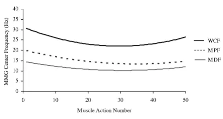
but linear decreases in MMG amplitude for the rectus fem-oris. At 180°·s-1, MMG amplitude decreased quadrati-cally for the vastus medialis muscle, but there were linear decreases in MMG amplitude for the rectus femoris and vastus lateralis [71]. The reductions in MMG amplitude at each velocity (60, 180, and 300°·s-1) were greater for the rectus femoris muscle than for the vastus lateralis and vas-tus medialis. In addition, at 60°·s-1, the decrease in con-centric isokinetic leg extension torque was best fit with a quadratic model, but at 180 and 300°·s-1, the patterns for torque were best fit with cubic models [71]. It was sug-gested that the decreases in MMG amplitude for each mus-cle at the three velocities may have been due to reduced muscle compliance and/or "muscle wisdom," in which the central nervous system reduces motor unit firing rates to compensate for fatigue-induced increases in muscle fiber relaxation times [74]. Theoretically, muscle wisdom allows for an economical activation of fatiguing muscle [75] and could reduce the number of pressure waves recorded per unit of time, thereby decreasing MMG ampli-tude [71,76]. In addition, it has been suggested [77-80] that during prolonged static and dynamic muscle actions, there are increases in muscle thickness, fluid content, and intramuscular pressure, all of which may contribute to reductions in the compliance (i.e. elastic properties) of the muscle. In theory, a decrease in muscle compliance could restrict the muscle fiber oscillations and/or attenu-ate the pressure waves that generattenu-ate the MMG signal, thereby decreasing MMG amplitude [71]. Søgaard et al. [81], however, recently suggested that MMG amplitude may not be influenced by changes in intramuscular pres-sure. Specifically, the authors [81] reported that during an isometric ramp muscle action of the forearm flexors from 10% to 60% MVC, MMG amplitude for the biceps brachii increased linearly with torque, even when intramuscular pressure was increased with a sphygmomanometer cuff. It was suggested that MMG amplitude may not be influ-enced by intramuscular pressure. This hypothesis should be tested, however, at higher torque levels and with differ-ent muscles to further examine the potdiffer-ential influence of intramuscular pressure on MMG amplitude. Furthermore, the greater decreases in MMG amplitude for the rectus femoris muscle than for the vastus lateralis and vastus medialis reported by Perry-Rana et al. [71] may have reflected a larger percentage of fast-twitch muscle fibers in the rectus femoris [60]. Beck et al. [72] examined the MMG amplitude and MPF responses from the biceps bra-chii during 50 consecutive maximal concentric isokinetic muscle actions of the forearm flexors at a velocity of 180°·s-1. The authors [72] found linear decreases in both MMG amplitude and MPF across the 50 repetitions. In addition, the decrease (approximately 70%) in mean fore-arm flexion torque was best fit with a cubic model [72]. It was suggested that the reductions in MMG amplitude and MPF may have been due to fatigue-induced decreases in Relationships for mechanomyographic (MMG) amplitude and
mean power frequency (MPF) versus torque during concen-tric isokinetic muscle actions
Figure 4
Relationships for mechanomyographic (MMG) ampli-tude and mean power frequency (MPF) versus
torque during concentric isokinetic muscle actions.
The top graph shows the linear relationship between
mechan-omyographic (MMG) amplitude (m·s-2) for the biceps brachii and concentric isokinetic torque. The bottom graph demon-strates the lack of a significant relationship between MMG mean power frequency (MPF, Hz) for the biceps brachii and concentric isokinetic torque. The data presented are from one subject, and the MMG signals were recorded with an accelerometer (Entran EGAS FT 10) during concentric isoki-netic forearm flexion muscle actions at a velocity of 30°·s-1.
y = 0.0032x + 0.1003 r2= 0.9682
0 0.1 0.2 0.3 0.4 0.5
0 20 40 60 80 100 %PT
0 5 10 15 20 25 30 35
0 20 40 60 80 100
%PT
MMG
MPF
(Hz
)
MMG
A
m
plit
ude
(m
·s
motor unit firing rates (i.e. muscle wisdom) and/or reduced compliance in the biceps brachii muscle. Beck et al. [72] also hypothesized, however, that de-recruitment of fast-twitch motor units [82,83], which have higher fir-ing rates than slow-twitch motor units [40,41,84], could, theoretically, result in decreases in both MMG amplitude and MPF. In addition, Perry-Rana et al. [73] examined the MMG amplitude responses from the rectus femoris, vastus lateralis, and vastus medialis during 25 consecutive maxi-mal eccentric isokinetic muscle actions of the leg exten-sors at a velocity of 120°·s-1. The authors [73] found that for the vastus lateralis and vastus medialis muscles, MMG amplitude decreased linearly across the 25 repetitions. For the rectus femoris muscle, however, the MMG amplitude pattern was best fit with a cubic model, in which MMG
amplitude increased slightly during repetitions 1–5, decreased during repetitions 5–20, and increased throughout the last 5 repetitions of the test. Interestingly, the pattern for eccentric leg extension torque was best fit with a cubic model, where torque increased during the first 10 repetitions, and then remained relatively stable from repetitions 11–25. The authors [73] hypothesized that the decreases in MMG amplitude for each muscle may have been due to the effects of muscle wisdom and/ or reduced muscle compliance. In addition, it was sug-gested that the muscle-specific differences in the MMG amplitude patterns (i.e. a cubic decline for the rectus fem-oris versus linear decreases for the vastus lateralis and vas-tus medialis) may have been due to differences among the three muscles in fiber type composition and/or architec-ture (unipennate versus bipennate).
Thus, the results from these studies [71-73] suggested that MMG amplitude and/or MPF could potentially provide information regarding the motor control strategies that are used during fatiguing concentric and eccentric isoki-netic muscle actions. Specifically, fatigue-induced decreases in MMG amplitude and MPF may reflect reduc-tions in motor unit firing rates (muscle wisdom) and/or de-recruitment of fast-twitch motor units [71-74,82]. In addition, MMG amplitude and/or MPF patterns during fatiguing isokinetic muscle actions may be influenced by fiber type composition and/or muscle architecture [71,73]. Finally, decreases in muscle compliance during repeated maximal isokinetic muscle actions [77-80] could attenuate the muscle fiber oscillations and/or pressure waves that generate the MMG signal.
MMG amplitude and MPF responses during cycle
ergometry
Several studies [29-32,85-87] have investigated the MMG amplitude and/or MPF responses during cycle ergometry. For example, Stout et al. [29] examined the patterns for
oxygen consumption rate (VO2) and MMG amplitude
from the vastus lateralis muscle during an incremental cycle ergometer test performed to exhaustion. The authors
[29] found that the MMG amplitude and VO2 versus
power output relationships were highly linear (r2 range = 0.79–0.99 and 0.97–0.99, respectively). In addition, for 20 of the 24 subjects that participated in the study, the lin-ear slope coefficients for the normalized MMG amplitude and VO2 versus power output relationships were statisti-cally equivalent. It was suggested that MMG could be use-ful for quantifying muscular activity and monitoring changes in exercise intensity during incremental cycle ergometry. Furthermore, the similar slope coefficients for
the normalized MMG amplitude and VO2 versus power
output relationships for the majority of the subjects indi-cated that there may be a close relationship between the metabolic (VO2) and mechanical (MMG) aspects of mus-Relationships for mechanomyographic (MMG) amplitude and
mean power frequency (MPF) versus torque during eccentric isokinetic muscle actions
Figure 5
Relationships for mechanomyographic (MMG) ampli-tude and mean power frequency (MPF) versus
torque during eccentric isokinetic muscle actions.
The top graph shows the linear relationship between
normal-ized mechanomyographic (MMG) amplitude and percent eccentric isokinetic peak torque (PT) for the vastus medialis muscle of one subject. The bottom graph demonstrates that for this particular subject (the same subject as in the top
graph), there was no relationship between normalized MMG
mean power frequency (MPF) for the vastus medialis muscle and percent eccentric isokinetic PT. The data presented are from one subject, and the MMG signals were recorded with a piezoelectric contact sensor during eccentric isokinetic mus-cle actions of the leg extensors at a velocity of 30°·s-1.
y = 0.9453x - 0.413
r2= 0.9426
0 10 20 30 40 50 60 70 80 90 100
0 20 40 60 80 100
% PT
N
or
m
al
iz
ed
M
M
G
A
mp
lit
ud
e
(%
max
)
0 10 20 30 40 50 60 70 80 90 100
0 20 40 60 80 100
%PT
N
or
m
al
iz
ed
M
M
G
M
P
F
(%
m
ax
cle activity during cycle ergometry. Shinohara et al. [30] also found that MMG amplitude for the vastus lateralis muscle increased linearly with power output during incre-mental cycle ergometry. In fact, MMG amplitude was more linearly related to power output than was EMG amplitude, which tended to increase curvilinearly at high power outputs. Perry et al. [31] reported similar results for the vastus lateralis muscle during incremental cycle ergometry. Specifically, for the majority of the subjects (7 out of 9), MMG amplitude increased linearly with power output, but EMG amplitude increased curvilinearly. The authors [31] also reported linear increases in normalized heart rate and ratings of perceived exertion (RPE) with power output, and the linear slope coefficients for these relationships were statistically equivalent to the slope coefficient for the linear normalized MMG amplitude ver-sus power output relationship. Thus, it was suggested that during incremental cycle ergometry, there may be a close relationship among the mechanical (MMG), heart rate, and perception of effort (RPE) aspects of muscular activ-ity.
The potential relationship between MMG frequency and motor unit firing rates [5,13,84] has suggested that the fre-quency domain of the MMG signal may provide informa-tion regarding the motor control strategies that are used during incremental cycle ergometry [32]. Theoretically, increases in MMG MPF could reflect modulation of motor unit firing rates with increasing power output. Perry et al. [32], however, reported no change in MMG MPF for the vastus lateralis muscle, but a linear increase in MMG amplitude with increases in power output during an incre-mental cycle ergometer test performed to exhaustion. It was suggested that during cycle ergometry, motor control strategies are examined across a narrow range of very low levels of force production. Specifically, Sjøgaard [88] reported that during cycle ergometry at 100% of VO2 max and a pedal cadence of 60 rev·min-1, the force exerted against the pedals represented approximately 16% of the isometric MVC. Large limb muscles (such as the vastus lat-eralis) typically rely heavily on recruitment for increasing isometric torque to about 50–80% MVC, above which, modulation of motor unit firing rates becomes progres-sively more important for increasing torque [33,70]. Thus, the lack of a significant change in MMG MPF for the vastus lateralis reported by Perry et al. [32] may have been due to the low force levels examined (relative to maximal capa-bilities) and recruitment as the primary motor control strategy for increasing power output, rather than firing rate modulation.
Several studies [85-87] have also examined the MMG amplitude responses during continuous cycle ergometry performed at constant, submaximal power outputs. For example, Housh et al. [87] investigated the patterns for
decrease in MMG amplitude for the vastus lateralis muscle during a 60-min cycle ergometer workbout at CP. It should be noted that different mathematical models have been used to estimate CP [91], and Housh et al. [92] found that CP was approximately 17% greater than the power output that could be maintained for 60-min. Thus, in some cases, CP may be a fatiguing workload, and it is possible that fatigue-related factors such as muscle wis-dom and/or reduced muscle compliance may have con-tributed to the decreases in MMG amplitude reported by Bull et al. [86] for the vastus lateralis muscle.
Collectively, the results from these studies provided sev-eral important pieces of information regarding the MMG amplitude and frequency responses during cycle ergom-etry. Specifically, the highly linear relationship between MMG amplitude and power output during incremental cycle ergometry (see Figure 6) [29-32] provided indirect support for the hypothesis of Bodor [44] that during max-imal isokinetic muscle actions, MMG amplitude may be more closely related to power output than force produc-tion. In addition, the lack of a significant change in MMG MPF for the vastus lateralis muscle reported by Perry et al. [32] suggested that during incremental cycle ergometry, firing rate modulation may not be as important as recruit-ment for increasing power output. Furthermore, the MMG amplitude responses during continuous, constant power output cycle ergometry are dependent on the relative workload that is being performed and may be related to the competing influences of fatigue-related decreases in motor unit firing rates and increases in motor unit recruit-ment. In particular, decreases in MMG amplitude for workloads below approximately 65% Wpeak may be due to fatigue-induced decreases in motor unit firing rates (muscle wisdom) and/or reduced muscle compliance, while an increase in MMG amplitude at 95% Wpeak may reflect a greater contribution of motor unit recruitment (which can increase MMG amplitude) than decreases in firing rates (which can decrease MMG amplitude) to the MMG signal [85-87]. Finally, the relatively stable MMG amplitude values during continuous cycle ergometry at 80% Wpeak may have been due to approximately equal influences of motor unit recruitment and decreases in fir-ing rates on the MMG signal [87].
Effects of dynamic resistance training on MMG
amplitude and frequency
Several studies [93-97] have investigated the potential for MMG to be used as a tool for examining the neuromuscu-lar adaptations that occur during a resistance training pro-gram. Resistance training results in increases in muscle fiber size (hypertrophy) [98,99] and muscle stiffness [100], and it has been suggested [94,95] that both of these factors may influence the amplitude and/or frequency contents of the MMG signal. For example, Evetovich et al.
[94] hypothesized that hypertrophy could decrease the distance between the muscle and the MMG sensor as the fibers press against the tissue layers superficial to the mus-cle (i.e. fascia, adipose tissue, etc.). The tissue between the muscle and the MMG sensor acts as a low pass filter for the muscle fiber oscillations and pressure waves that generate the MMG signal [4,25]. Thus, the close proximity of hypertrophied muscle fibers to the MMG sensor may result in less attenuation of the MMG signal by the tissue layers superficial to the muscle, resulting in greater MMG amplitude values [94]. In addition, it has been suggested [95] that training-induced increases in muscle stiffness could affect MMG frequency. Specifically, Barry and Cole [101] indicated that the MMG signal occurs at the "...res-onant frequency of the muscle," which is a function of the mass, length, topology, and stiffness of the muscle being investigated. It was suggested, however, that during an isometric muscle action, the changes in stiffness are greater than the changes in any of the other parameters [101]. In addition, both the resonant frequency and peak frequency of the MMG signal increased with force produc-tion during an electrically-stimulated tetanic contracproduc-tion of isolated frog gastrocnemius muscle [101]. Thus, these findings suggested that training-induced increases in mus-cle stiffness could potentially result in corresponding increases in the muscle's resonant frequency (and, by extension, MMG frequency).
The subjects in the training and control groups were tested for maximal concentric isokinetic leg extension PT at a velocity of 90°·s-1 prior to beginning the twelve week training program (week 0), and every four weeks thereaf-ter (weeks 4, 8, and 12, respectively). During each testing session, the MMG signal was recorded from the vastus lat-eralis muscle. The authors [94,95] found that there was a significant increase in leg extension PT across the twelve week training period for the training group, but not for the control group. There were, however, no changes in MMG amplitude or MPF for the vastus lateralis muscle from week 0 to week 12 for either group. It was suggested that the lack of a significant change in MMG amplitude and MPF following training may have been due to com-pression of hypertrophied muscle fibers in the vastus lat-eralis by the tendonous iliotibial band and/or training-induced adaptations in muscles other than the vastus
lat-eralis. Specifically, although muscle hypertrophy was not directly examined [94,95], increases in muscle fiber size could potentially stretch the iliotibial band, resulting in a taut layer of fascia that compresses the vastus lateralis and restricts the muscle fiber oscillations that generate the MMG signal. Theoretically, this could influence MMG amplitude and/or MPF. It is also possible, however, that training-induced adaptations in muscles other than the vastus lateralis may have contributed to increases in leg extension PT, with no changes in MMG amplitude and MPF. For example, reduced hamstring coactivation [102] and/or preferential hypertrophy of the other quadriceps femoris muscles [99] could have increased leg extension PT without affecting the MMG signal from the vastus lat-eralis [94,95].
Esposito et al. [96] recently examined the MMG ampli-tude and MPF versus isometric torque relationships for the vastus lateralis muscle of elderly men before and after an isokinetic strength training program. During this train-ing program, the subjects performed maximal concentric isokinetic muscle actions of the dominant leg extensors at velocities of 120 and 240°·s-1. The training was per-formed twice a week for twelve weeks. The authors [96] found that the training program had no effect on the MMG amplitude values for the vastus lateralis muscle at each relative torque level (20, 40, 60, 80, and 100% MVC). At 80% and 100% MVC, however, the training pro-gram resulted in significant increases in MMG MPF for the vastus lateralis muscle. In addition, the authors [96] reported that before training, the MMG power density spectrum for the vastus lateralis during an isometric MVC was "...unimodal, with a well-defined main peak at about 11 Hz." After training, however, the MMG power density spectrum during an MVC was bimodal, with a large peak at approximately 15 Hz and a smaller peak at about 30 Hz. It was suggested [96] that the training-induced increases in MMG MPF for the vastus lateralis muscle at 80% and 100% MVC, as well as the changes in the MMG power density spectrum, may have reflected a "retrieval" of fast-twitch motor units, which are lost in some muscles during the aging process [103]. In addition, Coburn [97] recently examined the effects of three days of velocity-spe-cific isokinetic training on the MMG amplitude and MPF values from the rectus femoris, vastus lateralis, and vastus medialis muscles. Thirty adults were randomly assigned to one of three groups: a) a control group, b) a slow velocity training group, or c) a fast velocity training group. The subjects in the two training groups performed three sepa-rate training sessions that consisted of maximal concentric isokinetic muscle actions of the nondominant leg exten-sors at a velocity of 30 (slow velocity training group) or 270°·s-1 (fast velocity training group). In addition, the subjects in the training groups were tested for maximal concentric isokinetic leg extension PT at velocities of 30, Relationships for mechanomyographic (MMG) amplitude and
mean power frequency (MPF) versus power output during incremental cycle ergometry
Figure 6
Relationships for mechanomyographic (MMG) ampli-tude and mean power frequency (MPF) versus power
output during incremental cycle ergometry. The top
graph shows the linear relationship between normalized
mechanomyographic (MMG) amplitude (%max) for the vastus lateralis muscle and normalized power output. The bottom graph demonstrates the lack of a significant relationship between normalized MMG mean power frequency (MPF, %max) for the vastus lateralis muscle and normalized power output. The data presented are from one subject, and the MMG signals were recorded with a contact sensor (HP 21050A) during an incremental cycle ergometer test to exhaustion.
y = 1.0202x + 0.3885
r2= 0.915
0 10 20 30 40 50 60 70 80 90 100
0 20 40 60 80 100
Normalized Power Output (%max)
N
or
m
al
ized
M
M
G
A
m
pl
itu
de
(%
m
ax
)
0 10 20 30 40 50 60 70 80 90 100
0 20 40 60 80 100
Normalized Power Output (%max)
N
or
m
al
iz
ed
M
M
G
M
P
F
(%
m
ax
150, and 270°·s-1 prior to, and following the training pro-gram. The subjects in the control group were also tested for leg extension PT at the same velocities, but did not per-form any training. Coburn [97] found training-induced increases in leg extension PT for the fast velocity training group at 270°·s-1 and for the slow velocity training group at 30, 150, and 270°·s-1. In addition, when compared to the control group, there were training-induced increases in MMG amplitude (averaged across the rectus femoris, vastus lateralis, and vastus medialis muscles) for the fast velocity training group at 270°·s-1 and for the slow veloc-ity training group at 150°·s-1. The isokinetic leg extension training did not, however, result in changes in MMG MPF for any muscle at any velocity. In addition, the training program had no effect on EMG amplitude for the rectus femoris, vastus lateralis, and vastus medialis muscles at any velocity. Thus, it was suggested that the increases in MMG amplitude for the rectus femoris, vastus lateralis, and vastus medialis muscles for the fast velocity training group at 270°·s-1 and for the slow velocity training group at 150°·s-1 may have been due to changes in muscle com-pliance. Specifically, although the EMG signal was not recorded from the hamstrings, it is possible that reduced coactivation in the biceps femoris, semitendinosus, or semimembranosus following the isokinetic training may have increased the net leg extension torque and affected compliance in the quadriceps femoris muscles [97]. As stated previously, muscle compliance could influence the muscle fiber oscillations and/or pressure waves that gen-erate the MMG signal [71-73,86,87].
Thus, the findings from these studies [93-97] provided conflicting evidence regarding the potential effects of dynamic strength training on the amplitude and fre-quency contents of the MMG signal. The training-induced increases in MMG frequency for the vastus lateralis and gastrocnemius muscles reported by Cerquiglini et al. [93] and Esposito et al. [96] suggested that the frequency domain of the MMG signal may provide information regarding functional changes in skeletal muscle following a training program, such as retrieval of fast-twitch motor units in elderly individuals. In contrast, Evetovich et al. [95] found that there was no change in MMG MPF for the vastus lateralis muscle following twelve weeks of concen-tric isokinetic leg extension training. It is possible that the discrepancies among the results from these investigations [93,95,96] may have been due to differences in the testing methods that were used and/or the characteristics of the subjects that participated in the studies. Specifically, Eve-tovich et al. [95] tested subjects during maximal concen-tric isokinetic muscle actions, while Cerquiglini et al. [93] and Esposito et al. [96] recorded MMG signals only during isometric muscle actions. In addition, Evetovich et al. [95] tested young untrained men, while Cerquiglini et al. [93] examined Olympic weightlifters and untrained
individu-als, and Esposito et al. [96] investigated elderly men. Fur-thermore, Coburn [97] found that three days of velocity-specific isokinetic training resulted in isolated changes in MMG amplitude for the rectus femoris, vastus lateralis, and vastus medialis muscles, but Evetovich et al. [94] reported that a twelve week isokinetic resistance training program had no effect on MMG amplitude for the vastus lateralis. There are several factors that may have contrib-uted to the differences between the results from these studies, the most obvious of which is the duration of the training program that was used (thirty-six training ses-sions over twelve weeks for Evetovich et al. [94] and three training sessions over approximately five to seven days for Coburn [97]). In addition, differences between the inves-tigations in the muscles that were examined and/or the velocities that were used for the testing and training may have influenced the results for MMG amplitude. Collec-tively, the findings from these studies [93-97] suggested that MMG could provide information regarding the adap-tations that occur during a resistance training program, but this possibility should be examined further in differ-ent muscles and/or subject groups, as well as following various types of training programs.
Acute effects of stretching on MMG amplitude
and MPF
Evetovich et al. [106] examined the acute effects of static stretching of the forearm flexors on PT, MMG amplitude, and EMG amplitude during maximal concentric isoki-netic muscle actions of the forearm flexors at velocities of 30 and 270°·s-1. The authors [106] found that the stretch-ing resulted in a significant decrease in forearm flexion PT (averaged across the 30 and 270°·s-1 velocities) and increases in MMG amplitude for the biceps brachii muscle at both velocities. The stretching had no effect, however, on the EMG amplitude values for the biceps brachii mus-cle at either velocity. It was suggested that the stretching-induced decreases in forearm flexion PT and increases in MMG amplitude for the biceps brachii may have been due to reduced muscle stiffness. Cramer et al. [105] examined the acute effects of static stretching of the dominant leg extensors on PT, mean power output, MMG amplitude, and EMG amplitude during maximal concentric isoki-netic leg extensions at velocities of 60 and 240°·s-1. The authors [105] reported that the stretching resulted in decreases in PT for the stretched limb at 60 and 240°·s-1, as well as for the unstretched limb at 60°·s-1. In addition, there were stretching-induced decreases in EMG ampli-tude for the rectus femoris and vastus lateralis muscles for both the stretched and unstretched limbs at 60 and 240°·s-1. It was suggested that the decreases in leg exten-sion PT and EMG amplitude following the static stretch-ing may have been due, at least partially, to reduced muscle activation in the rectus femoris and vastus lateralis muscles. The stretching had no effect, however, on mean power output and MMG amplitude for the rectus femoris or vastus lateralis muscles for either limb at 60 and 240°·s-1. Thus, these findings provided support for the hypothesis of Bodor [44] that MMG amplitude is more closely related to power output than PT during maximal isokinetic muscle actions. Marek et al. [110] recently examined the acute effects of static and proprioceptive neuromuscular facilitation (PNF) stretching on PT, mean power output, EMG amplitude, and MMG amplitude dur-ing maximal concentric isokinetic leg extensions at veloc-ities of 60 and 300°·s-1. PNF stretching is a technique that is designed to relax the stretched muscle(s) by activating Golgi tendon organs and reducing the stretch reflex, thereby allowing for less active resistance to muscle lengthening [111]. The authors [110] found that both static and PNF stretching resulted in decreases in PT, mean power output, and EMG amplitude for the rectus femoris and vastus lateralis muscles at 60 and 300°·s-1. In addi-tion, there was an increase in MMG amplitude following the static stretching, but only for the rectus femoris muscle at 60°·s-1. It was suggested that the stretching-induced decreases in PT, mean power output, and EMG amplitude for the rectus femoris and vastus lateralis may have been due to a combination of reduced muscle activation and decreases in muscle stiffness.
The acute effects of static stretching have also been exam-ined during maximal eccentric isokinetic muscle actions. Eccentric muscle actions may provide an interesting situa-tion for examining potential stretching-induced changes in PT and MMG amplitude, because Wilson et al. [112] found that unlike concentric and isometric muscle actions, maximal eccentric torque was not related to mus-culotendonous stiffness. Thus, decreases in muscle stiff-ness following stretching could, theoretically, result in increases in MMG amplitude, with no change in eccentric PT. Cramer [113] examined the acute effects of static stretching on PT, mean power output, EMG amplitude, EMG MPF, MMG amplitude, and MMG MPF during max-imal eccentric isokinetic muscle actions of the leg exten-sors at velocities of 60 and 180°·s-1. The results indicated that there were no meaningful changes in PT, EMG ampli-tude, EMG MPF, and MMG MPF following the static stretching. There were, however, stretching-induced decreases in mean power output and MMG amplitude for the rectus femoris muscle at 60 and 180°·s-1. It was sug-gested that the decreases in MMG amplitude for the rectus femoris muscle following stretching may have been due to a close relationship between MMG amplitude and mean power output.
Collectively, the results from these studies [105,106,110,113] indicated that the acute effects of stretching on MMG amplitude are muscle-specific as well as specific to the type of muscle action. For example, dur-ing maximal concentric isokinetic muscle actions, static stretching resulted in an increase in MMG amplitude for the biceps brachii [106], but not for the rectus femoris and vastus lateralis [105]. In addition, during maximal eccen-tric isokinetic muscle actions, static stretching actually decreased MMG amplitude for the rectus femoris and vas-tus lateralis [113]. The results from these studies also pro-vided support for the hypothesis of Bodor [44] that MMG amplitude is more closely related to power output than PT during maximal isokinetic muscle actions. Furthermore, the stretching-induced decreases in PT and EMG ampli-tude in both the stretched and unstretched limbs during maximal concentric isokinetic muscle actions [105] sug-gested that decreases in PT following static stretching may be partially due to neuromuscular factors such as altered motor control strategies and/or changes in reflex sensitiv-ity. Thus, simultaneous examination of EMG and MMG may provide information regarding the relative contribu-tions of neural (EMG) and mechanical (MMG) factors to stretching-induced decreases in isokinetic PT.
Processing MMG signals recorded during
dynamic muscle actions
units [4-6,114] and, in most cases, various sources of noise (e.g. movement artifact created by displacement of the muscle and/or limb) do not have a large influence on MMG amplitude or frequency. This is not always the case during dynamic muscle actions. For example, movement artifact is often present in biological signals such as the surface EMG [115-117], electroencephalogram (EEG) [118], and electrocardiogram (ECG) [119], and it has been suggested [120-122] that large limb and/or muscle movements may also influence the MMG signal. In addi-tion, changes in muscle length and the thickness of the tis-sue between the muscle and the MMG sensor could affect MMG amplitude and frequency during a dynamic muscle action [1,2,25]. Thus, the MMG signals recorded during dynamic activities are typically nonstationary [123] and may require different signal processing methodologies when compared to isometric muscle actions. Specifically, there are three factors that are particularly important when analyzing MMG signals recorded during dynamic muscle actions: a) filtering the signal to attenuate various sources of noise, b) choosing the length of the epoch(s) (i.e. window) to select from the recorded signal, and c) using the appropriate method to analyze the signal's fre-quency content.
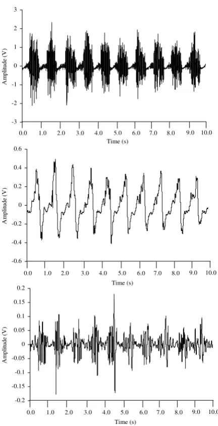
One of the most important considerations in filtering MMG signals is selecting the appropriate cutoff frequen-cies. Most previous studies have used analog and/or dig-ital bandpass filters with cutoff frequencies between 1 and 250 Hz [9,68,124-130] when processing MMG signals recorded during isometric muscle actions. Other investi-gations, however, have used filters with different cutoff frequencies to reduce various types of noise in the MMG signal. For example, Mealing et al. [84] used a 3 Hz high pass filter to attenuate arterial sounds, while Goldenberg et al. [83] used a high pass filter with a cutoff frequency of 14 Hz to reduce the influence of tremor on the MMG sig-nal. In addition, Weir et al. [131] used a 60 Hz notch filter to remove potential powerline interference from the MMG signal. Filters have also been used to isolate specific frequency components of the MMG power density spec-trum. For example, Petitjean and Bellemare [132] used a 30 Hz high pass filter to examine the high frequency com-ponents of MMG signals from the diaphragm muscle. It was suggested [132] that the filtered signals may provide information regarding the resonant frequency of the mus-cle being investigated [101]. During dynamic musmus-cle actions, however, filters have been used primarily to atten-uate movement artifact (see Figure 7). Movement artifact is a source of low frequency noise in many biological sig-nals [115-119]. For high frequency sigsig-nals, filtering the movement-related noise usually results in less attenuation of the original signal because the major portions of the spectra from the noise and the signal of interest do not overlap substantially [133]. For example, the influence of
movement artifact is often reduced in surface EMG signals by high pass filtering with a cutoff frequency between 20 and 30 Hz [115]. In most cases, this does not severely attenuate the original EMG signal because most of the power in surface EMG is between approximately 40 and 160 Hz [134]. For lower frequency signals (such as MMG), however, the low frequency cutoff should be care-fully selected to attenuate as much noise as possible, yet preserve the signal of interest. Petitjean et al. [64] used a bandpass filter with cutoff frequencies of 10 and 60 Hz to remove noise from MMG signals recorded during concen-tric DCER muscle actions of the biceps brachii and bra-chioradialis. Dalton and Stokes [26] used a bandpass filter with cutoff frequencies of 8 and 160 Hz when processing MMG signals from the biceps brachii recorded during concentric and eccentric DCER muscle actions of the fore-arm flexors. Most studies, however, have used a filter with a 5 Hz high pass cutoff frequency to attenuate movement artifact in MMG signals [120-122]. Theoretically, this technique reduces the influences of body and respiratory movements, as well as gross limb displacements [122].