R E S E A R C H
Open Access
Time-dependent post mortem changes in the
composition of intestinal bacteria using real-time
quantitative PCR
Sari Tuomisto
1,2*, Pekka J Karhunen
1,2and Tanja Pessi
1,2Abstract
Post mortem or even normal changes during life occurring in major gut bacterial populations are not known. We investigatedBacteroidessp.,Bifidobacteriumsp.,Clostridium leptum, Clostridium coccoides,Streptococcussp., Lactobacillussp. andEnterobacteriacaearatios in 7 fecal samples from healthy volunteers and in 61 autopsies rectum and cecum samples and studied the effect of post mortem time using quantitative real-time PCR. Bacterial ratios in stool samples from volunteers and rectum samples from autopsy cases were similar and did not change significantly up to 5 days post mortem. In cecum, significant post mortem time-dependent differences were observed in ratios ofBacteroidessp. (p = 0.014) andLactobacillussp. (p = 0.024). Our results showed that ratios of Bacteroidessp.,Bifidobacteriumsp.,Clostridium leptum,Clostridium coccoides,Streptococcussp.,Lactobacillussp. and Enterobacteriacaeacan be investigated in autopsy rectum samples up to 5 days after death.
Keywords:Forensic science, Post mortem microbiology, Fecal sample, Real-time quantitative polymerase chain
reaction, Bacterial relative amount, Time-dependent changes
Background
Basic knowledge on the composition of intestinal bacterial populations and changes occurring after death is lacking. Even the normal composition of intestinal microbiota in life is not fully known [1]. Only one study exists in which intestinal bacterial populations have been studied in three elderly women after death using PCR and sequencing [2].
Resident micro-organisms living in the intestinal tract influence host’s normal well-being and physiology includ-ing gut metabolism and the regulation of epithelial cell growth [3]. Intestinal microbiota functions as a physical barrier against invading pathogens. It has been suggested that gut microbiota may have a role on the development of diseases,e.g.alcoholic liver cirrhosis [4] and atheroscler-osis [5]. Detailed bacterial population studies on the in-testinal tract have mostly concentrated on fecal samples because they are easy to collect. Intestinal microbiota consists of a large and diverse community containing hun-dreds of commensal bacterial species [6]. From sequencing
libraries of 16S rRNA genes Durban et al. found that two dominant phyla, Firmicutes and Bacteroidetes accounted for nearly 85% of all sequences in stool samples [7]. Com-pared to these two major phyla,Bifidobacterium genus is present in eight to ten-fold lower numbers [8]. Although
Bacteroides sp., Bifidobacterium sp. and bacteria belong-ing to theClostridium coccoides–group (cluster XIVa) and
Clostridium leptum–group (cluster IV) dominate in colon [9,10] there is substantial inter- and intra-individual vari-ation in species composition and distribution [7,11].
This study aimed to investigate ratios of major intes-tinal bacterial populations in healthy volunteers and in rectum and cecum autopsy samples. Post mortem time-dependent changes were studied in order to see whether autopsy samples can be used for basic research concern-ing lifetime. Six species:Bacteroidessp. (phylum Bacter-oidetes), Clostridium sp. (Firmicutes), Streptococcus sp. (Firmicutes), Lactobacillussp. (Firmicutes),Bifidobacterium
sp. (Actinobacteria) andEnterobactericaea(Proteobacteria) were chosen since they represent the major intestinal bac-terial phyla [12].
* Correspondence:[email protected] 1
Department of Forensic Medicine, School of Medicine, University of Tampere, Medisiinarinkatu 3, Tampere 33014, Finland
2
Fimlab Ltd, Pirkanmaa Hospital District, Biokatu 4, Tampere 33520, Finland
Findings
Study design and results
This study comprises of 61 male cases collected in the Department of Forensic Medicine of the University of Tampere and 7 male volunteers. The selection criteria for the autopsies have been described elsewhere [13]. None of the controls or cases was reported to has been used antibiotics. Deceased had been stored in +4°C within 24 hours after death. Written consent was ob-tained from the volunteers.
Samples of the autopsy cases were taken from rectum and cecum. All samples were frozen immediately at−80°C until further processing. On the basis of time post mortem the cases were divided into groups: 1–3 days, 4–5 days and >5 days. Demographic characteristics of these groups are shown in the Table 1.
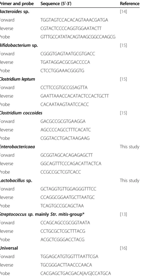
Fecal samples were weighed to be 150 mg (wet weight). Bacterial DNA was extracted from the samples using Zymo Fecal DNA Kit (Zymo Research Corporation, Irvine, California, USA). The bacterial ratios were deter-mined by RT-qPCR using specific primers and probes (Table 2). The primers and probes forEnterobacteriacaea
and Lactobacillus sp. were designed and confirmed by using BLAST (http://www.ncbi.nlm.nih.gov/) and Ribosomal Database Project (http://rdp.cme.msu.edu/probematch/ search.jsp). Specificity and cross reactivity of the de-signed primers and probes were tested using bacterial cultures from clinical samples [13]. PCR assays were performed with AbiPrism 7000 HT Sequence Detection System (Taqman, AppliedBiosystems, California, USA) with Taqman Environmental MasterMix. Endogen and DNA-free water was used as a negative control.
The comparative Ct method (ΔΔCt, ΔCt sample –
ΔCtreference sample)[17], was used where mean values from
healthy male volunteers were calculated and used as a reference to determine bacterial relative amount in rec-tum samples. The differences of the Ct values between the bacteria and the universal bacteria measurement (ΔCt) for each sample were calculated; the comparative Ct (ΔΔCt) for sample and reference samples was
Table 2 Used primers and probes
Primer and probe Sequence (5′-3′) Reference
Bacteroidessp. [14]
Forward TGGTAGTCCACACAGTAAACGATGA
Reverse CGTACTCCCCAGGTGGAATACTT
Probe GTTTGCCATATACAGTAAGCGGCCAAGCG
Bifidobacteriumsp. [15] Forward CGGGTGAGTAATGCGTGACC
Reverse TGATAGGACGCGACCCCA
Probe CTCCTGGAAACGGGTG
Clostridium leptum [15] Forward CCTTCCGTGCCGSAGTTA
Reverse GAATTAAACCACATACTCCACTGCTT
Probe CACAATAAGTAATCCACC
Clostridium coccoides [15] Forward GACGCCGCGTGAAGGA
Reverse AGCCCCAGCCTTTCACATC
Probe CGGTACCTGACTAAGAAG
Enterobactericaea This study Forward GCGGTAGCACAGAGAGCTT
Reverse GGCAGTTTCCCAGACATTACTCA
Probe CCGCCGCTCGTCACC
Lactobacillussp. This study Forward GCTAGGTGTTGGAGGGTTTCC
Reverse CCAGGCGGAATGCTTAATGC
Probe TCAGTGCCGCAGCTAA
Streptococcus sp.mainlyStr. mitis-group* [13] Forward CCAGCAGCCGCGGTAATA
Reverse CCTGCGCTCGCTTTACG
Probe ACGCTCGGGACCTACG
Universal [16]
Forward TGGAGCATGTGGTTTAATTCGA
Reverse TGCGGGACTTAACCCAACA
Probe CACGAGCTGACGACA[A/G]CCATGCA
*This was abbreviated asStreptococcussp. in the text.
Table 1 Demographic characteristics of the study subjects divided by post mortem time
Basic cause of death
N PM mean Age mean
(range)
BMI mean (range) Heart diseases %
Other diseases %
Violent deaths (suicide, accident, poisoning) %
Autopsy cases:
1–3 days 19 2.3 55 (18–79) 29.3 (20.4–42.1) 7 (37%) 4 (21%) 8 (42%)
4–5 days 21 4.5 58 (20–86) 28.4 (18.4–43.6) 10 (48%) 9 (43%) 2 (10%)
>5 days 21 6.5 61 (28–76) 30.7 (21.1–50.3) 15 (71%) 2 (10%) 4 (19%)
p-value 0.373 0.543 0.079 0.086 0.096
Control volunteers 7 45 (26–57) 27.1 (20.8–37.2)
PM mean = Post mortem mean time.
Tuomistoet al. Gut Pathogens2013,5:35 Page 2 of 5
calculated. To determine relative amounts of bacteria in cecum samples the rectal sample was used as an inner reference.
Two standard curves were used to determine the total amount of bacteria. Tenfold dilution series of between 33 ng/ml and 0.00033 ng/ml fromE. coligenomic DNA (ATCC 35401–5) as well as between 109and 105colony forming units (CFU) per milliliter from E .coli (ATCC 25922) were applied. The amount of CFU or bacterial DNA in the sample was calculated using values from
universal measurement and the equation y = slope log (X) + intercept [18].
Statistical analyses were performed with Kruskal-Wallis median test with PASW Statistical Software, ver-sion 18 (SPSS Ltd, Quarry Bay, Hong Kong). If P-value was less than 0.05 (considered significant) pairwise Post Hoc comparisons using Mann–Whitney U-test were done.
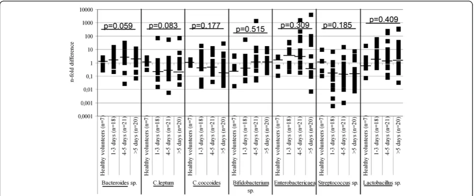
Median values of different bacteria in the stool of healthy controls and in post mortem rectum samples
Figure 1Relative amounts (n-fold difference) of measured bacteria (Bacteroidessp.,C. leptum,C. coccoides, Bifidobacteriumsp., Enterobactericaea,Streptococcussp. andLactobacillussp.) in fecal samples of controls and rectum of autopsy cases.Individual values are presented as boxes, median values with horizontal lines. Comparisons over the groups were calculated using non-parametric Kruskal-Wallis test.
Table 3 The relative amounts (n-fold difference) of measured bacteria in cecum samples compared to rectum samples over post mortem time
Bacterial Group
Bacteroidessp. C. leptum C. coccoides Bifidobacteriumsp. Enterobactericaea Streptococcussp. Lactobacillussp.
All Median 0.32 0.72 1.29 1.18 0.86 2.19 0.82
25th–75th 0.13–1.06 0.41–1.61 0.27–3.94 0.52–2.66 0.09–3.38 0.52–7.50 0.25–3.61
1–3 days
Median 0.15 0.59 0.64 2.03 1.37 3.56 1.25
25th–75th 0.01–0.43 0.31–2.94 0.20–4.55 0.84–35.63 0.26–14.77 0.85–35.32 0.46–7.11
4–5 days
Median 0.53 1.09 1.27 0.61 0.68 2.28 0.30
25th–75th 0.17–1.60 0.60–1.81 0.15–2.30 0.26–2.34 0.03–1.85 0.34–8.07 0.16–1.95
>5 days
Median 0.53 0.60 2.81 1.03 0.86 1.85 1.09
25th–75th 0.21–1.45 0.41–1.39 0.59–4.27 0.45–1.61 0.16–7.94 0.27–5.68 0.65–7.84
p-value 0.014 0.472 0.421 0.054 0.358 0.192 0.024
showed no statistically significant changes over post mortem time (Figure 1). In cecum, significant post mortem time-dependent differences were observed over the groups in the relative amounts of Bacteroides
sp. (p = 0.014) andLactobacillussp. (p = 0.024, Table 3). There were significantly more Bacteroides sp. (p = 0.012) and lessLactobacillussp. (p = 0.015) already in 4–5 days. Statistically significant differences in the total amount of bacterial DNA were seen in healthy volunteers and aut-opsy rectum samples (p = 0.044, Table 4). In autaut-opsy rec-tum, the amount of bacterial DNA remained quite stable with time elapsing post mortem except for a high increase observed after day 5 post mortem (p = 0.023). A slightly higher total amount of bacterial DNA (measured as a wet weight) in stool samples donated by the volunteers compared to autopsy rectum samples might be due to lower water concentration in stool compared to rectum without changes in bacterial ratios [19]. Inter-individual variation was great at all time points and in all bacterial measurements.
Conclusion
This study showed that relative amounts of major intes-tinal bacteria in rectum of autopsy cases were similar to stool donated by volunteers and remained quite stable over post mortem time up to 5 days, after which the total amount of bacteria started to increase. In contrast, in cecum significant post mortem time-dependent differ-ences were observed as increase in ratio of strictly anaerobic Bacteroides sp. and decrease of facultative
Lactobacillus sp. due to hypoxia after death. In cecum there is accumulation of undigested nutrients and me-tabolites produced by bacteria after death, which may be conducive to anaerobic bacterial growth. This study showed that autopsy rectum samples can be used to evaluate major intestinal bacterial populations concern-ing lifetime up to 5 days after death.
Competing interests
The authors declare that they have no competing interests.
Authors’contributions
ST performed experiments and analyses, helped in collection of the autopsy samples and wrote the manuscript. PK was the iniator of the project and group leader and participated in writing the script. TP was the guarantor of the microbiological part of the study, designed the sample collection and experiments, and participated in writing the manuscript. All authors read and approved the final manuscript.
Acknowledgements
The excellent technical assistance from the personnel of the Department of Forensic Medicine, especially Mervi Seppänen, Kari Mänttäri and Olli Penttilä, University of Tampere, Finland is gratefully acknowledged. We also appreciate the participation of the volunteers.
Funding
This study was financially supported by the Tampere Graduate Program in Biomedicine and Biotechnology (TGPBB), the Finnish Foundation for Alcohol Studies, the Competitive Research Funding of the Pirkanmaa Hospital District, the European Union 7th Framework Program grant number 201668 for the AtheroRemo Project, the Pirkanmaa Regional Fund of the Finnish Cultural Foundation.
Received: 30 August 2013 Accepted: 20 November 2013 Published: 25 November 2013
References
1. Peterson J, Garges S, Giovanni M, McInnes P, Wang L, Schloss JA, Bonazzi V, McEwen JE, Wetterstrand KA, Deal C, Baker CC, Di Francesco V, Howcroft TK, Karp RW, Lunsford RD, Wellington CR, Belachew T, Wright M, Giblin C, David H, Mills M, Salomon R, Mullins C, Akolkar B, Begg L, Davis C, Grandison L, Humble M, Khalsa J, Little AR,et al:The NIH human microbiome project. Genome Res2009,19:2317–2323.
2. Hayashi H, Takahashi R, Nishi T, Sakamoto M, Benno Y:Molecular analysis of jejunal, ileal, caecal and recto-sigmoidal human colonic microbiota using 16S rRNA gene libraries and terminal restriction fragment length polymorphism.J Med Microbiol2005,54:1093–1101.
3. Morris JA, Harrison LM, Biswas J, Telford DR:Transient bacteraemia: a possible cause of sudden life threatening events.Med Hypotheses2007, 69:1032–1039.
4. Chen Y, Yang F, Lu H, Wang B, Chen Y, Lei D, Wang Y, Zhu B, Li L: Characterization of fecal microbial communities in patients with liver cirrhosis.Hepatology2011,54:562–572.
5. Rosenfeld ME, Campbell LA:Pathogens and atherosclerosis: Update on the potential contribution of multiple infectious organisms to the pathogenesis of atherosclerosis.Thromb Haemost2011,106:858–867. 6. Eckburg PB, Bik EM, Bernstein CN, Purdom E, Dethlefsen L, Sargent M, Gill
SR, Nelson KE, Relman D:Diversity of the human intestinal microbial flora. Science2005,308:1635–1638.
7. Durbán A, Abellán JJ, Jiménez-Hernández N, Ponce M, Ponce J, Sala T, D'Auria G, Latorre A, Moya A:Assessing gut microbial diversity from feces and rectal mucosa.Microb Ecol2011,61:123–133.
Table 4 The total amount of bacterial DNA in fecal samples
N ng/g median* 25th-75th p-value1) p-value2)
Healthy volunteers Stool Control 7 26 9.2-36.7
Autopsy cases Rectum 1-3 days 18 8 2.0-53.6
4-5 days 21 8 1.7-41.4
>5 days 20 42 12.0-124.2 0.044 0.023
Autopsy cases Cecum 1-3 days 19 51 13-3-94.1
4-5 days 21 68 5.1-194.7
>5 days 21 48 6.5-113.6 0.982
*1 ng/g corresponds to 4.8x10¹⁰
colony forming units usingE. colias a standard. P-values (over the groups) for1)
healthy volunteers and autopsy cases, 2)
autopsy cases only. 25th
-75th
interquartile range. Non-parametric median, Kruskal-Wallis-test comparisons over the groups.
Tuomistoet al. Gut Pathogens2013,5:35 Page 4 of 5
8. Mariat D, Firmesse O, Levenez F, Guimarăes V, Sokol H, Doré J, Corthier G, Furet JP:The Firmicutes/Bacteroidetes ratio of the human microbiota changes with age.BMC Microbiol2009,9:123.
9. Lay C, Sutren M, Rochet V, Saunier K, Dore J, Rigottier-Gois L:Design and validation of 16S rRNA probes to enumerate members of the clostridium leptum subgroup in human faecal microbiota.Environ Microbiol2005, 7:933–946.
10. Orrhage K, Nord CE:Bifidobacteria and lactobacilli in human health. Drugs Exp Clin Res2000,26:95–111.
11. Turnbaugh PJ, Hamady M, Yatsunenko T, Cantarel BL, Duncan A, Ley RE, Sogin ML, Jones WJ, Roe BA, Affourtit JP, Egholm M, Henrissat B, Heath AC, Knight R, Gordon JI:A core gut microbiome in obese and lean twins. Nature2009,457:480–484.
12. Arumugam M, Raes J, Pelletier E, Le Paslier D, Yamada T, Mende DR, Fernandes GR, Tap J, Bruls T, Batto JM, Bertalan M, Borruel N, Casellas F, Fernandez L, Gautier L, Hansen T, Hattori M, Hayashi T, Kleerebezem M, Kurokawa K, Leclerc M, Levenez F, Manichanh C, Nielsen HB, Nielsen T, Pons N, Poulain J, Qin J, Sicheritz-Ponten T, Tims S,et al:Enterotypes of the human gut microbiome.Nature2011,12:174–180.
13. Tuomisto S, Karhunen P, Vuento R, Aittoniemi J, Pessi T:Evaluation of post mortem bacterial migration using culturing and real-time quantitative PCR.J Forensic Sci2013,58:910–916.
14. Brunk CF, Li J, Avaniss-Aghajani E:Analysis of specific bacteria from environmental samples using a quantitative polymerase chain reaction. Curr Issues Mol Biol2002,4:13–18.
15. Furet JP, Firmesse O, Gourmelon M, Bridonneau C, Tap J, Mondot S, Doré J, Corthier G:Comparative assessment of human and farm animal faecal microbiota using real-time quantitative PCR.FEMS Microbiol Ecol2009, 68:351–362.
16. Yang S, Lin S, Kelen GD, Quinn TC, Dick JD, Gaydos CA, Rothman RE: Quantitative multiprobe PCR assay for simultaneous detection and identification to species level of bacterial pathogens.J Clin Microbiol 2002,40:3449–3454.
17. Suzuki N, Yoshida A, Nakano Y:Quantitative analysis of multi-species oral biofilms by TaqMan real-time PCR.Clin Med Res2005,3:176–185. 18. Yoshida A, Suzuki N, Nakano Y, Kawada M, Oho T, Koga T:Development of
a 5′nuclease-based real-time PCR assay for quantitative detection of cariogenic dental pathogens streptococcus mutans and streptococcus sobrinus.J Clin Microbiol2003,41:4438–4441.
19. Geibel JP:Secretion and absorption by colonic crypts.Annu Rev Physiol 2005,67:471–490.
doi:10.1186/1757-4749-5-35
Cite this article as:Tuomistoet al.:Time-dependent post mortem changes in the composition of intestinal bacteria using real-time quanti-tative PCR.Gut Pathogens20135:35.
Submit your next manuscript to BioMed Central and take full advantage of:
• Convenient online submission
• Thorough peer review
• No space constraints or color figure charges
• Immediate publication on acceptance
• Inclusion in PubMed, CAS, Scopus and Google Scholar
• Research which is freely available for redistribution