Bridging Small-Gap Peripheral Nerve Defect Using Silicone Rubber
Chamber in the Rat Sciatic Nerve Transection Model
Saeed Azizi 1*
Rahim Mohammadi 1
Keyvan Amini 2 Roza Fallah 3 Kiana Karegar 4
1
Department of Clinical Sciences, Faculty of Veterinary Medicine, Urmia University, Urmia, Iran
2
Department of Veterinary Pathology, Western College of Veterinary Medicine, University of Saskatchewan, Saskatoon SK, Canada
3
DVM Student of Faculty of Veterinary Medicine, Tehran University, Tehran, Iran
4
DVM Student of Faculty of Veterinary Medicine, Urmia University, Urmia, Iran
Received: 11 August 2010, Accepted: 25 September 2010
Abstract
Despite promising results observed using silicone rubber chamber, no previous comprehensive work was performed on behavior of the conduit. Present study aimed at further functional, histomorphometrical and immunohistochemical assessment of nerve regeneration in the same animal along a 10-mm rat sciatic nerve gap. Fifty- four male Wistar rats were divided into three experimental groups (n = 18), randomly: Sham-operation (NC), Transected control (TC) and silicone conduit (SIL). In NC group after anesthesia left sciatic nerve was exposed through a gluteal muscle incision and after haemoestasis the muscle was sutured. In TC group left sciatic nerve was exposed the same way and transected proximal to the tibio-peroneal bifurcation leaving a 10-mm gap. In SIL group left sciatic nerve was transected the same way and proximal and distal stumps were each inserted into a silicone tube. Each group was subdivided into three subgroups of six animals each and were studied 4, 8, 12 weeks after surgery. Functional analysis showed significant improvement of nerve function in SIL group than in TC group (P < 0.05). Morphometric indices and immuohistochemistry indicated there were significant differences (P < 0.05) between SIL and TC groups 12 weeks after surgery. Silicone entubulation technique has offered the hope of providing a method for achieving the peripheral nerve regeneration in the least harmful way that is available, easily performed. Using silicone tubes in bridging of nerve defects could be promising because it is inert and does not induce extensive scarring or degeneration after implantation.
Key words: Peripheral nerve regeneration, Silicone rubber chamber, Rat
*
Corresponding author: Saeed Azizi, DVM, DVSc
Introduction
Peripheral nerves have self regeneration capacity after traumatic injury. In case of significant damage to nerve tissue, severed nerves do not spontaneously restore their function, and their continuity has to be first reestablished by microsurgical intervention such as suturing or interposition of a graft.1,2 Reconstructive surgical procedures are required following traumatic or iatrogenic damage to peripheral nerves or after excision of primitive neoplasms. Experimental studies and clinical reports indicate that insertion of a conduit could be an interesting alternative to direct end-to-end suturing of nerve stumps or interposition of an autograft.3-5 Nowadays, conduits are mainly made of non-bioabsorbable materials including silicone or bioabsorbable materials such as aliphatic polyesters, polyurethane, collagen, chitosan and excised artery or vein.1,6 The advantage of these conduits is the avoidance of sacrificing a segment of the donor nerve with subsequent loss of function and/or neuroma formation and also providing a microenvironment that is optimal for regeneration.7,8 It has been reported that using silicone tubes in bridging of nerve defects could be promising because it is inert and does not induce extensive scarring or degeneration after implantation.9 The advantages like no donor morbidity, availability, affordability and no foreign reactions make silicone rubber chamber an attractive alternative to other standard grafts.10 It has been demonstrated that silicone rubber tubes are well tolerated in humans even after 3 years of implantation.11 Silicone chambers are used as a standard experimental model to study cellular and molecular changes during the nerve regeneration process. Within the first hours of implantation, the chamber is filled by a fluid enriched with neurotrophic and neurotropic molecules and factors that stimulate migration and proliferation of Schwann cells.12 The
Schwann cells and its basal lamina are crucial components in the environment through which regenerating axons grow to reach their peripheral targets.13
We used well-established test systems because we aspired to examine comprehensive behavior of the silicon tube in the very same animal, an effort not attempted yet to the best of our knowledge. In light of promising clinical results obtained by silicone grafting technique, the present study aimed at comprehensive functional,
histomorphometrical and
immunohistochemical (Schwann cell detection by S-100 expression) assessments of rat sciatic nerve regeneration 4, 8, and 12 weeks after surgery.
Materials and Methods
Experimental Design. Fifty-four male Wistar rats weighing approximately 280g were divided into three experimental groups (n = 18), randomly: Sham-operation, normal control (NC), transected control (TC) and silicone conduit (SIL). Each group was further subdivided into three subgroups of six animals each (Fig. 1). Two weeks before and during the entire experiments, the animals were housed in individual plastic cages (50 × 40 × 20 cm) with an ambient temperature of 23 ± 3 ºC, stable air humidity, and a natural day/night cycle. The animals were handled on a regular daily basis for 2 weeks prior to the study in order to acclimatize them with testing area and experiments. The rats had free access to standard rodent laboratory food and tap water.
Committee and the University Research Council approved all experiments.
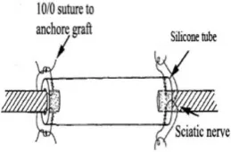
Following surgical preparation in the sham-operation group (NC), the left sciatic nerve was exposed through a gluteal muscle incision and after careful haemoestasis the muscle was sutured with 4/0 Vicryl (Ethicon, Norderstedt) and the skin with 3/0 nylon (Dafilon, B/Braun, Germany). In transected control group (TC) the left sciatic nerve was exposed the same way, transected proximal to the tibio-peroneal bifurcation where a 7 mm segment was excised, leaving a gap about 10 mm due to retraction of the nerve ends. The proximal and distal stumps were fixed in the adjacent muscle with 10/0 nylon epineurial suture. No conduit was placed between the stumps. In silicone conduit group (SIL), proximal and distal stumps were each inserted 2 mm into the conduit and two 10/0 nylon sutures were placed at each end of the cuff to fix the tube in place and leave a 10-mm gap between the stumps (Fig 1). After surgery had carried out animals were housed in groups of six per cage under the same conditions mentioned above. The animals of each group were anesthetized by intraperitoneal administration of ketamine-xylazine (see above) and were perfused via left cardiac ventricle with a fixative containing 2 % paraformaldehyde and 1 % glutaraldehyde buffer (pH = 7.4) at 4 (n = 6), 8 (n = 6) and 12 weeks (n = 6) after surgery.
Fig 1. End-to-end anastomosis of silicone tube to the stumps of transected sciatic nerve. Proximal and distal stumps were each inserted 2 mm into the graft and two 10/0 nylon sutures were placed at each end of the cuff to fix the graft in place and leave a 10-mm gap between the stumps.
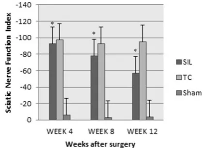
Functional assessment of nerve regeneration. Walking track analysis was performed 4, 8 and 12 weeks after surgery based on Bain et al.,14 The lengths of the third toe to its heel (PL), the first to the fifth toe (TS), and the second toe to the fourth toe (IT) were measured on the experimental side (E) and the contralateral normal side (N) in each rat. The Sciatic Function Index (SFI) in each animal was calculated by the following formula:
SFI= - 38.3 × (EPL-NPL)/NPL + 109.5 × (ETS-NTS)/NTS + 13.3 × (EIT-NIT)/NIT-8.8
In general, the SFI oscillates around 0 for normal nerve function, whereas around −100 SFI represents total dysfunction. The SFI was assessed based on the NC group and the normal level was considered as 0. The SFI was a negative value and a higher SFI meant the better function of the sciatic nerve.
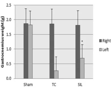
Muscle mass. Recovery assessment was also indexed using the weight ratio of the gastrocnemius muscles 12 weeks after surgery. Immediately after sacrificing of animals, gastrocnemius muscles were dissected and harvested carefully from intact and injured sides and weighed while still wet, using an electronic balance. All measurements were made by two blinded observers unaware of the analyzed group.
Histological preparation and
Immunohistochemical analysis. In this study, anti-S-100 (1:200, DAKO) was used as marker for myelin sheath. Specimens prior to immunohistochemistry were post fixed with 4% paraformaldehyde for two hours and embedded in paraffin. After non-specific immunoreactions were blocked, sections were incubated in S-100 protein antibody solution for one hour at room temperature. They were washed three times with PBS and incubated in biotynilated anti-mouse rabbit IgG solution for one hour. Horseradish peroxidase-labelled secondary antibody was developed by the diaminobenzidine method. The results of Immunohistochemistry were examined under a light microscope.
Statistical analysis. Experimental results were expressed as means ± SD. All data were analyzed by one-way analysis of variance (ANOVA) to assess statistical significance between experimental groups (SPSS 17.0 for Windows). Dunnett's test for pairwise comparisons was used to examine the effect of time and treatments. The differences were considered significant when P < 0.05.
Results
Recovery of sciatic nerve function.
Figure 2 shows sciatic function index (SFI) values in all three experimental groups. Prior to surgery, SFI values in all groups were near zero. After sciatic nerve transection, the mean SFI decreased to -100 due to the complete loss of sciatic nerve function in all animals. Four weeks after surgery mean SFI was -93.82 ± -3.24 in SIL group, compared to -97.3 ± -0.78 in group TC, with insignificant change in the NC. Eight weeks after surgery the improvement in SFI was observed in SIL, indicating that some regenerating axons have passed through the silicon graft and eventually into the target organ, whereas in group TC, no comparable SFI value was obtained after 8 weeks. After 12 weeks, animals of group SIL achieved a mean
value for SFI of -57.1 ± 3.10, i.e. an approximate improvement of 52 %, whereas in group TC, a mean value of -95.2 ± 0.97, i.e. an approximate improvement of 5 %, was found. Recovery of nerve function was not observed in the TC throughout 12 weeks post operation. The statistical analyses revealed that the recovery of nerve function was significantly (P < 0.05) different between SIL and TC groups and interposition of the silicone conduit significantly promoted functional recovery in the course of time.
Fig 2. Diagrammatic representation of effects on the sciatic nerve function index (SFI). Entubulation with silicone tube gave better results in functional recovery of the sciatic nerve than in TC group. Data are presented as mean ± SD. * P < 0.05 vs TC group.
Muscle mass measurement. The mean ratios of gastrocnemius muscles weight were measured. There was statistically significant difference between the muscle weight ratios of TC and SIL groups (P < 0.05). The results showed that in SIL group muscle weight ratio was bigger than TC group and weight loss of the gastrocnemius muscle was ameliorated by silicone tube entubulation (Fig 3).
Histological and morphometric
Fig 3. Measurement of gastrocnemius muscle. The gastrocnemius muscle of both sides (injured left and intact right) was removed and weighted in experimental groups 12 weeks after surgery. Data are presented as mean ± SD. * P < 0.05 vs TC group.
regenerated nerve fibers were present within the silicone guide and regenerated nerve fibers could be confirmed after 4 weeks without any foreign body reaction. In TC group, four animals presented lower number of nerve fibers at distal stumps after 8 weeks. The other two showed degenerated distal stumps. Sham-operation group presented significantly greater nerve fiber and axon diameter, and myelin sheath
thickness compared to SIL and TC animals. Although both TC and SIL presented regeneration patterns, the number of nerve fibers in SIL both after 8 and 12 weeks was significantly higher than TC (Fig 4-6). The mean diameter of the nerve fibers in the SIL (8.45 ± 0.72) was significantly larger than that of TC (4.11 ± 0.22) (P < 0.05). The myelin sheath thickness in SIL (2.21 ± 0.24) was significantly larger than in TC (0.83 ± 0.02) (P < 0.05) (Table 1)
Immunohistochemistry.
Immunoreactivity to S-100 protein was extensively observed in the cross sections of regenerated nerve segments. The expression of S-100 protein signal was located mainly in the myelin sheath. The axon also showed a weak expression indicating that Schwann cell-like phenotype existed around the myelinated axons (Fig 7). In the SIL group, the structure and function of regenerated axons and myelin sheath were far more similar than TC group to those of normal nerve. In TC, the expression of S-100 was dispersed and the findings resembled those of the histological evaluations.
Table 1. Morphometric analyses of regenerative nerves for each of the experimental groups: values are given as mean ± SD
N: Number of fibers D: Diameter of fibers (µm) d: Diameter of axon (µm) T: Thickness of myelin sheath (µm) †Results were significantly different from those of sham-operated (NC) animals (P < 0.05)
‡ Results were significantly different from those of sham-operated (NC) animals (P < 0.001)
Sham TC SIL
Weeks 4 8 12 4 8 12 4 8 12
N 8124 ± 385 8379 ± 446 8028 ± 404 0‡ 1003 ± 295† 1131 ± 219† 1654 ± 301† 3026 ± 285† 3674 ± 272†
D 12.01 ± 0.01 11.93 ± 0.17 12.06 ± 0.23 0‡ 3.98 ± 0.55† 4.11 ± 0.22† 3.57 ± 0.83† 8.03 ± 0.21† 8.45 ± 0.72†
d 7.03 ± 0.02 6.97 ± 0.39 7.06 ± 0.46 0‡ 2.38 ± 0.36† 2.44 ± 0.63† 2.68 ± 0.53† 4.21 ± 0.38† 4.78 ± 0.34†
Fig 4. Line graph shows the quantitative results of fiber counting. The mean number of nerve fibers in sham-operated group was nearly 8177 ± 411 (mean ± SD). Both groups of SIL and TC showed the lower number of fibers than the sham-operated group even at the end of the study. From 4 to 8 weeks, SIL group had significantly more nerve fibers than TC group and this kind of significant difference increased in favor of SIL group at the end of the study period. *P < 0.05, SIL group is compared with TC group.
Fig 5. Line graph shows the quantitative results of mean diameter of nerves fibers. The mean diameter of nerve fibers in sham-operated group was nearly 11.6 ± 0.13 (mean ± SD). Both groups of SIL and TC showed the lower mean diameter of nerve fibers than the sham-operated group even at the end of the study. From 4 to 8 weeks, SIL group had significantly more mature nerves than TC group. *P < 0.05, SIL group is compared with TC group.
Fig 6. Line graph shows the quantitative results of myelin thickness. The mean myelin thickness in sham-operated group was nearly 2.52 ± 0.01 (mean ± SD). Both groups of SIL and TC showed the lower mean myelin thickness than the sham-operated group even at the end of the study. From 4 to 8 weeks, SIL group had significantly more mature axons than TC group and this kind of significant difference increased in favor of SIL group in the later period. Mean myelin thickness in SIL group did not show significant increase after 8weeks. *P < 0.05, SIL group is compared with TC group.
Fig 7. Immunohistochemical analysis of the
Discussion
In the present study, we analyzed and compared functional, histomorphometrical and immunohistochemical results in bridging small-gap peripheral nerve defect using a silicone rubber chamber in the rat sciatic nerve transection model.
Walking track analysis has frequently been used to reliably determine functional recovery following nerve repair in rat models.16 In our study as observed histologically, morphometrical values did not differ significantly between 8 and 12 weeks in entubulated defects. However, recovery of nerve function improved significantly in the course of time in SIL group. This study again supports the idea that the walking track analysis (SFI) is more comprehensive than histomorphometrical methods.17,18 Our possible explanation for improvement in function is that regenerating nerve fibers easily grow out throughout the silicone tube.
As the posterior tibial branch of the sciatic nerve regenerates into the gastrocnemius muscle, it will regain its mass proportional to the amount of axonal reinnervation.19,20 In the present study 12 weeks after surgery the muscle mass was found in both experimental groups. SIL group showed significantly greater ratios of the mean gastrocnemius muscle weight than TC group indicating indirect evidence of successful end organ reinnervation in SIL group.
In the histological studies, the number of nerve fibers regenerated after transection appeared to be higher when silicone tube was used. In the present study a lower number of myelinated fibers were counted by the week 4 after surgery in SIL group. Nerve fiber diameter and myelin thickness were also lower in SIL group and TC group than in NC group. Regenerating axonal sprouts tended to be smaller than those from uninjured axons.
The expression of axon and myelin sheath special proteins was evident in NC
group which indicates the normal histological structure. The location of positive reactions to S-100 further implied that both regenerated axon and Schwann cell-like cells existed when bridging with silicone tube was performed, and were accompanied by the process of myelination and the structural recovery of the regenerated nerves.
The silicone rubber chamber as a conduit has been utilized to repair segmental nerve tissue loss which proved to be supportive conduit for peripheral nerve axonal regeneration and maturity.4,21 Others reported that smooth and rigid wall of the silicone tube has potential for deformation and, therefore, the subsequent compression of a regenerated nerve segment occurs.12 The functional, histological and immunohistochemical examinations of the present study demonstrated that entubulation of transected nerve ends enhanced rat sciatic nerve regeneration and the wall of the conduit was not problematic in regenerated nerves. We demonstrated in this proof of principle study that the tube can support axonal regrowth across a one cm gap in an adult rat sciatic nerve. We used well-established test systems because we aspired to examine comprehensive behavior of the conduit, an effort not attempted yet to the best of our knowledge. Both stumps of the severed nerve were fixed and sealed into the ends of the conduit. Neurotrophic and neurotropic substances produced by cells of the distal stump are accumulated inside the tube and create an environment stimulating growth of axons from proximal nerve stump. This offers an advantage of over use of the tube because there are no other influences restricting regenerating axons in their growth. The newly regenerated axons are probably "navigated" to the appropriate peripheral stump fascicles by neurotropic factor.22
recovery, immunohistochemical and morphometrical indices.
It is known from previous studies that regeneration process in rats would not have been completed by 12 weeks, a phenomenon which has been reported in a variety of experimental models since the introduction of the entubulation as a research tool.25,26 Quantitatively, our results are consistent with these findings. However, a 12-week experimental period is sufficient for evaluation of regeneration process because in rats functional recovery after repair of a transected peripheral nerve occurs during this timeline.17,27,28
Silicone entubulation technique has offered the hope of providing a method for achieving the peripheral nerve regeneration in the least harmful way that is available, easily performed and affordable. Using silicone tubes in bridging of nerve defects could be promising because it is inert and does not induce extensive scarring or degeneration after implantation.
Acknowledgements
The authors like to thank Dr. Mahdi Behfar, Department of clinical sciences, and Mr. Jaafary, Urmia Pathobiology Center, for their expert technical help. We also express our special appreciations to Dr. Bahram Dalir-Naghadeh for his contribution in statistical analyses.
References
1. Pfister LA, Papaloizos M, Merkle HP, et al. Nerve conduits and growth factor delivery in peripheral nerve repair. J Peripher Nerv Syst 2007; 12: 65-82. 2. Schmidt CE, Leach JB. Neural tissue
engineering: strategies for repair and regeneration. Annu Rev Biomed Eng 2003; 5: 293-347.
3. Doolabh VB, Hertl M, Mackinnon SE. The role of conduits in nerve repair: a review. Rev Neurosci 1996; 7: 47-84.
4. Belkas JS, Shoichet MS, Midha R. Peripheral nerve regeneration through guidance tubes Neurological Research 2004; 26: 151-160.
5. Lundborg G, RosenB, Dahlin L, et al. Tubular versus conventional repair of median and ulnar nerves in the human forearm: early results from a prospective randomized clinical study. J Hand Surg [Am] 1997; 22: 99-10. 6. Itoh S, Shinomiya K, Samejima H, et al.
Experimental study on nerve regeneration through the basement membrane tubes of the nerve, muscle, and artery. Microsurgery 1996; 17: 525-534.
7. Nicoli Aldini N, Fini M, Rocca M, et al. Guided regeneration with resorbable conduits in experimental peripheral nerve injuries. International Orthopaedics (SICOT) 2000; 24: 121-125.
8. Kelleher MO, Al-Abri RK, Eleuteirio ML, et al. The use of conventional and invaginated autologous vein grafts for nerve repair by means of entubulation. Br J Plast Surg 2001; 54: 53-57.
9. Cnpolat L, Kukner A, Canpolat I, et al. Ultrtastructural and morphometric analysis of peripheral nerve reganaration within silicone tubes. Tr J of Medical Sciences 1999; 29: 203-209. 10. Chen YS, Hsieh CL, Tsai CC, et al. Peripheral nerve regeneration using rubber chambers filled with collagen, laminin and fibronectin. Biomaterials 2000; 21: 1541-1547.
11. Lundborg G, Rosen B, Abrahamson SO, et al. Tubular repair of the median nerve in the human forearm. Preliminart findings. J hand Surg (Br) 1994; 198: 273-276.
12. Svizenska I, Dubvoy P, Stastna M. Immunohastochemical study of the extracellular matrix formed during peripheral nerve regeneration through a knitted prosthesis. Scripta Medica (Brno) 2001; 74: 221-230.
autologous Scwann cells improve nerve regeneration. Exp Neurol 2000; 162: 571-574.
14. Bian JR, Mackinnon SE, Hunter DA. Functional evaluation ofcomplete sciatic, peroneal, and posterior tibial nerve lesions in the rat. Plast Reconstr Surg 1989; 83: 129-136.
15. Geuna A, Gigo-Benato D, Rodrigues AC. On sampling and samplig errors in histomorphometry of peripheral nerve fibers. Microsurgery 2003; 23: 72-76. 16. De Medinaceli L, Freed WJ, Wyatt RJ.
An index of the functional condition of rat sciatic nerve based on measurements made fromwalking tracks. Exp Neurol 1982; 77: 634-643.
17. Castaneda F, Kinne RKH. Omental graft improves functional recovery of transected peripheral nerve. Muscle Nerve 2002; 26: 527-532.
18. Munro CA SJ, Mackinnon SE, Midha R. Lack of association between outcome measures of nerve regeneration. Muscle Nerve 1998; 21: 1095-1097.
19. Hou Z, Zhu J. An experimental study about the incorrect electrophysiological evaluation following peripheral nerve injury and repair. Electromyogr Clin Neurophysiol 1998; 38: 301-304.
20. Evans GR, Brandt K, Widmer MS, et al. In vivo evaluation of poly (L-lactic acid) porous conduits for peripheral nerve regenerationA. Biomaterials 1999; 20: 1109-1115.
21. Lundberg G, Dahlin LB, Danielsen N, et al. Nerve regeneration in silicone chambers: Influence of gap length and distal stump contents. Exp Neurol 1982; 76: 361-375.
22. Hasegava J, Shibata M, Takahashi H. Nerve coaptation studies with and without a gap in rabbits. J Hand Surg 1996; 21: 259-265.
23. Schroder JM, May R, Weis J. Perineurial cells are the first to traverse gaps of peripheral nerves in silicone tubes. Clin Neurol Neuroserg 1993; 95 (suppl): 78-83.
24. Meyer RS, Abrams RA, Botte MJ, et al. Functional recovery following neurorrhaphy of the rat sciatic nerve by epineurial repair compared with tubulization. J Orthop Res 1997; 5: 664-669.
25. Gattuso JM, Glasby MA, Gschmeissner SE, et al. A comparison of immediate and delayed repair of peripheral nerves using freeze-thawed autologous skeletal muscle grafts – in the rat. Br J Plast Surg 1989; 42: 306-313.
26. Glasby MA, Gattuso J, Huang CL-H. Recovery of peripheral nerves after surgical repair with treated muscle grafts: Physiological assessment. Neuro-Orthopedics 1988; 5: 59-66. 27. Frerichs O, Fansa H, Ziems P, et al.
Regeneration of peripheral nerves after clenbuterol treatment in a rat model. Muscle Nerve 2001; 24: 1687-1691. 28. Koka R, Hadlock TA. Quantification

![Evaluation of an evidence-based guidance on the reduction of physical restraints in nursing homes: a cluster-randomised controlled trial [ISRCTN34974819]](data:image/gif;base64,R0lGODlhAQABAIAAAP///wAAACH5BAEAAAAALAAAAAABAAEAAAICRAEAOw==)