isolate a-type or
α
-type of yeast cells
spontaneously emerging from
MAT
a/
α
diploids
Fukuda and Honda
R E S E A R C H
Open Access
Development of growth selection systems to
isolate a-type or
α
-type of yeast cells
spontaneously emerging from
MAT
a/
α
diploids
Nobuo Fukuda and Shinya Honda
*Abstract
Background:Manufacture ofMATa andMATαyeast cells is required for crossbreeding, a procedure that permits hybridization and the generation of new heterozygous strains. Crossbreeding also can be performed with a- and
α-type of cells, which have the same mating abilities asMATa andMATαhaploid cells, respectively. Results:In this work, we describe a method to generate a- andα-type of cells via the naturally-occurring chromosomal aberration in parentalMATa/αdiploids. We successfully designed suitable genetic circuits for expression of theURA3selection marker gene to permit isolation of a- andα-type of cells, respectively, on solid medium lacking uracil. Furthermore we succeeded in generation of zygotes by mating of both the manufactured a- andα-type of yeast cells.
Conclusions:This process does not require exposure to mutagens such as UV irradiation, thereby avoiding the accumulation of undesirable mutations that would detract from the valuable traits that are under study. All the genetic modifications in the current study were introduced into yeast cells using plasmids, meaning that these traits can be removed without altering the genome sequence. This approach provides a reliable and versatile tool for scientific research and industrial yeast crossbreeding.
Keywords:Yeast, Biotechnology, Gene regulation, Transcription factors, Promoters
Background
Crossbreeding is an effective approach used to improve and combine traits of yeast strains by zygosis of cells of opposite mating types (MATa andMATα) [1-3]. Generally, parental diploid and polyploid strains used for industrial application are never able to mate directly, and hence isolation of mating strains (typically, haploid strains) via sporulation is a prerequisite for crossbreeding. Yet clas-sical crossbreeding can be problematic because numerous industrial yeast strains sporulate poorly or not at all [4-8].
TheMATgenes locate on chromosome III that is the most unstable one among 16 chromosomes of Saccha-romyces cerevisiae[9]. Naturally, a- andα-type of yeast cells emerge fromMATa/αdiploids with quite low fre-quency, due to chromosomal aberration during mitotic division. These cells can be used as alternative mating
strains, possessing the same mating ability asMATa and MATαhaploids generated via sporulation, respectively.
There are several kinds of chromosomal aberration such as loss of heterozygosity (LOH) and mitotic chromosome loss. LOH is a natural event that generates homozygous loci via chromosomal rearrangement in heterozygous loci [10-13], and LOH occurring at the MAT locus within a MATa/αcell produces either a MATa/a or aMATα/αcell. The spontaneous frequency of LOH is below 1 × 10⁻⁴[14]. Mitotic chromosome loss is also a naturally-occurring event that polyploid cells lose single or multiple chromo-somes [15]. The frequency of loss of chromosome III in yeast diploid cells was reported to be 5 × 10⁻⁵[9], and yeast cells having lost one of two chromosomes III containing the MATa or MATα gene acquire either a- or α-type of mating ability. Unfortunately, however, it can be difficult to isolate the generated a- andα-type of cells, since the spon-taneous frequencies of such events are quite low.
Ultraviolet (UV) irradiation of yeast diploid cells has been used successfully to increase the frequency of LOH
* Correspondence:[email protected]
Biomedical Research Institute, National Institute of Advanced Industrial Science and Technology (AIST), Higashi, Tsukuba, Ibaraki 305-8566, Japan
© 2013 Fukuda and Honda; licensee BioMed Central Ltd. This is an open access article distributed under the terms of the Creative Commons Attribution License (http://creativecommons.org/licenses/by/2.0), which permits unrestricted use, distribution, and reproduction in any medium, provided the original work is properly cited.
to about 30% [16]. According to other report, chemicals such as benzonitrile and methyl ethyl ketone increase the frequency of chromosome loss [17]; however, these treatments have the potential to randomly induce un-desirable mutations at loci other than theMATlocus or undesirable loss of chromosomes other than chromo-some III, which might prevent the generated a- or α -type of cells from inheriting the desired properties of the parental strains when used in crossbreeding.
Here we designed genetic circuits to express theURA3 gene as a selection marker, permitting isolation of the rare a- orα-type of cells arising via spontaneous chromosomal aberration (Figure 1). Furthermore, we introduced a previ-ously constructed expression system to prevent autopoly-ploidization between a- andα-type of cells generated from the same parental strain. Specifically, we used the a1-α2 complex [18], a repressor of haploid-specific genes (hsg) that endow yeast cells with the ability to mate [19]. In this work, we show the feasibility of this approach and its po-tential to provide mating strains for yeast crossbreeding.
Results
General strategy
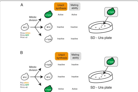
An outline of the experimental design for production of a- andα-type of cells is shown in Figure 1. Spontaneous chromosomal aberration during cultivation provide low numbers of both a- and α-type of cells in cultures con-taining large numbers of parentalMATa/αcells. The in-clusion in our system of the a1-α2 complex (which represses expression of hsg) suppresses mating by the rare a- andα-type of cells, which therefore can grow in-dependently in the cell mixture without forming non-hybrid (autopolyploid) cells [18]. Here we usedURA3as the selection marker gene for a- (Figure 1A) orα-type of cells (Figure 1B), permitting isolation by selection on solid medium lacking uracil (SD–Ura plates).
All the genetic modifications in the current study were introduced into yeast cells using plasmids, permitting complete removal of the modification following isolation of the target cells. Final plasmid sets suitable for isola-tion of a- or α-type of cells were defined by sequential
PSTE2-URA3
PSTE2-GFP
PPGK1-a1
α-type
a-type
a/α
Active Mitotic
division
PHO-URA3
PSTE3-GFP
PSTE2-α2
Inactive Uracil synthesis
Mating ability
Inactive
Active
Inactive
Inactive
SD - Ura plate
Active Inactive
Uracil synthesis
Mating ability
Inactive
Active Inactive Inactive
SD - Ura plate
B
A
a/α
a/α
a/α
a-type
a-type
α-type
α-type
Mitotic division
Figure 1Schematic outline of experimental design. (A)Isolation of a-type of cells generated via spontaneous chromosomal aberration. PPGK1-a1 suppresses mating ability ofα-type of cells to prevent autopolyploidization (mating between a- andα-type of cells derived from the
same parental strain). TheURA3gene is expressed only in a-type cells, and a-type of cells would be selected on SD solid medium lacking uracil (SD–Ura plates).(B)Isolation ofα-type of cells generated via spontaneous chromosomal aberration.PSTE2-α2 suppresses mating ability of a-type
trial and error. The process is described below in a step-by-step manner.
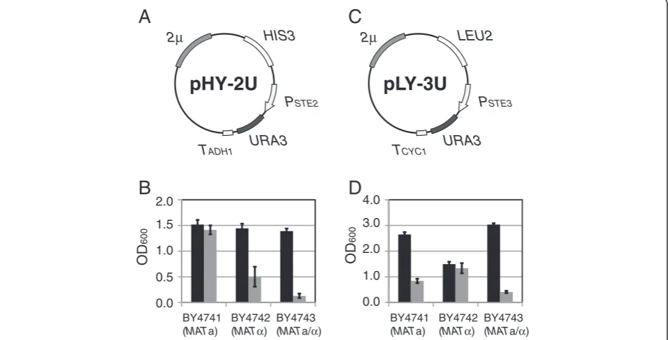
Trial 1: expression of theURA3gene via YEp-type plasmids Two kinds of YEp-type (multi-copy) plasmids were con-structed to express theURA3gene in a- orα-type of cells (Figure 2). Plasmid pHY-2U (Figure 2A) places the selec-tion marker under control ofPSTE2, a promoter of a-type-specific genes (asg), so that only MATa cells can express the URA3 gene. On the other hand, plasmid pLY-3U (Figure 2C) places the selection marker under control of PSTE3, a promoter of α-type-specific genes (αsg), so that only MATαcells can express the URA3gene. Growth of each transformant was evaluated by monitoring the OD600. Only MATa cells possessing pHY-2U grew without uracil at the same level as with uracil (Figure 2B). Al-though there were obvious differences in growth ability between MATa cells and others, MATα and MATa/α cells harboring pHY-2U exhibited some growth in the absence of uracil. As shown in Figure 2D, MATa and MATa/α cells harboring pLY-3U exhibited levels of growth equivalent to that ofMATαcells harboring pLY-3U when cultured on SD - Ura. These results suggest that leaky expression of the URA3gene via YEp-type of plasmids would preclude isolation of target cells on SD -Ura plates.
Trial 2: expression of theURA3gene via YCp-type plasmids To reduce leakyURA3gene expression, DNA fragments containingPSTE2-URA3 orPSTE3-URA3 were transferred into YCp-type (single-copy) plasmids to yield pLS-2U (Figure 3A) or pHS-3U (Figure 3C), respectively. To make detailed examination of changes in growth ability of yeast cells, we tracked OD600 values over time, and then plotted growth curves for each transformant with or without uracil. Whereas pLS-2U permitted MATa cells to grow without uracil, MATα and MATa/α cells harboring pLS-2U exhibited only minimal growth in the absence of uracil (Figure 3B). These results suggested that pLS-2U is suitable for isolation of a-type of cells.
Among cells transformed with pHS-3U, MATa and MATa/αgrew poorly without uracil compared toMATα (Figure 3D). Although the leakiness of URA3 gene ex-pression was significantly reduced by altering plasmid type from YEp to YCp, further optimization was needed to establish an isolation method forα-type of cells.
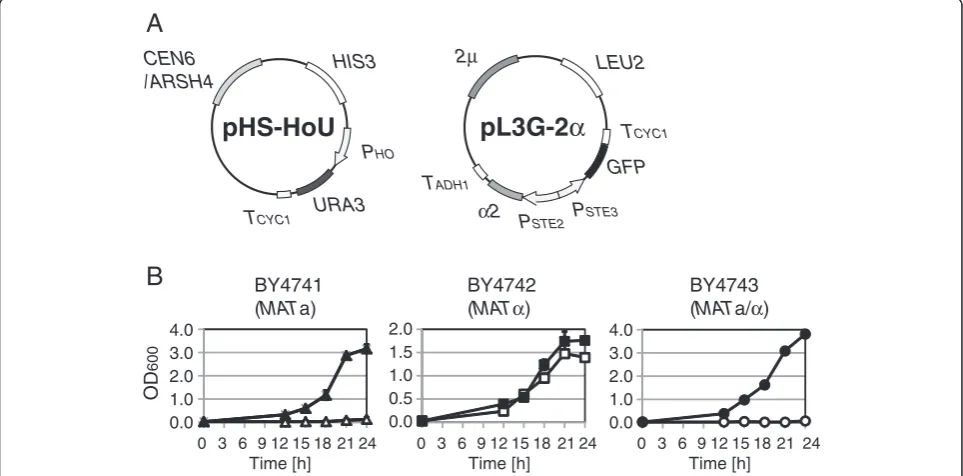
Trial 3: use of the a1-α2 repressor complex combined withPHO, a promoter ofhsg, forα-type-specificURA3 gene expression
Two plasmids shown in Figure 4A were used in an im-proved strategy for α-type-specific URA3 gene expres-sion. The plasmid pL3G-2α that contains PSTE2-α2
2μ HIS3
PSTE2
TADH1 URA3
pHY-2U
2μ LEU2
PSTE3
TCYC1 URA3
pLY-3U
A
C
D
B
2.0
1.5
1.0
0.5
0.0
OD
600
4.0
3.0
2.0
1.0
0.0
OD
600
BY4741 (MATa)
BY4742 (MATα)
BY4743 (MATa/α)
BY4741 (MATa)
BY4742 (MATα)
BY4743 (MATa/α)
Figure 2Growth of yeast transformants harboring YEp-type plasmids. (A)Plasmid map of pHY-2U containing2μorigin of replication (providing cellular retention of multicopy plasmids) andPSTE2-URA3construct (activated in a-type yeast cells).(B)The OD600values of cultures of
pHY-2U transformants at 18 h cultivation. Black bars indicate cultivation with uracil, and gray bars indicate cultivation without uracil. Values are presented as means ± standard deviations from three independent experiments.(C)Plasmid map of pLY-3U containing2μorigin of replication andPSTE3-URA3construct (activated inα-type yeast cells).(D)The OD600values of cultures of pLY-3U transformants at 18 h cultivation. Symbols
are as in B.
Fukuda and HondaJournal of Biological Engineering2013,7:27 Page 3 of 10
suppresses the mating ability ofMATa cells by artificially forming the a1-α2 repressor complex [18]. Because the a1-α2 complex can directly repress gene expression under the control of promoters of hsg, we selectedPHO (a hsg promoter) as an alternative promoter to express the URA3 gene. Hence, as shown in Figure 1B, PHO
-URA3 is expected to be activated only in MATα cells,
where PSTE2-α2 can drive expression off the HO promoter.
Figure 4B shows growth curves of transformants in the improved strategy. As expected, whileMATα trans-formants grew without uracil, MATa and MATa/α transformants exhibited minimal or no growth with-out uracil. These results suggest that use of pHS-HoU
A
C
CEN6 /ARSH4
LEU2
PSTE2
TADH1 URA3
pLS-2U
HIS3
PSTE3
TCYC1 URA3
pHS-3U
CEN6 /ARSH4
D
B
BY4741(MATa)
BY4742 (MATα)
BY4743 (MATa/α)
BY4741 (MATa)
BY4742 (MATα)
BY4743 (MATa/α) 4.0
3.0 2.0 1.0 0.0
OD
600
2.0 1.5 1.0 0.5 0.0
OD
600
4.0 3.0 2.0 1.0 0.0
2.0 1.5 1.0 0.5 0.0
2.0 1.5 1.0 0.5 0.0 2.0
1.5 1.0 0.5 0.0 0 3 6 9 12 15 18 21 24
Time [h]
0 3 6 9 12 15 18 21 24 Time [h]
0 3 6 9 12 15 18 21 24 Time [h]
0 3 6 9 12 15 18 21 24 Time [h]
0 3 6 9 12 15 18 21 24 Time [h]
0 3 6 9 12 15 18 21 24 Time [h]
Figure 3Growth of yeast transformants harboring YCp-type of plasmids. (A)Plasmid map of pLS-2U containingCEN6/ARSH4origin of replication (providing cellular retention of single-copy plasmids) andPSTE2-URA3construct (activated in a-type yeast cells).(B)The growth curves
of pLS-2U transformants. Closed symbols indicate cultivation with uracil, and open symbols indicate cultivation without uracil. Values are presented as means ± standard deviations from three independent experiments.(C)Plasmid map of pHS-3U containingCEN6/ARSH4origin of replication andPSTE3-URA3contruct (activated inα-type yeast cells).(D)The growth curves of pHS-3U transformants. Symbols are as in B.
HIS3
PHO
TCYC1 URA3
pHS-HoU
CEN6 /ARSH4
2μ LEU2
TADH1
α2 PSTE2
pL3G-2
α
PSTE3
TCYC1
GFP
A
B
BY4741(MATa)
BY4742
(MATα)
BY4743
(MATa/α)
4.0 3.0 2.0 1.0 0.0
OD
600
2.0 1.5 1.0 0.5 0.0
4.0 3.0 2.0 1.0 0.0 0 3 6 9 12 15 18 21 24
Time [h]
0 3 6 9 12 15 18 21 24 Time [h]
0 3 6 9 12 15 18 21 24 Time [h]
Figure 4Improved growth selection system forα-type of cells. (A)Plasmids used forα-type-specificURA3gene expression. The plasmid pHS-HoU containsCEN6/ARSH4origin of replication (providing cellular retention of single-copy plasmids) andPHO-URA3construct (activated in
yeast haploid cells). The plasmid pL3G-2αcontainsPSTE2-α2, a construct that repressesURA3gene expression in a-type yeast cells.(B)The growth
combined with pL3G-2α is suitable for isolation ofα -type of cells.
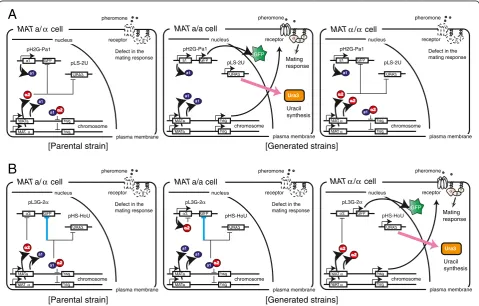
Artificial regulation network designed for isolation of a- andα-type of cells
As described above, the plasmid pLS-2U (constructed in trial 2) was preferred for isolation of a-type of cells, and the combination of plasmids pHS-HoU and pL3G-2α(as described in trial 3) was preferred for isolation of α-type of cells. A model that describes how our artificial gene network can function in isolation of a- orα-type of cells is shown in Figure 5.
For isolation of a-type of cells, we introduced plasmid pLS-2U into the parental strain in combination with plasmid pH2G-Pa1 (see Additional file 1: Figure S1A and B), which prevents autopolyploidization (Table 1 and Figure 5A). The a1-α2 complex represses expression ofhsgthat are required for mating signal transduction in α-type of cells generated via chromosomal aberration, as
well as in parental MATa/α cells. In this system, only a-type of cells can survive on SD–Ura plates, because of a-type-specific URA3 gene expression. To confirm the mating-type of yeast cells isolated on SD – Ura plates using this method, we used a GFP reporter gene under the control of the a-type-specific promoter [15]. GFP-fluorescence was observed in these yeast isolates, verifying the a-type, and confirming that this system can distinguish positives from false-positives in the scheme proposed in Figure 1A.
Next, a plasmid set composed of pHS-HoU and pL3G-2α was introduced into the parental strain for isolation of α-type of cells. pL3G-2α also provides the autopoly-ploidization prevention function (Table 1 and Figure 5B). The resulting a1-α2 complex represses expression of hsg in a-type of cells as well as in the parentalMATa/αcells. In this system, only α-type of cells can survive on SD– Ura plates, because of α-type-specific URA3 gene ex-pression. To confirm the mating-type of isolated yeast
α βγ
a1
chromosome pH2G-Pa1
URA3 GFP MATa/α cell
pLS-2U
plasma membrane nucleus
pheromone
Defect in the mating response ] s n i a r t s d e t a r e n e G [ ] n i a r t s l a t n e r a P [ E a1 α2
a1α2
receptor Mating response GFP hsg MATα a1 hsg MATa a1 chromosome pH2G-Pa1 URA3 GFP MATa/a cell
pLS-2U plasma membrane nucleus pheromone a1 receptor hsg MATa a1 hsg MATa a1 Ura3 Uracil synthesis Mating response GFP α2 chromosome pL3G-2α URA3 GFP pHS-HoU plasma membrane nucleus pheromone receptor hsg MATα hsg MATα Ura3 Uracil synthesis
MATα/α cell
α2
chromosome pL3G-2α
URA3 GFP MATa/α cell
pHS-HoU
plasma membrane nucleus
pheromone
Defect in the mating response
α2
a1α2
receptor hsg MATα a1 hsg MATa α2 α2 a1 chromosome pH2G-Pa1 URA3 GFP pLS-2U plasma membrane nucleus pheromone
Defect in the mating response
a1
α2
a1α2
receptor
hsg MATα
hsg MATα
MATα/α cell
α2 E α2 chromosome pL3G-2α URA3 GFP MATa/a cell
pHS-HoU
plasma membrane nucleus
pheromone
Defect in the mating response
a1α2
receptor hsg MATa a1 hsg MATa a1 α2
A
B
] s n i a r t s d e t a r e n e G [ ] n i a r t s l a t n e r a P [α βγ
α βγ α βγ
α βγ
α βγ α βγ
Figure 5Schematic outline of yeast mating-type regulation and our engineered approach. (A)Engineering for isolation of a-type of cells. TheMATa/αparental strain indicates BY4743 harboring pH2G-Pa1 and pLS-2U (see Table 1).(B)Engineering for isolation ofα-type of cells. The MATa/αparental strain indicates BY4743 harboring pL3G-α2 and pHS-HoU (see Table 1). Chromosomal aberration regarding theMATlocus induces generation of two kinds of yeast cells,i.e., a- andα-type of cells. Here we described yeast cells possessing two sets ofMATgenes (generated via LOH) as an example. Then, spontaneous or engineered formation of the a1-α2 complex suppresses the mating response by repressing expression of haploid-specific genes (hsg), which encode components of the mating signaling pathway. Target cells expressing theURA3selection marker gene can be isolated on SD–Ura plates in the absence of autopolyploidization. The target cells also express theGFPreporter gene. Regulation colored with cyan indicates indirect gene repression; the a1-α2 complex prevents expression of transcriptional activatorα1 that is required for gene expression under the control ofPSTE3. Note thatURA3gene is directly repressed by a transcriptional repressor in non-target cells.
Fukuda and HondaJournal of Biological Engineering2013,7:27 Page 5 of 10
cells, we used a GFPreporter gene under the control of theα-type-specific promoter [18]. GFP-fluorescence was observed in these yeast isolates, verifying theα-type, and confirming the validity of the scheme proposed in Figure 1B.
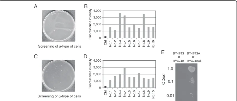
Isolation of a- andα-type of cells generated via spontaneous chromosomal aberration
As shown in Figure 1A, MATa/α cells possessing both plasmids pLS-2U and pH2G-Pa1 were washed and spread on SD – Ura plates after overnight cultivation with uracil. From approximately 1 × 10⁷ cells spread on the SD–Ura plate, 28 colonies were formed (Figure 6A). Ten of these colonies were randomly selected, and fluor-escence intensity was measured. Values were compared to that of the parental MATa/α cells (harboring
pH2G-Pa1), which were used as a negative control (labeled as
“Ctrl”in Figure 6B). All of the isolates derived from the SD–Ura plates exhibited higher fluorescence than Ctrl, suggesting that the proposed selection system for a-type of cells rarely produced false-positives. We selected the colony (No. 3) with the highest fluorescence intensity and performed another round of colony isolation to re-move the cells of all plasmids. The resulting strain was designated BY4743A (Table 1).
Next,MATa/αcells possessing both plasmids pHS-HoU and pL3G-2α were washed and spread on the SD–Ura plates after overnight cultivation with uracil, as shown in Figure 1B. From approximately 1 × 10⁶cells spread on the SD–Ura plate, 23 colonies were formed (Figure 6C). Ten of these colonies were randomly selected, and fluorescence intensity was measured. Values were compared to that of the parental MATa/α cells (harboring pL3G-2α), which were used as a negative control (labeled as “Ctrl” in Figure 6D). All of the colonies isolated on the SD –Ura plates exhibited higher fluorescence than Ctrl, suggesting that the proposed selection system for α-type of cells rarely produced false-positives. We selected the colony (No. 4) with the highest fluorescence intensity and per-formed another round of colony isolation to remove the cells of all plasmids. The resulting strain was designated BY4743AL (Table 1).
To verify the mating ability of BY4743A and BY4743AL, mating assays [20,21] were carried out as follows. Because BY4743A and BY4743AL have the same auxotrophy as BY4743, pH2G-Pa1 (containing HIS3 marker) was introduced into BY4743A (a-type) and BY4743 (MATa/α; parental strain), and pL3G-2α (con-taining LEU2 marker) was introduced into BY4743AL (α-type) and BY4743. This allowed us to select zygotes using solid medium lacking histidine and leucine. The mating assays revealed successful mating between BY4743A and BY4743AL, but not between the two kinds of BY4743 transformants (Figure 6E). These results con-firmed the validity of our proposed scheme. We believe that our approach will find application in manufacturing mating strains for yeast crossbreeding.
Discussion
The aim of this study was to establish a versatile method, using growth-based selection, for the isolation of mating strains for yeast crossbreeding (Figure 1). Al-though mating strains are traditionally prepared by sporulation from MATa/α yeast strains used for indus-trial purposes, there are numerous strains that have ex-treme difficulty sporulating [7]. Alternative approaches using HO endonuclease have been developed in order to provide mating strains without sporulation [18,22]. Un-fortunately, however, these approaches cannot be applied to yeast strains containing the“stuck”mutation (a single Table 1 Yeast strains and plasmids used in this study
Name Description Reference source
Yeast strains
BY4741 MATahis3Δ1 ura3Δ0 leu2Δ0 met15Δ0 Brachmann
et al., [24]
BY4742 MATαhis3Δ1 ura3Δ0 leu2Δ0 lys2Δ0 Brachmann
et al., [24]
BY4743 MATa/αhis3Δ1/his3Δ1 leu2Δ0/leu2Δ0 LYS2/ lys2Δ0 met15Δ0/MET15 ura3Δ0/ura3Δ0
Brachmann
et al., [24]
BY4743A MATahis3Δ1 leu2Δ0 LYS2 MET15 ura3Δ0 Present study
BY4743AL MATα/αhis3Δ1/his3Δ1 leu2Δ0/leu2Δ0 LYS2/ lys2Δ0 met15Δ0/MET15 ura3Δ0/ura3Δ0
Present study
Plasmids
pRS313 Yeast expression vector;CEN6/ARSH4ori and
HIS3marker
NBRP*
pRS315 Yeast expression vector;CEN6/ARSH4ori and
LEU2marker
NBRP
pRS316 Yeast expression vector;CEN6/ARSH4ori and
URA3marker
NBRP
pHY-2GA Yeast expression vector;2μori,HIS3marker andPSTE2-EGFP
Fukudaet al., [18]
pLY-3GC Yeast expression vector;2μori,LEU2marker andPSTE3-EGFP
Fukudaet al., [18]
pH2G-Pa1
PPGK1-a1in pHY-2GA Fukudaet al.,
[18]
pL3G-2α PSTE2-α2in pLY-3GC Fukudaet al.,
[18]
pHY-2U Yeast expression vector;2μori,HIS3marker andPSTE2-URA3
Present study
pLY-3U Yeast expression vector;2μori,LEU2marker andPSTE3- URA3
Present study
pLS-2U PSTE2-URA3in pYO315 Present study
pHS-3U PSTE3-URA3in pYO313 Present study
pHS-HoU PHO-URA3in pYO313 Present study
*
base substitution) at the MAT locus [23]. Here we fo-cused on chromosomal aberration such as LOH and chromosome loss, alternative processes that produce a-and α-type of cells from parental MATa/α cells. Al-though chromosomal aberration is remarkably tolerant of slight differences in the base sequences at the MAT locus, spontaneous frequencies of such events are less than 1 × 10⁻⁴.
Hashimoto et al. used UV irradiation to increase the frequency of LOH [16], and Whittaker et al. used 12 kinds of chemicals to increase the frequency of chromo-some loss [17]; however, these treatments are likely to randomly induce additional mutations and loss of other chromosomes. Because the purpose of yeast crossbreed-ing is to combine favorable traits of parental strains, these undesirable changes would be a concern for the resulting a- orα-type of cells. To isolate a- andα-type of cells generated via spontaneous chromosomal aberration in the absence of mutagens, we describe here the devel-opment of mating-type-dependent URA3 gene expres-sion systems (Figure 1).
Generally, chromosomal stability becomes diminished by an increase in ploidy (e.g. less stable in triploid and tetraploid than in diploid cells). According to past report, the frequency of losing chromosome VII was approxi-mately 10,000-fold higher in tetraploid than in diploid cells of S. cerevisiase [15]. In principle, mating-type-dependent screening cannot exclude triploid, tetraploid
and other higher polyploid cells that may emerge via chromosome aberration following autopolyploidization (mating between the a- andα-type of cells generated from the same parental strain). Although these cells would pre-sumably be available for yeast crossbreeding, chromosome stability of the hybrids should inevitably become dimin-ished due to unneeded increase in ploidy. Hence, we uti-lized artificial formation of the a1-α2 complex [18] to remove the potential risk of autopolyploidization.
To develop growth selection systems for mating strains, we initially adoptedPSTE2(for isolation of a-type of cells) andPSTE3(for isolation ofα-type of cells as the promoters to express theURA3selection marker gene. By the use of YCp-type of vector, PSTE2-URA3 successfully permitted only a-type of yeast cells to grow without uracil. Compared to PSTE2, regulation of gene expression under the control ofPSTE3was leaky, a fact that had not been noted in a pre-vious study using theGFPreporter gene [18]. Although we attempted to reduce leakiness of the URA3 gene expres-sion by incluexpres-sion of the artificially formed a1-α2 complex (see Additional file 1: Figure S1C and D), little change was seen in the OD600value ofMATa andMATa/αcells after 24 h cultivation (see Additional file 1: Figure S1D; compare to Figure 3D).
The difference in leakiness between PSTE2 and PSTE3 may reflect the distinct regulatory mechanisms control-lingasgandαsgpromoters. InMATαandMATa/αcells, expression of asg is suppressed by the α2 repressor
A
C
D
B
Screening of a-type of cells
Screening of α-type of cells
Fluorescence intensity
Fluorescence intensity
4,000
3,000
2,000
1,000
0
4,000
3,000
2,000
1,000
0
E
OD
600
1.0
0.1
0.01 Ctrl No.1 No.2 No.3 No.4 No.5 No.6 No.7 No.8 No.9
No.10
Ctrl
No.1 No.2 No.3 No.4 No.5 No.6 No.7 No.8 No.9 No.10
BY4743
BY4743 BY4743A
BY4743AL
Figure 6Isolation of a- orα-type of cells generated via spontaneous chromosomal aberration. (A)Direct images of colony formation in isolation of a-type of cells. Cell suspensions containing approximately 1 × 10⁷yeast cells (1 mL of suspension at OD6001.0) were spread on selective
solid medium (SD–Ura plate).(B)Fluorescent reporter assay to quantify expression of theGFPreporter gene in yeast transformants possessing plasmid pH2G-Pa1 (PSTE2-GFP). Ctrl indicates the parentalMATa/αtransformant (negative control).(C)Direct images of colony formation in isolation ofα-type of
cells. Cell suspensions containing approximately 1 × 10⁶yeast cells (1 mL of suspension at OD6000.1) were spread on selective solid medium.
(D)Fluorescent reporter assay to quantify expression of theGFPreporter gene in yeast transformants possessing plasmid pL3G-2α(PSTE3-GFP). Ctrl
indicates the parentalMATa/αtransformant (negative control).(E)Mating assays to investigate the mating abilities of yeast cells. Each spot corresponds to 10μL of suspension at the indicated OD600.
Fukuda and HondaJournal of Biological Engineering2013,7:27 Page 7 of 10
encoded by the MATαallele [24]. TheMATαallele also encodes theα1 activator, which facilitates the expression ofαsg. WhereasMATa cells rarely expressαsgdue to ab-sence of the α1-coding gene within the MAT locus, MATa/αcells are unlikely to expressαsgbecause theα 1-coding gene is repressed by the a1-α2 complex (a1 is encoded by the MATa allele) [19]. Unlike promoters of asg, there is no component that can directly repress pro-moters of αsg, which might permit a low level of basal (leaky) expression. In the present work, we succeeded in tight α-type-specific URA3 gene expression by utilizing PHO, a promoter of hsg that is directly repressed by the a1-α2 complex (Figure 4).
While use of auxotrophic markers has an advantage in transformation efficiencies, the utility of them is limited within laboratory strains or genetically modified strains because industrial yeast strains usually do not have auxo-trophic mutations such asura3, his3 and leu2. However, in principle, our system can adopt drug-resistance markers which are available for industrial yeast strains. Instead of the URA3marker, we attempted to use kanMX4 marker (see Additional file 2) that gives resistance for geneticin (G418), and succeeded in growing only target cells in cul-tivation medium containing G418 (see Additional file 3: Figure S2).
To evaluate the availability of the established growth selection systems, we manufactured a- and α-type of cells as shown in Figure 1. The appearance frequencies of a- and α-type of cells were 2.8 × 10⁻⁶and 2.3 × 10⁻⁵, respectively. Because the frequency of chromosomal ab-erration generally varies by yeast strain, genetic modifi-cations might have some effect on the frequency of chromosomal aberration in the parental BY4743 strain. Next, we verified the mating-type of colonies isolated on SD–Ura plates usingGFPreporter gene expression. All colonies had the expected mating-type, suggesting the utility of isolating a- and α-type of cells via the con-structed growth-based selection systems.
We performed another round of colony isolation to re-move all plasmids from yeast cells, yielding BY4743A and BY4743AL strains, respectively. To identify the event generating BY4743A or BY4743AL strain, we car-ried out quantitative analysis for the number of chromo-some III (containing MATgenes) using real-time PCR (see Additional file 4: Figure S3). BY4743A strain has only 1 set of chromosome III, suggesting that it was generated via chromosome loss. On the other hand, BY4743AL strain has 2 sets of chromosome III, suggest-ing that it was generated via LOH.
Furthermore, to confirm the stability of their mating ability, we carried out mating assays after serial passage in culture (see Additional file 5: Figure S4). Even after three passages, BY4743A and BY4743AL continued to exhibit equivalent levels of mating. We expect that the
mating abilities of the generated a- and α-type of cells will remain extremely stable, given that additional chromosomal rearrangements are unlikely to occur at theMATloci.
Conclusion
We have established a new approach to manufacture a-and α-type of cells from parental MATa/α cells using growth selection systems. Use of spontaneous chromo-somal aberration is quite beneficial in acquisition of mating strains inheriting desirable properties of industrially-used strains. We showed that yeast strains generated via spon-taneous chromosomal aberration have stable mating ability, providing hybrid strains for use in yeast crossbreeding. Use of our method should help promote further advances of yeast-based biosynthesis approaches and in other experi-mental areas of research such as quantitative trait locus analysis and genome-wide association studies that permit the linking of phenotypic traits and genotypic data.
Methods
Strains and media
Detailed information about Saccharomyces cerevisiae strains BY4741, BY4742, and BY4743 [25], as well as other strains used in this study, is shown in Table 1. Yeast cells were grown in YPD medium (1% yeast ex-tract, 2% peptone and 2% glucose) or in SD medium (0.67% yeast nitrogen base without amino acids (Becton Dickinson and Company, Franklin Lakes, NJ, USA) and 2% glucose). A final concentration of 2% agar was added to make solid media.
Construction of plasmids
The sequences of oligonucleotides used in this study are shown in Table 2. The plasmids shown in Table 1 were made as follows. Using pRS316 (provided by the National BioResource Project (NBRP) of the MEXT, Japan) as a template, theURA3gene was amplified with oligonucleo-tide pair o1 and o2, and inserted in place of theEGFPat the NotI-BamHI sites of pHY-2GA [18], yielding a YEp-type plasmid designated pHY-2U. Similarly, using pRS316
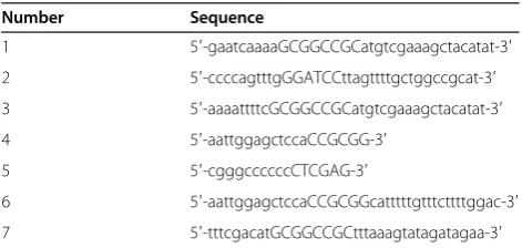
Table 2 Sequences of oligonucleotides used to construct plasmids
Number Sequence
1 5′-gaatcaaaaGCGGCCGCatgtcgaaagctacatat-3′
2 5′-ccccagtttgGGATCCttagttttgctggccgcat-3′
3 5′-aaaattttcGCGGCCGCatgtcgaaagctacatat-3′
4 5′-aattggagctccaCCGCGG-3′
5 5′-cgggccccccCTCGAG-3′
6 5′-aattggagctccaCCGCGGcatttttgtttcttttggac-3′
as a template, the URA3 gene was amplified with oligo-nucleotide pair o3 and o2, and inserted in place of the EGFPat theNotI-BamHI sites of pLY-3GC [18], yielding a YEp-type plasmid designated pLY-3U.
The YCp-type plasmids used to express theURA3gene were constructed as follows. DNA fragments containing PSTE2-URA3 were amplified from pHY-2U using oligo-nucleotide pair o4 and o5, and inserted into theSacII-XhoI sites of pRS315 (provided by NBRP), yielding the plasmid pLS-2U. DNA fragments containing PSTE3-URA3 were amplified from pLY-3U using oligonucleotide pair o4 and o5, and inserted into the SacII-XhoI sites of pRS313 (provided by NBRP), yielding the plasmid pHS-3U. The haploid-type-specific promoter, PHO, was amplified using oligonucleotide pair o6 and o7 from genomic DNA de-rived from strain BY4741, and inserted in place ofPSTE3at theSacII-NotI sites of 3U, yielding the plasmid pHS-HoU. Each plasmid was introduced into yeast cells using the lithium acetate method [26].
Investigation of cell growth characteristics
Each yeast transformant was grown in 500 μL of SD medium without or with 20 mg/L uracil at 30°C, setting ini-tial optical density at 600 nm (OD600) at 0.03. The OD600 values of cultures were monitored using a UV/visible spec-trophotometer (Ultrospec 3100 pro; GE Healthcare Japan Corporation, Tokyo, Japan).
Fluorescent reporter assay
TheEGFPgene was used as a fluorescent reporter to indi-cate mating type. Reporter-containing cells were incubated at 30°C for 18 h, harvested and washed with distilled water. The cells then were resuspended in 100μL of dis-tilled water to an OD600 of 5.0. GFP fluorescence inten-sities were measured using an Infinite 200 fluorescence microplate reader (Tecan Japan Co., Ltd., Kawasaki, Japan). For detection of the GFP signal, the excitation wavelength was set at 485 nm with a bandwidth of 20 nm, and the emission wavelength was set at 535 nm with a bandwidth of 25 nm. The gain was set at 50.
Mating assay
Evaluation of mating ability was performed as follows. Yeast diploid cells were cultivated with the mating part-ner in 1 ml of YPD medium at 30°C for 1.5 h, with an initial OD600 of 0.1. After cultivation, yeast cells were harvested, washed, and resuspended in distilled water. Starting from an initial OD600of 1, 0.1, or 0.01, 10μl of cell suspensions were spotted on SD solid medium with-out the appropriate amino acids for growth selection of zygotes. After incubation at 30°C for 2 days, the image data was recorded for colonies on solid medium.
Isolation of yeast cells with target mating-type
Parental MATa/α cells were grown in 500 μL of SD media with 20 mg/L uracil at 30°C for 18 h, and then harvested and washed with distilled water. The cells then were resuspended in distilled water. Cell suspensions were spread on SD–Ura plates.
Additional files
Additional file 1: Figure S1.Alternative growth selection systems for isolation of a-type orα-type yeast cells by formation of the a1-α2 complex. (A) Plasmids used for a-type-specificURA3gene expression. The plasmid pLS-2U was used in combination with pH2G-Pa1, which suppresses the mating ability ofα-type cells. (B) The OD600values of
cultures of double transformants (harboring both plasmids pLS-2U and pH2G-Pa1) at 24 h cultivation. Black bars indicate cultivation with uracil, and gray bars indicate cultivation without uracil. (C) Plasmids used for α-type-specificURA3gene expression. The plasmid pHS-3U was used in combination with pL3G-2α, which is required for suppressing the mating ability of a-type cells. (D) The OD600values of cultures of double
transformants (harboring both plasmids pHS-3U and pL3G-2α) at 24 h cultivation. Black bars indicate cultivation with uracil, and gray bars indicate cultivation without uracil.
Additional file 2:Supporting information for Materials and Methods. Table S1.Sequences of oligonucleotides used to construct plasmids.
Additional file 3: Figure S2.Growth of yeast transformants harboring kanMX4selection marker gene. (A) Plasmid map of pLS-2 K containing CEN6/ARSH4origin of replication (providing cellular retention of single-copy plasmids) andPSTE2-kanMX4construct (activated in a-type
yeast cells). (B) The growth curves of pLS-2 K transformants. Closed symbols indicate cultivation without G418, and open symbols indicate cultivation with G418. (C) Plasmid map of pHS-HoK-2αcontaining CEN6/ARSH4origin of replication andPHO-kanMX4construct combined
withPSTE2-α2 construct (activated inα-type yeast cells). (D) The growth
curves of pHS-HoK-2αtransformants. Symbols are as in B.
Additional file 4: Figure S3.Ploidy analysis using real-time PCR. The normalized copy number of thePGK1gene is an indicator of ploidy (A) for BY4743A and (B) for BY4743AL strains. Standard deviations of three replicates are presented.
Additional file 5: Figure S4.Investigation of stability of the mating abilities of yeast cells after serial passage of cultures. Up to three serial passages were carried out, and then the resulting BY4743, BY4743A, and BY4743AL transformants were used for mating assays.
Abbreviations
EGFP:Enhanced green fluorescent protein; LOH: Loss of heterozygosity;MAT locus: Mating-type locus;hsg: Haploid-specific genes;asg: a-type-specific genes;αsg:α-type-specific genes.
Competing interests
We declare that all authors are inventors on a pending patent using aspects of this system.
Authors’contributions
NF designed the study, conducted experiments, analyzed data, and co-wrote the manuscript. SH analyzed data and co-wrote the manuscript. Both authors read and approved the final manuscript.
Acknowledgements
The plasmids pRS313, pRS315, and pRS316, were provided by the National BioResource Project (NBRP) of the MEXT, Japan. This work was supported by JSPS KAKENHI Grant Number 25820406.
Received: 8 July 2013 Accepted: 12 November 2013 Published: 21 November 2013
Fukuda and HondaJournal of Biological Engineering2013,7:27 Page 9 of 10
References
1. Higgins VJ, Bell PJL, Dawes IW, Attfield PV:Generation of a novel Saccharomyces cerevisiaestrain that exhibits strong maltose utilization and hyperosmotic resistance using nonrecombinant techniques.
Appl Environ Microbiol2001,67:4346–4348.
2. Kishimoto M:Fermentation characteristics of hybrids between the cryophilic wine yeastSaccharomyces bayanusand the mesophilic wine yeastSaccharomyces cerevisiae.J Ferment Bioeng1994,77:432–435. 3. Shinohara T, Mamiya S, Yanagida F:Introduction of flocculation property
into wine yeasts (Saccharomyces cerevisiae) by hybridization.J Ferment Bioeng1997,83:96–101.
4. Benitez T, Gasent-Ramirez JM, Castrejon F, Codon AC:Development of new strains for the food industry.Biotechnol Prog1996,12:149–163.
5. Guijo S, Mauricio JC, Salmon JM, Ortega JM:Determination of the relative ploidy in differentSaccharomyces cerevisiaestrains used for fermentation and‘flor’film ageing of dry sherry-type wines.Yeast1997,13:101–117. 6. Maráz A:From yeast genetics to biotechnology.Acta Microbiol Immunol
Hung2002,49:483–491.
7. Gunge N:Breeding of bakers’yeast-determination of the ploidy and an attempt to improve practical properties.Japan J Genet1966,41:203–214. 8. Tsuboi M, Takahashi T:Genetic analysis of the nonsporulating phenotype
of brewer’s yeasts.J Ferment Technol1988,66:605–613.
9. Kumaran R, Yang SY, Leu JY:Characterization of chromosome stability in diploid, polyploid and hybrid yeast cells.PLoS One2013,8:e68094. 10. Andersen MP, Nelson ZW, Hetrick ED, Gottschling DE:A genetic screen for
increased loss of heterozygosity.Saccharomyces cerevisiae2008,179:1179–1195. 11. Takagi Y, Akada R, Kumagai H, Yamamoto K, Tamaki H:Loss of
heterozygosity is induced inCandida albicansby ultraviolet irradiation.
Appl Microbiol Biotechnol2008,77:1073–1082.
12. Alvaro D, Sunjevaric I, Reid RJ, Lisby M, Stillman DJ, Rothstein R:Systematic hybrid LOH: a new method to reduce false positives and negatives during screening of yeast gene deletion libraries.Yeast2006,23:1097–1106. 13. Daigaku Y, Endo K, Watanabe E, Ono T, Yamamoto K:Loss of
heterozygosity and DNA damage repair inSaccharomyces cerevisiae.
Mutat Res2004,556:183–191.
14. Hiraoka M, Watanabe K, Umezu K, Maki H:Spontaneous loss of heterozygosity in diploidSaccharomyces cerevisiaecells.Genetics2000,156:1531–1548. 15. Mayer VW, Aguilera A:High levels of chromosome instability in polyploids
of Saccharomyces cerevisiae.Mutat Res1990,231:177–186.
16. Hashimoto S, Aritomi K, Minohara T, Nishizawa Y, Hoshida H, Kashiwagi S, Akada R:Direct mating between diploid sake strains ofSaccharomyces cerevisiae.Appl Microbiol Biotechnol2006,69:689–696.
17. Whittaker SG, Zimmermann FK, Dicus B, Piegorsch WW, Resnick MA, Fogel S:
Detection of induced mitotic chromosome loss in Saccharomyces cerevisiae–an interlaboratory assessment of 12 chemicals.Mutat Res 1990,241:225–242.
18. Fukuda N, Matsukura S, Honda S:Artificial conversion of the mating-type ofSaccharomyces cerevisiaewithout autopolyploidization.ACS Synth Biol 2013. in press.
19. Gelfand B, Mead J, Bruning A, Apostolopoulos N, Tadigotla V, Nagaraj V, Sengupta AM, Vershon AK:Regulated antisense transcription controls expression of cell-type-specific genes in yeast.Mol Cell Biol2011,31:1701–1709. 20. Fukuda N, Ishii J, Tanaka T, Kondo A:The competitor-introduced Gγ
recruitment system, a new approach for screening affinity-enhanced proteins.FEBS J2010,277:1704–1712.
21. Fukuda N, Ishii J, Kondo A:Gγrecruitment system incorporating a novel signal amplification circuit to screen transient protein-protein interactions.FEBS J2011,278:3086–3094.
22. Kanai K, Kobayashi O:Method for imparting mating ability to yeast.Japanese Kokai Tokkyo: Koho patent publication number JP 2010-220481. Japanese Kokai Tokkyo Koho Application number JP 2009–068013, Date 2009.03.19. 23. Ray BL, White CI, Haber JE:Heteroduplex formation and mismatch repair
of the“stuck”mutation during matingtype switching in Saccharomyces cerevisiae.Mol Cell Biol1991,11:5372–5380.
24. Botstein D:Ira Herskowitz: 1946–2003.Genetics2004,166:653–660. 25. Brachmann CB, Davies A, Cost GJ, Caputo E, Li J, Hieter P, Boeke JD:
Designer deletion strains derived from Saccharomyces cerevisiae S288C: a useful set of strains and plasmids for PCR-mediated gene disruption and other applications.Yeast1998,14:115–132.
26. Gietz D, St Jean A, Woods RA, Schiestl RH:Improved method for high efficiency transformation of intact yeast cells.Nucleic Acids Res1992,20:1425.
doi:10.1186/1754-1611-7-27
Cite this article as:Fukuda and Honda:Development of growth selection systems to isolate a-type orα-type of yeast cells
spontaneously emerging fromMATa/αdiploids.Journal of Biological En-gineering20137:27.
Submit your next manuscript to BioMed Central and take full advantage of:
• Convenient online submission
• Thorough peer review
• No space constraints or color figure charges
• Immediate publication on acceptance
• Inclusion in PubMed, CAS, Scopus and Google Scholar
• Research which is freely available for redistribution