Original Article
Development a New Human Skin Continuous Cell Line Sensitive
to Mumps Virus
Mahmudi-Gharoeie N1, Mohammadi A2*, Saffar B1, Esna-Ashari F2, Foroghi A2 Alirezaee B2, Ghorbani R2, Sedigh ZA2
1. Genetic Department, Shahrekord University Faculty of Science, Shahrekord, Iran.
2. Human Viral Vaccine Department, Razi Vaccine and Research Institute, Alborz Karaj, Iran.
Abstract
Background and Aims: During the last years a new Foreskin cell line strain (RFSC) has been isolated, characterized and stored in a representative cell bank stock in liquid nitrogen as a part of HCB (Human Cell Bank) development program in Razi institute.
Materials and Methods: The cell population was serially passaged for mass production purpose based on WHO schedule .The controls on pool samples were carried out upon our sampling method and international recommendations.
Results: The data concerning parameters as well as the result of compulsory controls on Kario logy oncogenicity, cross contaminations, species specificity, no oncogenicity, and mycoplasma free of a human cell line that contained heteroploid chromosomal characterizations are performed.
Conclusion: Kariological examination found an obvious discrepancy in the frequency of aberrations due to high passage level that produced a heteroploid continuous cell line. Virus susceptibility shows RFSC-1 more sensitive to RS#12 Mumps virus strain. This cell line is completely sterile and has no any cross contamination to other pathogen and can be used for production and control of Mumps vaccine.
Keywords: Primary culture; Cell Characterization; Human Cell Bank; RFSC-1
Introduction
major obstacle of isolating the viral causative agent has been the lack of convenient and sensitive in vitro cell culture systems. Viruses are obligatory intracellular parasites and their in vitro replication requires permissive cell lines. In addition to specific cell-tropism requirements, some viruses can be propagated only in cell lines derived from the same host species. There has been an interest in developing cell lines from several mammalian species such as
human nonhuman and other animals for vaccine manufacturer.
The current widespread and intensive development of cells from numerous animal sources especially for biological purpose has resulted in in the development a cell line by Endores and his coworkers in 1949 (1). He demonstrated that Cell lines are generally employed for virus isolation and propagation. Regardless of numerous viruses (as well as other microorganisms) that derived from the cells till now, the overall attempt is developing better and more susceptible cell line system. In this study we describe the successful establishment and characterization of a foreskin cell line (RFSC-1) derived from 4-8 week old boys. Human cells were chosen because of their phylogenetic relationship to
A
*Corresponding author: Ashrf Mohammadi, PhD. Human Viral Vaccine Department, Razi Vaccine and Research Institute, Alborz Karaj, Iran.
Email: [email protected]
human natural cells. The purpose was to determine whether or by using cells from human, a broader susceptibility for human viruses. It was also thought at the initiation of these studies that use of human cells might eliminate the potential of non human harboring indigenous viruses.
Methods
Sources of cells
Foreskin samples collected from three different 4-8 week old healthy boys. Tissue samples were incubated for 2hr in antibiotic containing medium consisting of DMEM, 1000µg/ml streptomycin 1000IU/ml penicillin. Following two additional washes in the medium (15 min/wash), tissue sample were minced with sterile instrument, and 20-30 tissue fragment were planted in 25-cm2 Nunc tissue culture flask. In addition minced tissue fragments were digested with 0.25% trypsin at 30°C for 20 minute. Dissociated cells were then washed once with phosphate buffer saline (PBS) and approximately 1 to 2×10⁶ cells were transferred into each of several 25 cm2 Nunc tissue culture flasks. Primary cell culture were initially maintained in the Dulbecco's Eagle Minimum Essential (DMEM) medium supplemented with 8% fetal bovine serum (FBS) according to the previously described method (9). Cell cultures were incubated at 37°C and when monolayers were confluent, cell were sub cultivated at a ratio of 1:4 every 3 to 5 days with 0.25% trypsin solution (fluka chemical co). Concentration of FBS was reduced by half after 10 h passage. After cloning and several sub passage cells were seeded into 32 oz bottles. Subsequent subcultures were made twice per week. As positive and negative control the other human cell line (MRC-5) and a Rabbit kidney (RK-13) used in this study .Cell culture were maintained either by sub culturing in 32 oz bottles or were preserved in the frozen state suspended in the Dulbecco's Eagle Minimum Essential Medium (DMEM) containing 10% dim ethyl-sulfoxide and 20% fetal calf serum. Preservation step made when the cells placed in 2ml cryotube, which
completely sealed, gradually frozen and then stored in liquid nitrogen (9).
Media
Growth media consisted of DMEM plus 5-8% bovine serum. Maintenance medium was the same medium containing lower (2%) serum concentration.
Viruses Susceptibility
Viruses used in this study (Mumps, Measles, Rubella, Polio Vesicular stomatities virus) were part of the collection of Human Viral Vaccine department of Razi vaccine and serum research institute. Following adaptation of virus, inoculation (0.1 ml/25 cm2 Nunc tissue culture flask or 16mm well in 96 well plastic micro plate) were done. The culture vessels were held for periods up to14 days at 33-35°C, when virus cytopathic effect (CPE) was observed supernatant fluid was harvested. The virus titer was determined by inoculating 10 fold dilution in a similar method and were expressed as log10 TCID50/0.1ml. The final result calculated by the method of Read and Munch (2).
Chromosomal analysis
Chromosomal studies were done by treating cells with a hypotonic solution, fixation, flame spreading and Giemsa staining, according to the method of Moorhead et.al (3). A total of 500 selected metaphase spreads were counted with phase-contrast microscopy. These procedures were repeated 3 times.
Isoenzyme Assay
For detection of interspecies cross contamination Isoenzyme analysis method was used. In this method polymorphic enzymes can be visualized as electrophoresis variant. The pattern of electrophoresis giving rise specific for each species O’Brien (4), Bright (5), Stratton (6), Nims (7) was observed.
PCR-RFLP
A Polymerase Chain Reaction (PCR) Restriction Fragment Length Polymorphism (RFLP) assay was optimized, based on a pair of primers anneal to a portion of cytochrome b gene and a panel of six restriction enzymes. The derived pattern of RFLP was resolved on 3% agarose gel. Each species has specific pattern for restriction enzyme panel. By this pattern not only the species but also cross
contamination between different cell lines can be distinguished. Losi FS, et al (8), Pun KM AC (9).
Adventitious agent testing
Verification the sterility of the cell line with other different bacteria and viruses were made by the protocol of WHO (TRS: 842) (10).
Results
Cell morphology
RFSC-1 cell line was established from fore skin tissues of 4-8 week old healthy boy. This cell line has been passages more than 135 times since its initiation based on the methods of Hay flick and Moorhead (1961). The methods we used for clonally isolation of RFSC-1 have been previously reported by Fereshney (11, 12). In primary cloned cultures both fibroblastic and epithelial cell shape were obtained. Then fibroblastoid clones grew more rapidly in all of cultures flask. In next subcultures RFSC-1 showed fibroblast like appearance in its morphology although derived from skin (Fig. 1). Growth medium was Dulbecco's Eagles Basal Medium (DMEM) supplemented with 5-8% serum. 4-ozflasks were seeded each with 6ml of cells at concentration of 105cells per milli liter. After 3 days incubation in 37C a confluent sheet of fibroblastoid cells (Fig. 1) was formed. Cell were sub-cultured every 3 days, split ratio was 1:4 .Up to 135th subculture this features were constant. When the passage number reaches to 5, cells were harvested and frozen in the presence of preservative (10% dimethyl sulfoxide in DMEM). Attempt was made for every 5 passage stocking and producing enough working seed from this cell line.
Viral susceptibility
Viruses used for evaluation the RFSC-1
susceptibility were vesicular stomatites virus (New jersey serotype), Measles virus (AIK strain) Mumps virus (Hoshino&RS#12 strain), Rubella (Takahashi strain). Two different Subcultures were selected for these studies. Different stock viruses were first adopted by one or two passages and then were seeded in 4-oz flasks containing a confluent sheet of RFSC-1. The supernatant fluid of cultures were harvested when cytopathic effect (CPE) were sufficiently progressed and potency test were made in sensitive cells depending on the type of virus. Results are reflected in (Table 1). The virus sensitivity spectrum of this cell line showed a good sensitivity for mumps virus strain (RS#12). This sensitivity was positive also during Heamadsorption against Guinea pig RBC.
Chromosomalanalysis
The RFSC-1 Chromosomal analysis is shown in (Fig. 2). This analysis was performed at population doubling level 57. At each population doubling 500 cells spread were examined and counted. This procedure was repeated independently 3 times (Fig. 2). The chromosomes were arranged according to decreasing size for pair 1to 19. The sex chromosomes were placed at the end of the Karyotype as normal shape in comparison with other species.
Iso enzyme and PCR-RFLP Assay
The result of isoenzyme assay and RFLP-PCR were shown in (Table 2 & Fig. 3 & 4). The results of isoenzyme assay test show that RFSC-1 cell line has the human origin. In this test as positive and negative control MRC-5 (Human fetal lung) and RK-13 (Rabbit Kidney) were used. Also the result of RFLP-PCR (Fig. 4) showed that there is not any cross contamination with another cell line.
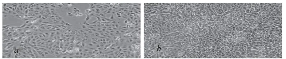
Fig. 1. Comparative picture of confluent (a) and sub confluent (b) RFSC-1 cell line.
Adventitious agent testing
Adventitious agent testing of RFSC-1 showed that there was not any cross contamination with other viruses and this cell line was completely free of contamination.
Discussion
Today the entire virological laboratories are in urgent need to have cell bank from different cell line. This need comes from different degree of sensitivity that a virus shows to different cell substrate. Although tissue culture was started at the beginning of the twentieth century (13, 14) as a technique for studying the behavior of animal cells, but at present is the most important tool in production of antigen (15) biological product, vaccine (16), isolation and detection of viruses (17, 18). The primary explants of tissue dominated the field of 'tissue culture’’ for more than 50 years (19, 20). Despite the fact that most of the explosive expansion in the field of virology, was made possible by the use of dispersed cells in the
second half of the twentieth century. These cells taken from original tissue as primary culture, cell line or cell strain, following enzymatic, mechanical, or chemical disaggregation (21). Dulbecco's was the first who demonstrated the outgrowth disaggregation of explanted cells and subsequent plating out of the dispersed cells. Although the term of cell strains generated by Fischer, Carrel, and others often using for that cell line that access by surgical subdivision of the culture. The first cloned cell strain, isolated by capillary cloning from mouse L-cells was L929 (22). It was not until the 1950s that trypsin became more generally used for subculture, following procedures described by Dulbecco to obtain passaged monolayer cultures for viral plaque assays (23) and generation of a single cell suspension by trypsinization, which facilitated the further development of single cell cloning. Establishment a foreskin line (RFSC-1) derived will greatly enhance attempts to isolate and characterize the Mumps viruses. This cell line replicated well under the described condition, was easy to maintain, and has high viability following storage in liquid nitrogen. Razi vaccine and serum research institute has the history of more than 90 years of biological and about 50 years in human viral vaccine production.
Fig. 2. Haploid karyotype of RFSC-1 shows more than one pair for each chromosome. The number of chromosome has variation during each passage.
Fig. 3. Distribution of parvovirus B19 IgG antibody means (IU/mL) showed in vertical axis according to age groups of women showed in horizontal axis.
Table 1: Taxonomic distribution of questions MRC-5/MUMPS
RFSC-1/MUMPS Cell
passage
5.5-6
5.5-6*
17
5.5-6 5.5-6
45
Establishment of cell substrates from human tissue as a part of our project on human cell line may be accomplished with relative ease. Outgrowths in continuous culture vary and much of the effort in developing selective conditions has been aimed at suppressing fibroblastic overgrowth, which not only dominates other cells in the culture but may actively inhibit them (24). A foreskin line (RFSC-1) derived during this study and was maintained more than 135 sub passages using 1-2 split twice weekly. The virus sensitivity spectrum of this line showed not only in susceptibility but in titer there is a closer relationship between MRC5 and RFSC-1. The RFSC-1, like their human counterpart MRC-5 supported the propagation of Mumps virus strain. Also of importance was the ability of (RFSC-1) to continue in vitro for a greater period of time than MRC-5 cells .These cells could be sub-passaged 138 till now in contrast with the 40-50 passage seen with human lung
cells MRC-5. The results of chromosome analyses of RFSC-1 are presented in fig. 3. The RFSC-1 remained heteroploid for over138 passage and also an extra-large metacentric chromosome observed in these cells. These feature demonstrated developing of a new continuous cell line. In the 1950s and 1960s many continuous lines were unknowingly cross contaminated with other cell lines including Hella cells. In 1970s and 1980s as many as one in three cell lines deposited in cell banks were contaminated (25).
Comprehensive testing regimens for detection of adventitious agents in vaccine cell substrate are designed to minimize the risk of virus and cell to cell cross contamination in vaccines and assure product safety. This contamination comes from different material such as viruses', bacteria, and other cell line. There are different test for detection of cross contamination such as Using standardized STR or DNA bar-coding (26, 27). In this way, it is important to retain Fig. 4. Electophoretic profile on 3%agarose gel of PCR-RFLP on DNA from (a)RK-13 as negative control (b)MRC-5 as positive control(c)RFSC-1 the new human skin cell line. For each agarose gel shown, the loading order of restrictions was AluI, HinfIII, HaeIIi,TagI, RsaI, MboI. Molecular weight marker were 100 -lane1 and 20bp lane 8.
Table 2: Bioinformatic restriction profile of cell culture belonging to RFSC-1 in compare of negative (RK-13) and positive (MRC-5) control
MboΙ RsaΙ
TaqΙ HaeΙΙΙ
HinfΙ AluΙ
Species
192 115 51 358
217,141 231,106,21
198 160 358
RFSC-1
192 115 51 358
217,141 231,106,21
198 160 358
MRC
243,115 358
358 153,128,45, 32
236,122 358
RK-13
tissue or DNA from the donor and to do comparative DNA STR profiling or DNA bar-coding (28, 29) between the new cell line and its presumed source as soon as it becomes established. In this paper two test isoenzyme assays and RFLP-PCR used for safety verification of RFSC-1. The result of two test conducted suggest that this new cell line belong to human origin and also had no any cross contamination to other cell line or any other pathogens.
The authors describe briefly studies on development of continuous cell line from human tissue .The main criteria to be used for evaluation of RFSC-1 establishment were its infinite life potential, it's maintenance in cell culture medium as determined by cytogenetic analysis ,it's sensitivity to human virus . Related criteria included the testing of a qualifying line for proof of species of origin, for freedom from any type of contamination, and the capability of storage in frozen state or liquid nitrogen.
Acknowledgements
This work has been supported by the Razi Vaccine and serum research institute. The authors wish to thank to all of personnel working in human viral vaccine department for their kindly cooperation.
References
1. Enders JF, Weller TH. Cultivation of Lansing strain of poliomyelitis virus in culture of various human embryonic. Tissue Science. 1949;109:85-7. 2. Reed J, Munch H. Simple method of estimating 50 percent endpoints. Amr j.hyg. 1938; 27:493-49.
3. Moorhead D,Nowell C. Chromosome
preparations of leukocytes cultured from human peripheral blood. EXP Cell Res. 1960;20:613-6. 4. O’Brien S, Shannon J, Gail M. Molecular approach to the identification and individualization of human and animal cells in culture: Isozyme and
allozyme genetic Signatures. In Vitro.
1980;16:119–35.
5. Stratton MR, Darling J, Pilkington GJ,
Lantos PL, Reeves BR, Cooper CS.
Characterization of the human cell line TE671. Carcinogenesis. 1989;10:899–905.
6. Bright RK, Lewis JD. Long-term culture of normal and malignant human prostate epithelial cells. Culture of human tumor cells, Hoboken, NJ: Wiley-Liss. 2004;125–44.
7. Nims RW, Shoemaker A, Rec L, Harbell J. Sensitivity of isoenzyme analysis for the detection of interspecies cell line cross-contamination. In Vitro Cell Dev Biol Anim. 1998;34:35–9.
8. Nims J. Cell line authentication using
isoenzyme analysis.Biopharm International.
2005;18:76-82 .
9. Freshney RI. Culture of animal cells. 6th edn: Hoboken, NJ: Wiley-Liss chs. 2010.
10. Losi FS, Sossi E. Cell line identification and interspecies cross-contaminations: cytochrome b PCR-RLFP analysis.In Vitro Cellular & Developmental Biology - Animal. 2008;44:321-9. 11. Pun C, Castella V, Fumagalli L. Species identification in mammals from mixed biological samples based on mitochondrial DNA control region length polymorphism. Electrophoresis. 2009;30:1008-14.
12. WHO (TRS: 842) www.WHO.org.
13. Harrison R. Observations on the living developing nerve fiber. Proc Soc Exp Biol Med. 1907;4:140–3.
14. Carol A. On the permanent life of tissues outside the organism. J Exp Med. 1912;15:516–28. 15. Zohammadi A. Preparation of Dual purpose cell culture Mumps virus antigen for HI and CF test. Ach of Razi. 1994:44:73-8.
16. Rules and Guidance for Pharmaceutical
Manufacturers and Distributors. 2007.
www.mhra.gov.uk/Publications/Regulatoryguidanc e/Medicines/index.htm.
17. Fischer A. Tissue culture. Studies in experimental morphology and general physiology of tissue cells in vitro. 1925; London: Heinemann 18. Parker C. Methods of tissue culture. 1961;3rd ed. London: Pitman Medical.
19. Jones A. A method of obtaining suspensions of living cells from the fixed tissues, and for the plating out of individual cells. J Exp Med. 1916;23:555.
20. Sanford E, Likely D. The growth in vitro of single isolated tissue cells. J Natl Cancer Inst. 1948;9:299.
21. Dulbecco R. Production of plaques in monolayer of tissue Cultures by single particles of an animal virus. Proc Natl Acad Sci USA. 1952;38:747.
22. Halaban R. Culture of melanocytes from normal, benign, and malignant lesions. Culture 25-cm2Nunc tissue culture flask of human tumor cells. Hoboken, NJ: Wiley-Liss. 2004; 289–318.
23. Capes A, Theodosopoulos G, Atkin I, Drexler HG, Kohara A. Check your cultures! A list of cross-contaminated or misidentified cell lines. Int J Cancer. 2010;127:1–8.
24. Freshney I , Freshney G , In Freshney I. Culture of epithelial cells, 2nd ed. Hoboken, NJ: Wiley-Liss chs. 2002;chapter 6, 10-13.
25. Aliabadian M, Kaboli M, Nijman V, Vences M. Molecular identification of birds: performance of distance-basedDNA bar-coding in three genes to
delimit parapatric species. PLoS ONE.
2009;4(1):4119.
26. Espi neira M , Gonz´alez-Lav´ın N , Vieites J M , Santaclara F. Development of a method for
the genetic identification of commercial bivalve species based on mitochondrial 18S rRNA sequences. J Agric Food Chem. 2009 ;57:495–502 27. Cooper K, Syke G, King S, Cottrill K, Ivanova V ,
28. Hanne R, Ikonomi P. Species identification in cell Culture: A two-pronged molecular approach. In Vitro Cell. Dev Biol Anim. 2007;43:344–51. 29. Romano P, Manniello A, Aresu O, Armento M, Cesaro M, Parodi B. Cell line data base: Structure and recent improvements towards molecular authentication of human cell lines. Nucl. Acids Res. 2009;37:925–932.